
Phaenicocleus sabahensis, Štys, Pavel & Ř, Petr Ba Ň A, 2009
|
publication ID |
https://doi.org/ 10.5281/zenodo.186281 |
|
DOI |
https://doi.org/10.5281/zenodo.6224261 |
|
persistent identifier |
https://treatment.plazi.org/id/03C26853-FF9E-1945-FF55-1F5FA0375246 |
|
treatment provided by |
Plazi |
|
scientific name |
Phaenicocleus sabahensis |
| status |
sp. nov. |
P. sabahensis View in CoL n. sp.
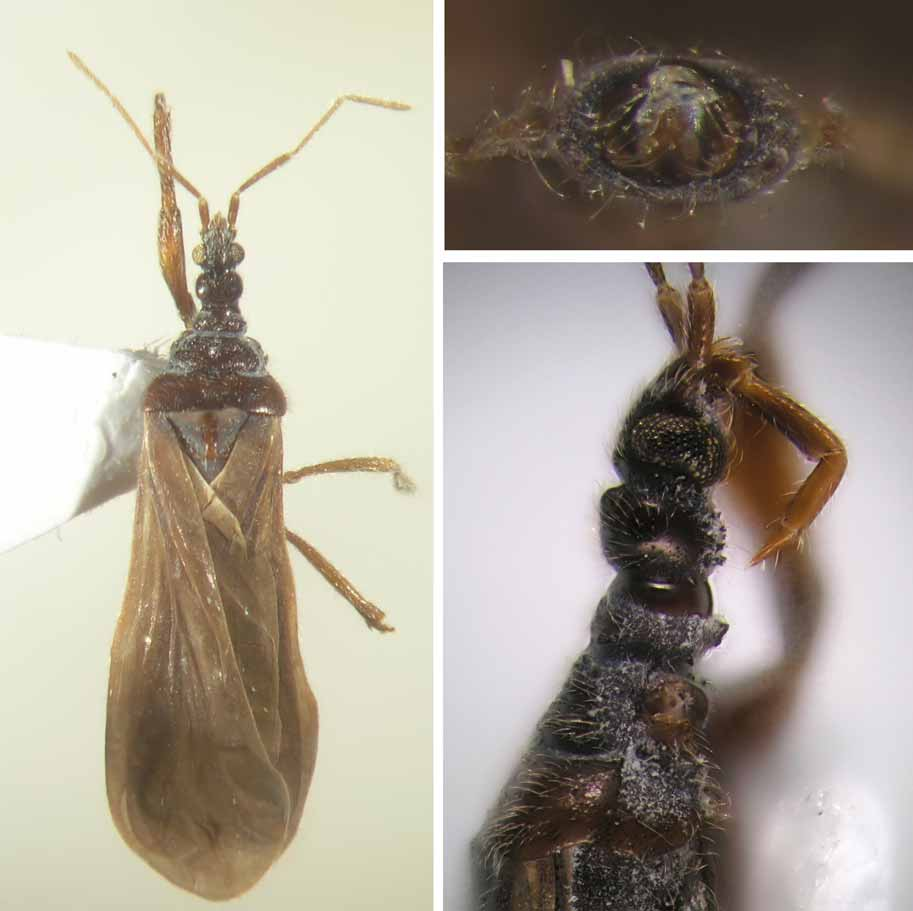
( Figs. 1–3 View FIGURES 1 – 3 , 9–13 View FIGURES 9 – 14. 9 – 13 , 16, 19, 22 View FIGURES 15 – 23 , 25, 28 View FIGURES 24 – 29. 24 – 26 )
Etymology: toponymical adjective derived from Sabah (terra typica).
Type material. Holotype, ɗ, ‘27a SABAH (Tambunan distr.): Crocker / Range, 1560–1650m á proximité du / col (route Kota Kinabalu-Tambunan), / forêt de Lithocarpus - Castanopsis , tamisage / de bois pourri, feuilles mortes et mousses / 16.v.1987, leg. D.Burckhardt et I.Löbl’. Specimen is cardmounted, right foreleg mounted separately. The same pin bears locality label and red label: ‘ HOLOTYPE / Phenicocleus sabahensis n. gen. & n. sp. / Štys & Baňař det. 2009’. Holotype is deposited in the collections of Museum of Natural History, Geneva.
Habitat. Collected in a humid mountain forest of Lithocarpus and Castanopsis .
Method of collecting. The specimen was collected by sieving rotten wood, moss, and dead leaves.
Total length: 5.2 mm. For other measurements see Table 1.
Distribution of setigerous tubercles on body: epicranium and the entire posterior lobe covered by numerous minute setigerous tubercles all over (rather inconspicuous marginally).
Lateroventral margin of buccular bridge (ventral view) with two large pointed tubercles directed towards the basis of the antennifer.
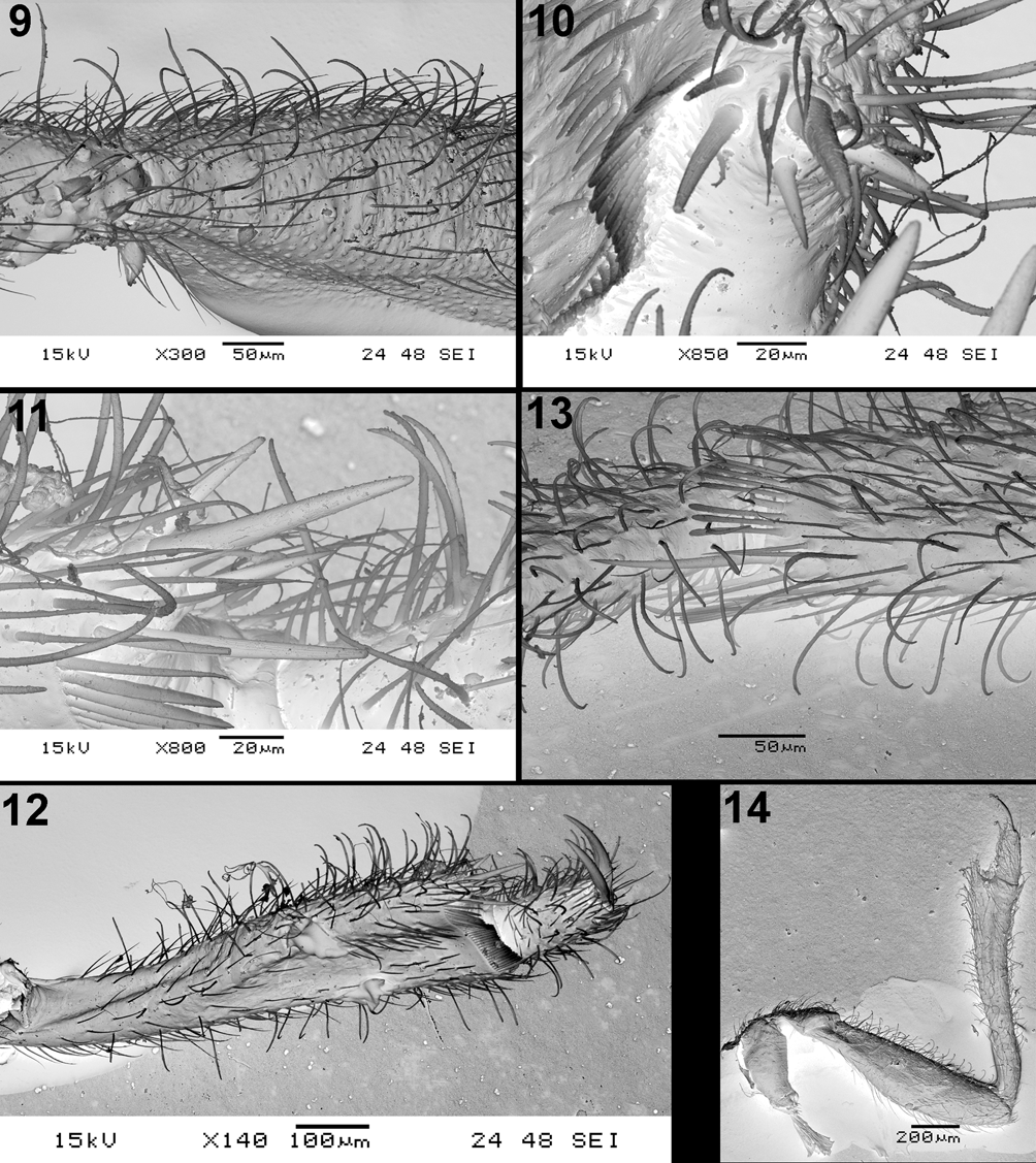
Distribution of setigerous tubercles on forelegs. Forecoxa on distal half of ventral face with irregularly distributed 10 sharp tubercles, foretrochanter on ventral face with 14 such tubercles, situated approximately in a curved double-row. Basal half of ventral face of forefemur with 9 setigerous tubercles ( Fig. 9 View FIGURES 9 – 14. 9 – 13 ), situated in irregular double-row. Foretibia and foretarsus without tubercles.
Coloration. Mostly yellowish brown to brown; head, antennal segment I, pronotum and ventral part of thorax strongly lustrous, brownish black; forewings brownish grey (both veins and membrane). Fore knees concolorous with other parts of forelegs.
Head. Cuticle on dorsum rugulose. Maximum width of eye 0.79 times width of dorsal synthlipsis, 1.38 times width of ventral synthlipsis. Posterior lobe laterally regularly rounded, widest in the middle, 1.29 times as wide as long, its median formed by broad, shallow, percurrent concavity. Ocelli large, situated sublaterally, interocellar distance 1.4 as long as shortest distance ocellus – eye. Antennal formula (longest segment first) III> IV, II, I.
Labium. Ratio length segment II: III 1.33, II: (III + IV combined) 0.84.
Pronotum. Collum rather flat, with no median, apparently protruding far laterad because of presence of protruding large lateral, subhorizontal, sharp setigerous tubercles.
Posctollar constriction deep. Midlobe lateral margins rounded, diverging posterad, not at all embraced by posterior lobe (not at all extending anterad); posterior margin moderately convex laterally and medially, the medial convexity obtusely subangular. Posterior lobe: lateral margins in anterior half moderately convex and diverging, in posterior half subparallel, nearly straight, posterolateral angles subrectangular; posterior margin straight in lateral sectors, broadly and shallowly, subangularly excised in mesal sector. Midlobe without median, with an extensive central fossette, hindlobe with a percurrent medial linear ridge (sic!) ( Fig. 16 View FIGURES 15 – 23 ). Midlobe – width: medial length 2.7; hindlobe – ditto 4.7.
Proepimeral lobes fully closing fore acetabula from behind. Prosupracoxale meeting mesally onto an euprosternal keel ( Fig. 19 View FIGURES 15 – 23 ). Posterior bars of eumesosternal apodemes separately rounded and surrounding posterior parts of sternum separately convex. Median wedge of eumetasternum strikingly narrow, terminating far from posterior margin of sternum, and anteriorly provided with transverse bars ( Fig. 22 View FIGURES 15 – 23 ).
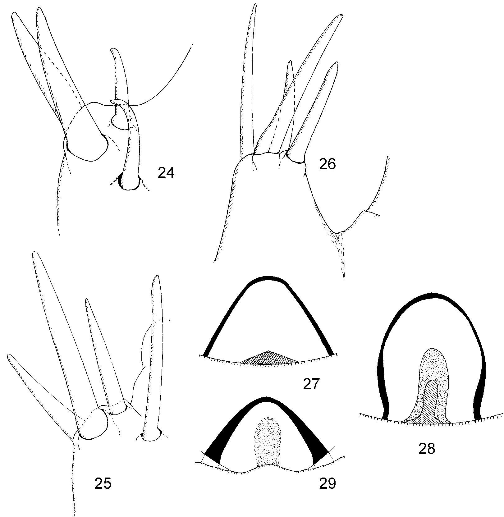
Foreleg. Posteroventral face of forecoxa, posteroventral face of trochanter and forefemur, with the exception of dorsal face with numerous, lens-like, nonsetigerous tubercles; for sharp setigerous tubercles see above. Foretrochanter robust, the same length as forecoxa. Forefemur three times as long as wide, proximal part slender, widening distad, maximum width in the middle of its length. Foretibia ( Fig. 12 View FIGURES 9 – 14. 9 – 13 ) 3.2 times as long as wide, without cuticular tubercles. Apex of foretibia protruding as a short process only. Apicitibial armature consists of four spiniform setae ( Figs. 10–11 View FIGURES 9 – 14. 9 – 13 , 25 View FIGURES 24 – 29. 24 – 26 ), three ventralmost in a short row and the shortest one dorsally, similar to P. minor . Bristle comb long, consisting of approximately 38–40 setae, two dorsalmost much stouter and longer. Foretarsus sligtly shorter than foretibia maximum width (ratio 0.8), posttarsus formed from two well developed claws, subequal in length. Tarsal armature similar to P. minor .
Guide ( Fig. 28 View FIGURES 24 – 29. 24 – 26 ) inversely U-shaped; its internal sclerite moderately long, tongue-shaped, strongly sclerotized, and surrounded by much larger, little sclerotized but similarly shaped structure resembling its hallo.
Associated enicocephalids in the sample. Holotype of a macropterous n. gen., n. sp. (female) to be described later.
Differential diagnosis. See the key and illustrations of diagnostic characters. This species in most character states (excepting shape of the guide) more similar to P. minor than to P. schwendingeri , but differing from the former by its larger size and less abundant, smaller, and more rounded setigerous tubercles.

No known copyright restrictions apply. See Agosti, D., Egloff, W., 2009. Taxonomic information exchange and copyright: the Plazi approach. BMC Research Notes 2009, 2:53 for further explanation.