
Phaenicocleus granulosus, Baňař, Petr & Štys, Pavel, 2011
|
publication ID |
https://doi.org/ 10.5281/zenodo.202491 |
|
DOI |
https://doi.org/10.5281/zenodo.6187666 |
|
persistent identifier |
https://treatment.plazi.org/id/83318788-FFF1-B318-BEBE-FF03B282228D |
|
treatment provided by |
Plazi |
|
scientific name |
Phaenicocleus granulosus |
| status |
sp. nov. |
Phaenicocleus granulosus View in CoL sp. n.
( Figs. 1–18 View FIGURES 1 – 6 View FIGURES 7 – 14 View FIGURES 15 – 21. 15 – 18 , 22–25 View FIGURES 22 – 25 )
Etymology. granulosus (lat.) = granulate, provided with granules.
Type material. Holotype, Ƥ, ‘27a SABAH (Tambunan distr.): Crocker / Range, 1560–1650m proximit du / col (route Kota Kinabalu-Tambunan), / forêt de Lithocarpus - Castanopsis , tamisage / de bois pourri, feuilles mortes et mousses / 16.v.1987, leg. D.Burckhardt et I.Löbl’. The specimen is mounted on triangular card, forelegs and right hindleg mounted separately. Claws of left foretarsus lost. The same pin bears locality label and red label: ‘HOLO- TYPE / Phaenicocleus granulosus sp. n. / Baňař & Štys det. 2011’. The holotype is deposited in the collections of Museum of Natural History, Geneva.
Habitat. Collected in a humid mountain forest of Lithocarpus and Castanopsis .
Method of collecting. The specimen was collected by sieving rotten wood, moss and dead leaves.
Description. S pecies-specific characters shown in Tab.1 form an integral part of the description and taken as a whole represent diagnosis and comparative diagnosis of the new species.
Measurements (in mm). Total body length —3.64. Head (without neck). Total length—0.49; posterior lobe, length—0.23, posterior lobe, width—0.30; distance of eye to apex of antennifer—0.12; diatone (maximum width across eyes)—0.31; dorsal synthlipsis (minimum interocular distance)—0.17; ventral synthlipsis (minimum interocular distance)—0.13; eye, length—0.10, eye, width—0.08; interocellar distance—0.14. Labium. Total length—0.69; segment I, length—0.10; segment II, length—0.29, segment II, maximum thickness—0.10; segment III, length—0.18, segment III, maximum thickness—0.07; segment IV, length—0.12. Antenna. Total length—1.40; segment I, length—0.22; segment II, length—0.41; segment III, length—0.37; segment IV, length—0.40, segment II, basal width—0.03, segment II, distal width—0.06. Pronotum. Total length (maximum)—0.72; collum, length (median)—0.13, maximum width—0.38; midlobe, length (median)—0.29, midlobe, width (maximum)—0.67; hindlobe, length (maximum)—0.24, hindlobe, length (mediane)—0.17, hindlobe, width (maximum)—0.76. Foreleg. Femur, length—0.70, femur, maximum width—0.28; tibia, length—0.62, tibia, maximum width—0.21; tarsus, length—0.16, tarsus, maximum width—0.08; anterior and posterior foreclaws, length—0.14. Forewing. Maximum length—2.29.
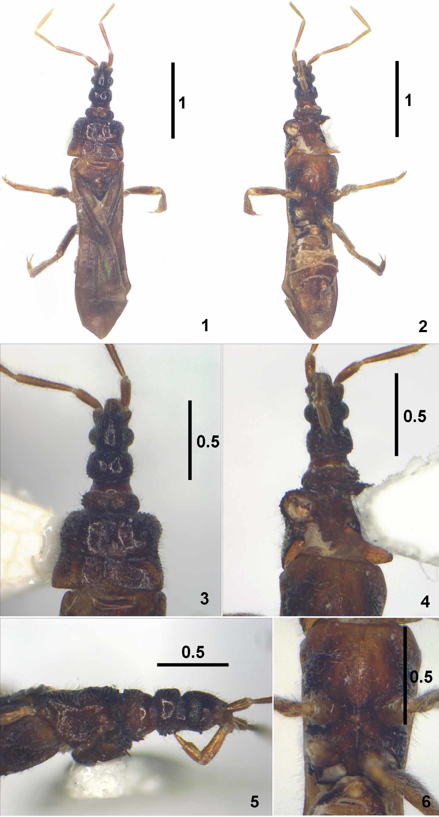
Coloration ( Figs. 1–6 View FIGURES 1 – 6 ). Essentially monochromatic, without patterns, uniformly lustrous (except for pronotal constrictions, wings and abdomen). Head dark piceous, nearly blackish. Antennae and labium dark yellowish (except scape - dark piceous, and pedicel - graduately piceous to light brown distad). Thorax brown, midlobe of pronotum darker (humeral angles blackish) and mesoscutellum paler than the rest. Legs light brown, femora piceous; ‘knees’ slightly paler. Forewings and abdomen pale brown.
Vestiture whitish to yellowish, straight to slightly curved, semierect to strongly diagonal, moderately long and dense. Head covered with semierect setae (= macrotrochia), on all faces directed anteriad, denser on anterior lobe. Antennae with semierect, regularly distributed setae of identical length, apex of the second segment and segments III and IV also with sparser and longer, more outstanding setae. Pronotum with semierect, sparsely distributed setae, denser on dorsal and lateral parts of collum. Venter of abdomen with short, appressed setae, becoming longer caudad. Forewing membrane bare, veins with sparse, short setae, only costal margin with long, outstanding setae, directed caudad.
Foreleg. Dorsal and posterior faces of coxa, trochanter and femur covered with semierect setae. Distal half of ventral face of coxa, ventral face of trochanter and proximal two thirds of ventral face of femur with long, erect setae arising from sharp setiferous tubercles, mixed with shorter semierect setae and trichobothrium-like setae (= long, thin, erect or semierect setae, of probably constant position and resembling trichobothria). Anterior face of coxa, trochanter and femur nearly bare, with a few short, semierect setae. Tibia covered with long, semierect setae (sparsely on anterior face), mixed with numerous trichobothrium-like setae (20–25 on ventral, dorsal and posterior faces).
Midlegs and hindlegs. Trochanters and femora densely covered with simple, oblique and moderately long setae interspersed with a few semierect trichobothria-like setae on ventral face of trochanters, ventral proximal part and distal dorsal and ventral parts of femora. Midtibiae and hindtibiae with dense cover of long and dense oblique setae on all faces, particularly dense and forming a continuous hairy fringe on ventral faces; some of the oblique ventral seate longer than others and representing trichobothrium-like setae; the sparse erect trichobothrium-like setae present on both dorsal and ventral faces but particularly common on dorsal face of hind femur. Also midtarsus and hindtarsus fully covered with short oblique setae, with a few trichobothrium-like setae on venter of segment 1, dorsum of 2 and ventral apex of 2.
Distribution of setigerous tubercles on body. Ventral face of anterior lobe of head with 4 sharp, strongly outstanding tubercles, ventral face of posterior lobe of head approximately with 15 tubercles. Dorsolateral and ventral faces of collum, dorsolateral faces of both midlobe and hindlobe of pronotum, ‘proepimeral lobes’ and mesepisternal and metepisternal parts of thorax with numerous, large tubercles.
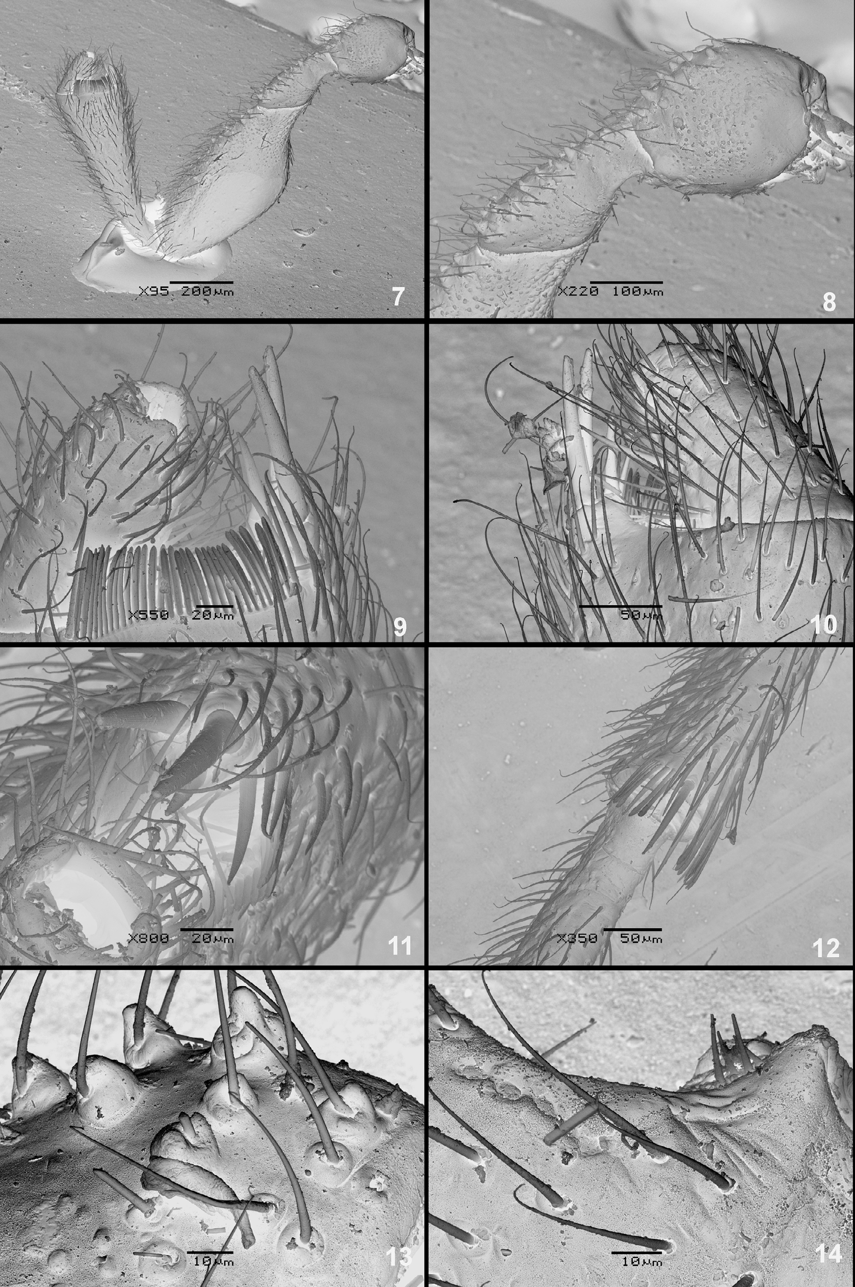
Distribution of setigerous tubercles on forelegs. Forecoxa anteroventrally with about 14 tubercles ( Figs. 8, 13 View FIGURES 7 – 14 ), foretrochanter with about 24 tubercles regularly distributed on ventral face ( Fig. 8 View FIGURES 7 – 14 ) and forefemur with 10–12 tubercles in 2 irregular rows on ventral face. The sharp setigerous tubercles often intermixed with or replaced by much lower, semispherical granules, not bearing setae and not prominent ( Fig. 7 View FIGURES 7 – 14 ). Foretibia without tubercles.
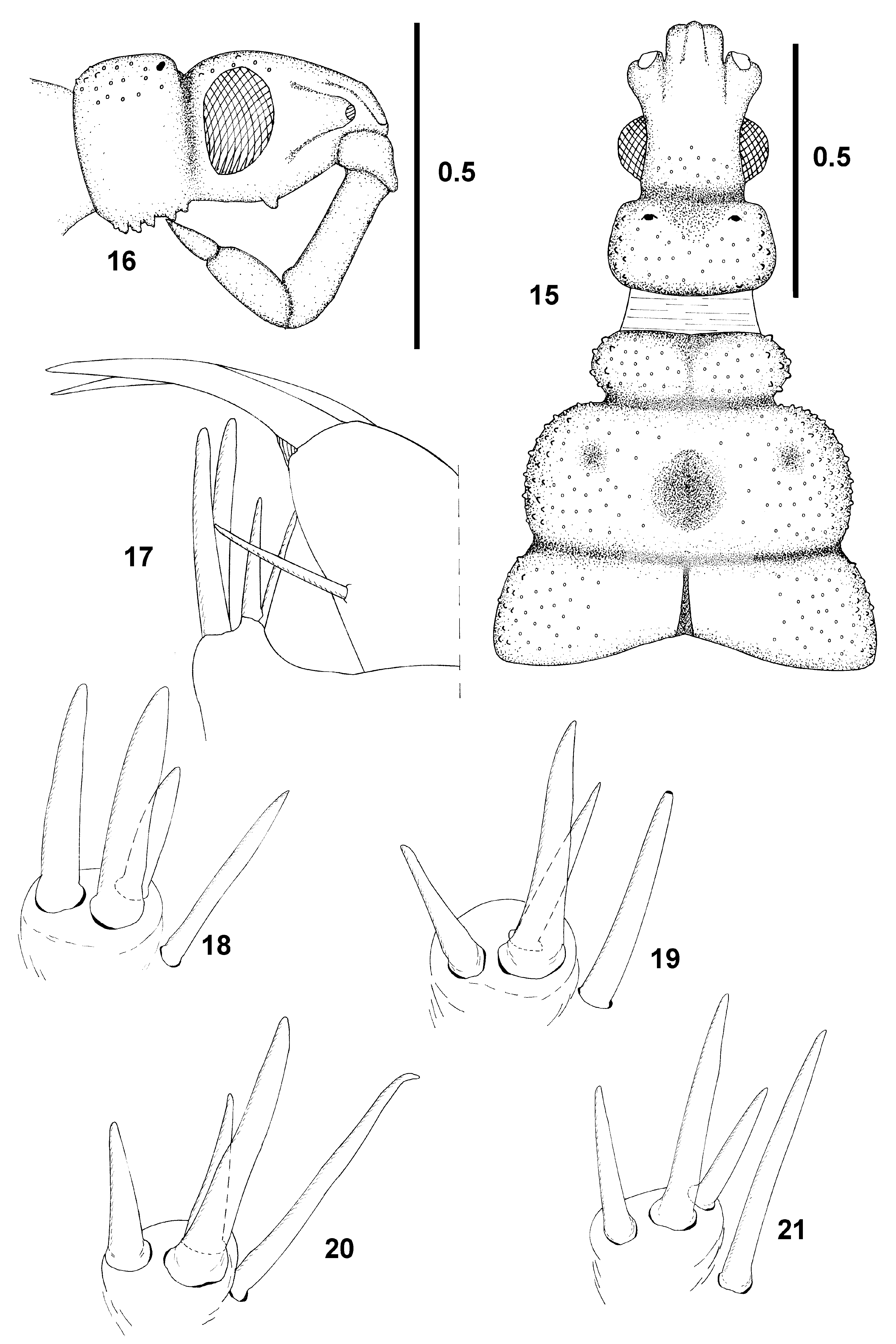
Head ( Figs. 3–5 View FIGURES 1 – 6 , 15–16 View FIGURES 15 – 21. 15 – 18 ). Cuticle of dorsum lustrous, posterolateral parts of posterior lobe of head with small, non-setigerous tubercles. Anterior lobe short, antenniferous tubercles moderately diverging immediately in front of eyes, no part of genae visible in dorsal view; epicranium strongly convex. Maximum width of eye 0.47 times width of dorsal synthlipsis, 0.61 times width of ventral synthlipsis. Eyes short and high, in lateral view narrowly kidneyshaped, distinctly closer to dorsal than to ventral margin of head; close to a strikingly deep postocular constriction (short tempora parallel-sided). Facets numerous, separately convex. Posterior lobe much shorter than anterior lobe, strikingly transverse, widest subproximally, 1.3 times as wide as long, subrectangular (though with anterolateral angles and particularly the posterolateral ones broadly rounded), lateral margins otherwise nearly straight, slightly diverging caudad, surface of the lobe with an extensive, shallow, anteromedial subtriangular arcuate depression, without a structurally marked median. Ocelli very small, interocellar distance more than twice (2.3) as long as the shortest distance ocellus-eye. Dorsum nearly flat. Posterior lobe sharply separated from the neck, ratio of its length to maximum width 0.77.
Antenna. Pedicel terete, gradually incrassate distad, segments 2–4 subequal in length; segments 3 and 4 long, not markedly thinner than proximal part of pedicel. Antennal formula (longest segment first): II: IV: III: I.
Labium. Thin, geniculate, directed posterad, apex reaching basis of posterior lobe of head. Labial formula: II: III: IV: I, typical for genus; segment 2 much longer than segment 3 ( Figs. 5 View FIGURES 1 – 6 , 16 View FIGURES 15 – 21. 15 – 18 ); ratio length segment II: III 1.61, II: (III+IV combined) 0.96. In lateral view, segment II parallel-sided, segment III slightly widening distad.
Pronotum ( Figs. 1, 3, 5 View FIGURES 1 – 6 , 15 View FIGURES 15 – 21. 15 – 18 , 22 View FIGURES 22 – 25 ). Cuticle finely rugose, all faces with minute, non-setigerous tubercles; sharp setigerous tubercles as above. Collum short, ring-shaped, without subventral tubercles; subdivided in precollum and collum (distinct in alcohol-preserved specimen only), the median vaguely and broadly concave; constriction separating collum from the midlobe deep, broad, matt and finely sculptured. Midlobe strongly transverse, widest behind the middle, lateral margins moderately rounded. Disc of the midlobe with a broad, shallow, vaguely subcircular depression not interrupting its posterior margin and a pair of very shallow lateral pits. Lateral sectors of the constriction between mid- and hindlobe deep and pruinose, medial sector shallow and shiny, nearly obliterate.
Ratio midlobe width to medial length 2.3, widest in its posterior 2/3. Ratio hindlobe width to medial length 4.5, ratio hindlobe width to maximum length 3.17. Hindlobe longer than collum, ample, rather short, widest posteriorly, less than 1.2 times as wide than midlobe, its lateral sides little divergent, posterior margin broadly and shallowly convex, posterolateral angles somewhat obtusangular, their apices rounded, not prominent. Median with a narrow, longitudinal ridge.
Mesoscutellum triangular, obtusely mucronate, with an anchor-shaped elevation.
Ventral and lateral parts of thorax 1(ventral parts described as seen in strictly ventral view).
Prothorax. Notopleural sulcus distinct over the bases of supracoxalia, but pleuron externally not developed except for supracoxalia.
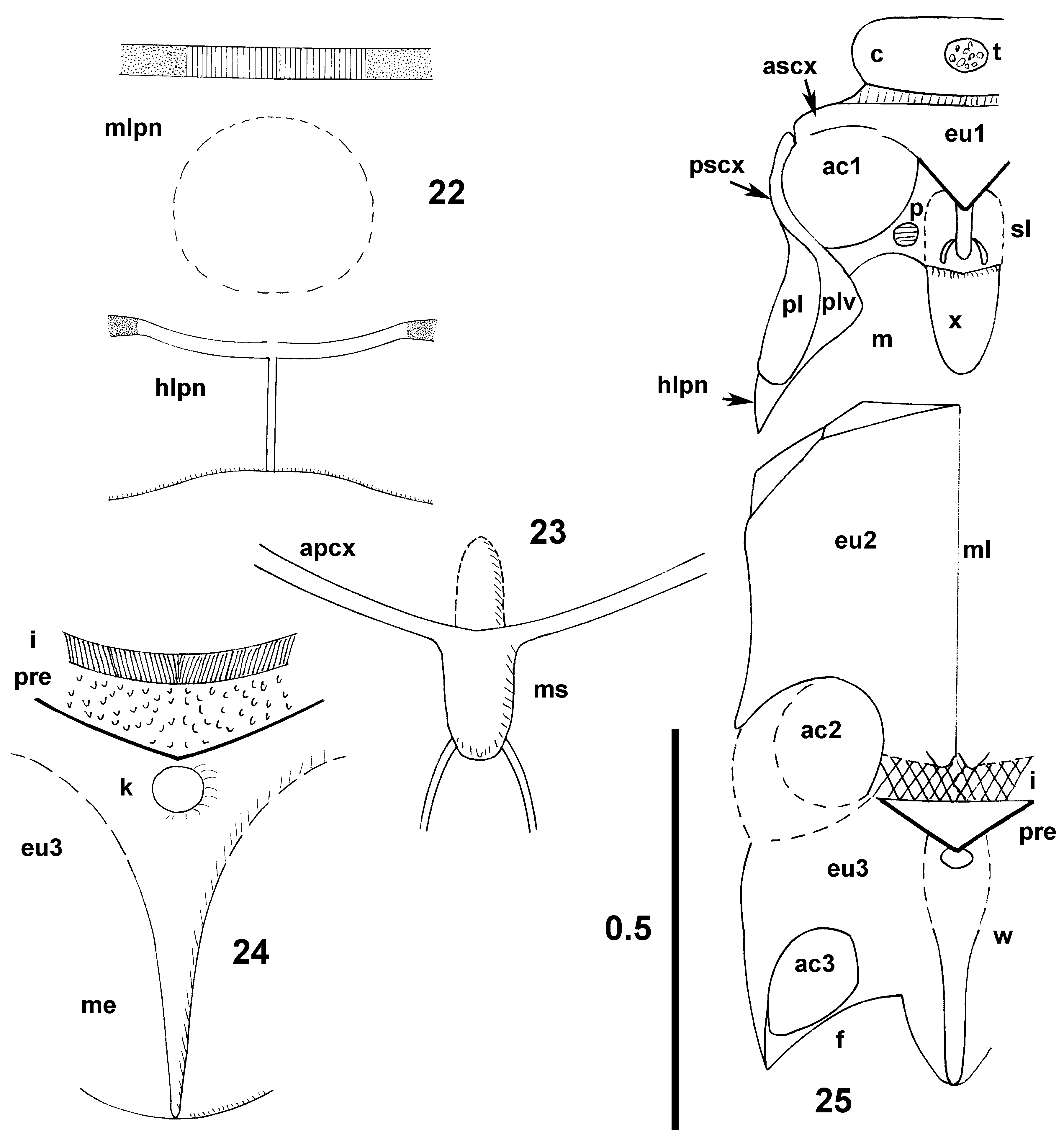
Collum ring-shaped, convex, with a complex, irregular, sharply demarcated medioventral protuberance; collareusternal constriction deep, laterally with a paired fosettes. Eusternum basically pre-acetabular, subtriangular, moderately convex, its median indistinctly keeled, the keel distally forming a distinct, sharply delimited mucronate ridge situated already on sternellum. Anterior borders of eusternum formed by postcollar constriction, posterior ones coinciding with jointly fused ventral margins of anterior supracoxalia ( Figs. 23, 25 View FIGURES 22 – 25 ), lateral borders not developed owing to fusion of eusternum with anterior supracoxale.
1. The following description of lateral and ventral parts of the thorax is provided for diagnostic reasons (partly utilized by Štys & Baňař (2009) for diagnosis of Phaenicocleus View in CoL species). The characters described are probably useful at clade level as well but they have been so far unexploited and are unknown in most genera. The terminology applied here is tentative.
Postsupracoxale and ‘proepimeral lobes’ modal, the latter reaching to 2/3 of posterior margin of fore acetabulum.
Sternellum longitudinally subdivided in (a) ‘eusternellum’, situated between the pro-acetabula and directed posterodorsally, and (b) ‘proxiphus’, situated posteriad to proacetabula and directed horizontally to posteroventrally. Eusternellum subdivided by longitudinal sulci in a convex mesal part with a strong median ridge (continuing from the eusternum) reaching two thirds of its length and lateral parts contacting inner acetabular frames. Prosternal pits situated just at the lateral meeting place of eusternellum and proxiphus. Proxiphus flat, tongue-shaped, strikingly long, with straight lateral margins converging caudad, and the apex rounded.
Pterothorax ( Figs. 6 View FIGURES 1 – 6 , 24, 25 View FIGURES 22 – 25 ). Mesosternum huge, strongly convex, not subdivided, with a percurrent, linear, impressed median; apex slightly produced, truncate; laterally forming long and broad mesoprecoxale. Mesosternum and metasternum (none subdvided in eusternum and sternellum) fused, the apparently original intersegmental area relatively large, melanized, slightly compressed, with no median structure. Anterior part of meteusternum (? presternite) small, continuously covered by minute granules, laterally moderately compressed, without structurally distinct median; caudally delimited by a black arcus associated with meteusternal wedge. Metasternum laterally merging with anterior metapresupracoxale, the latter continuous with metaprecoxal bridge. Metasternum strongly compressed, with a keel-like median wedge ( Figs. 24-25 View FIGURES 22 – 25 ), the keel being proximally somewhat rounded and wide (and provided with an anteromedial knob), distally strikingly sharp; reaching the posterior margin of metasternum; the apex of metasternum rounded, produced between metacetabula, contacting mesal parts of circumcoxal frames.
Foreleg ( Figs. 7–11, 13–14 View FIGURES 7 – 14 , 17–18 View FIGURES 15 – 21. 15 – 18 ) robust; anterior and ventral faces of coxa, trochanter and femur with numerous, non-setigerous, lens-like cuticular granules. Trochanter simple, long and slender. Femur in the middle of dorsal face extremely gibbose, twice as wide as basal width, ratio of its length to maximum width 2.5. Tibia without cuticular granules, strongly and regularly widening distad, ratio length to maximum width 2.9. Apex of foretibia with a small process bearing three smooth spiniform setae; the two distal ones subequal in length, the proximal seta shorter ( Figs. 17–18 View FIGURES 15 – 21. 15 – 18 ). One closely situated, still more proximal seta inserted outside the process ( Fig. 18 View FIGURES 15 – 21. 15 – 18 ) much stronger than the rest and possibly belonging to the specialized apicitibial complex. Tarsus broad, twice longer than wide, with one long, unpaired spiniform seta ( Figs. 10–11 View FIGURES 7 – 14 , 17 View FIGURES 15 – 21. 15 – 18 ). The two claws of the same length and shape.
Midleg and hindleg rather short and stout, from trochanter up to tarsus moderately compressed in anteroventral plane. Ventral outline of trochanter convex, exceeding that of femur. Femora moderately incrassate, slightly compressed. Tibiae strikingly compressed, not stick-shaped but widening distad, particularly on midlegs. Midleg and hindleg combs very short, paired, situated on anteroventral and posteroventral apicitibial edges respectively, separated by strong, adjacent spiniform seta ( Fig. 12 View FIGURES 7 – 14 ), only slightly longer than 6–7 spatulate comb-forming setae. Tarsus thick, 2-segmented, first segment very short, well distinct under SEM only. Claws gently curved, isomorphic, the anterior one slightly thinner.
Forewing ( Fig. 1 View FIGURES 1 – 6 ). Considerably exceeding apex of abdomen. Venation of forewing (partly seen in Fig. 1 View FIGURES 1 – 6 ): basal cell absent, large discal cell closed, costal fracture absent. Pterostigma not developed. Discal cell long and strikingly narrow, particularly in its proximal part; crossveins entering the cell widely distant mutually (r-m situated much more distally than cu-an). Claval veins (terminology follows Štys & Baňař, 2008): AA1+2 complete, AA3+4 distinct, sclerotized and convex only up to the level of forewing overlap, its distal sector indistinct, consequently the closed claval cell not developed; the basal space between AA shortly fully sclerotized, marginal AP distinct up to about the level of termination of the intervenal sclerotization.
Abdomen. Venter (proximal ventrites 1–5 strikingly complex). Ventrites 6 and 7 with small and large lateral sclerites, respectively, and correspondingly extensive or narrow mesal membranous part. Ventrite 8 fully and strongly sclerotized, strongly vaulted, and forming a large subgenital plate, narrowing caudad but with broadly rounded posterior margin (subgenital plate incompletely bissected by a linear, shallow median, visible in alcoholpreserved specimen only). Ventrite 9 simple, very short, transverse, sclerotized, fringing the apex of subgenital plate.
Key to the genera of Enicocephalinae with closed discal cell and absent basal cell in forewing (M=male; F=female)
As indicated, some genera are known from one sex only, and the unknown sex may not fit the key. The conspicuous autapomorphies and synapomorphies are indicated by ‘(!)’. Body length is to be understood as from the apex of anteclypeus to apex of forewings in resting position.
1 Posterior lobe of pronotum minute, its posterolateral angles acute, often projecting laterad (!). (Body and legs without a striking array of setigerous tubercles. Labial segment II always much shorter than III). American genera.......................... 2
- Posterior lobe of pronotum small to large, its posterolateral angles rounded, if subacute, then projecting posteriad and body and legs with many long setigerous tubercles. Eastern Hemisphere, including Oceania...................................... 3
2 Foretarsus lacking spiniform setae (!). Collum of pronotum long, subconical (!). Body length 2.9–4.1 mm. California...................................................................... Urnacephala Wygodzinsky & Schmidt, 1991. M, (? F)
- Foretarsus with 4 spiniform setae. Collum of pronotum simply transverse. Body length 3.2–3.5 mm. Texas and Mexico (Baja California)................................................... Lysenicocephalus Wygodzinsky & Schmidt, 1991, M, (? F)
3 Posterior margin of pronotum nearly straight. Postocular lobe of head strikingly transverse, subrectangular. Body length over 3.5 mm. Madagascar.............................................................. (Shenchiella Villiers, 1969), (?M), F Notes. Forewing venation unknown, only one female deprived of wings available. Possibly belonging to another group of genera. The name Shenchiella was formed by Villiers (1969; etymology not provided) as an obvious anagram of Henschiella View in CoL . The name was inadvertently misspelled Schenchiella by Štys as incorrect subsequent spellings (1978: 248; 1992: 349, 352, correctly four times in the key).
- Posterior margin of pronotum moderately to deeply concave, or at least medially emarginate. If posterior lobe of head transverse, then its lateral sides rounded, or straight and diverging caudad.....................................................4
4 Labial segment II as long or longer than segment III (!). Claval vein AA3+4 distinct to half its length only, consequently the claval cell indistinct (!). Apicitibial armature of foreleg of 3+1 spiniform setae (!). Foretarsal armature formed by 1 unpaired spiniform seta (!). Head, thorax and foreleg with setigerous tubercles, the latter provided with minute semispherical tubercles as well. Antennal segment II markedly incrassate distad. (Head short, gena absent, tempora very short. Males of the usual enicocephaline facies, female elongate, flat, strikingly covered by setigerous tubercles, with midlobe of pronotum very short, but widest in the middle, with lateral sides rounded. Prothoracic acetabula open to closed; none of the other autapomorphies of Usingeriella View in CoL and Nesenicocephalus View in CoL present.) Body length 3.6–5.2 mm. Sabah............................. Phaenicocleus Štys & Baňař, 2009 View in CoL , M, F
- Labial segment II distinctly shorter than III. Claval vein AA3+4 and claval cell distinct. Apicitibial armature of fore leg of 7 spiniform (or spiniform and spatulate) setae. Armature of foretarsus of 2+2 spiniform (or spiniform and spatulate) setae. Setigerous tubercles present to absent. Antennal segment II not markedly incrassate distad........................................ 5
5 Setigerous tubercles on head, thorax and forelegs dense, large and high, markedly affecting outlines of these body parts. Body flat, nearly parallel-sided, elongate. Fore acetabula posteriorly closed by proepimeral lobes (!). Head strikingly elongate (!), genae nearly as long as antennifers in dorsal view, tempora nearly as long as eyes. Posterior lobe of head strikingly elongate (!), widest in the middle. Midlobe of pronotum subrectangular, equally wide throughout, lateral margins straight, slightly indented in the middle. Hindlobe of pronotum minute, shorter than the collum (!). Body length 4.5 mm. Australia................................................................................................ Usingeriella Wygodzinsky, 1950 View in CoL (?M), F Note. Usingeriella View in CoL and a related undescribed genus from Australia include a number of undescribed species (PŠ, unpubl.). Not all of them would fit all the above key theses but none of them would fit those introduced under 4a, 5b, 6, 7.
- Setigerous tubercles on head, thorax and forelegs absent, or minute and inconspicuous. Body of modal shape; if flat, then not markedly elongate. Fore acetabula posteriorly open. Gena and tempora very short to absent. Hindlobe of head transverse, widest proximally. Midlobe of pronotum not subrectangular, its sides laterally rounded, without indentations. Hindlobe of pronotum longer than collum. Less than 3 mm long species...................................................................6
6 Midlobe of pronotum without a structurally distinct median (!). Ambient vein along the posterior margin of forewing (ca in the sector anteriad and posteriad to cu-an) truly marginal. (!) (Females caducous!). Gena absent in dorsal view. Body length 2.0– 2.9 mm. Hawaii Is, Caroline Is, Philippine Is ( Taiwan, New Guinea and Australia - PB, PŠ, unpubl.).................................................................................................... Nesenicocephalus Usinger, 1939 View in CoL M, F
- Midlobe of pronotum with a structurally distinct median. Ambient vein along the posterior margin of forewing (ca in the sector anteriad and posteriad to cu-an) distant from wing margin. (Females not caducous or unknown). Gena short but distinctly visible in dorsal view. Body length 1.3–2.8 mm. Madagascar............................... Pseudohenschiella Villiers, 1958 M, F
No known copyright restrictions apply. See Agosti, D., Egloff, W., 2009. Taxonomic information exchange and copyright: the Plazi approach. BMC Research Notes 2009, 2:53 for further explanation.