
Pasipha mbya, Negrete, Lisandro & Brusa, Francisco, 2016
|
publication ID |
https://doi.org/ 10.11646/zootaxa.4137.2.2 |
|
publication LSID |
lsid:zoobank.org:pub:94661073-A6EB-4134-B709-BF4ED7AF50EA |
|
DOI |
https://doi.org/10.5281/zenodo.6070969 |
|
persistent identifier |
https://treatment.plazi.org/id/03F24A30-FFC7-FF82-98FA-EF19092FFD30 |
|
treatment provided by |
Plazi |
|
scientific name |
Pasipha mbya |
| status |
sp. nov. |
Pasipha mbya View in CoL sp. nov.
( Figs. 10–14 View FIGURE 10 View FIGURE 11 View FIGURE 12 View FIGURE 13 View FIGURE 14 )
Pasipha sp. 1: Negrete et al. 2014a
Type material. Holotype. MLP-He 6476. Urugua-í Wildlife Reserve, Misiones Province, Argentina, 21.VIII.2009, L. Negrete, coll.; cephalic region and anterior region at level of ovaries: transverse sections on 42 slides (8 µm); pre-pharyngeal region: transverse sections on 12 slides (8 µm); pharynx and copulatory apparatus: sagittal sections on 30 slides (8 µm).
Paratype. MLP-He 6978. Iguazú National Park, Misiones Province, Argentina, 13.IV.2013, L. Negrete coll.; cephalic region: transverse sections on 36 slides (6–8 µm); anterior region at level of ovaries: sagittal sections on 28 slides (6–8 µm); pre-pharyngeal region: transverse sections on 5 slides (6 µm); pharynx and copulatory apparatus: sagittal sections on 35 slides (7 µm).
Diagnosis. Dorsum dark grey with a yellow median longitudinal band, and thin black para-median stripes. Eyes dorsal with clear halos. Glandular margin present. Pharynx cylindrical. Prostatic vesicle extrabulbar, globose, with folded walls, and proximally forked. Male atrium richly folded, with small folds in its proximal part, 2.5–4 times longer than the female atrium. Ovaries anterior to the anteriormost testes. Ovovitelline ducts ventral to the female atrium, joining behind it. Common glandular ovovitelline duct vertical, and female genital canal almost horizontal. Female atrium without folded walls.
Etymology. The specific name is devoted to the Mbyá indigenous community, member of the Guaraní Tribe, native people of the rainforests of Misiones Province, Argentina.
Type locality. Urugua-í Wildlife Reserve (25° 59’ S, 54° 05’ W), Misiones Province, Argentina.
External morphology. The body is elongate, with parallel margins. The anterior tip is slightly rounded and the posterior end is sharply pointed. The dorsal surface is dark grey with a longitudinal yellow median band which arises a few millimetres from the tip and extends along the body ( Fig. 10 View FIGURE 10 ). The yellow band, the width of which is approximately one quarter of the body width; it is slightly mottled with black spots, mostly concentrated at the level of pharynx and copulatory apparatus. Thin black para-median stripes border the median band ( Figs. 10 View FIGURE 10 , 11 View FIGURE 11 ).
After fixation, the ground colour becomes paler, and para-median stripes are no longer distinguishable. The ventral surface is light grey. The eyes are initially distributed in a single row around the anterior tip. Posteriorly, the eyes are located uniserially on the body margins along 2 mm and biserially for 2 mm. Then they spread dorsally, forming 4–5 irregular rows on each side of the body. The dorsal eyes are surrounded by clear halos. At the level of the pharynx, the eyes are less numerous and at the level of the copulatory apparatus they are again uniserial and scattered on the body margins, reaching the posterior end ( Fig. 11 View FIGURE 11 ). The holotype was 40 mm long and ~2.5 mm wide when crawling. After fixation, body length of study specimens ranged from 28 mm (holotype) to 46.2 mm, maximum width was 3–3.2 mm, and maximum height 1.3–1.5 mm. The mouth and the gonopore were located at a distance of 70–72% and 83–84% from the anterior tip, respectively ( Table 4 View TABLE 4 ).
Internal morphology. Epidermis, secretions and musculature in the cephalic region. Dorsal epidermis (20–25 µm height) receives abundant rhabditogen secretion and amorphous erythrophil secretion. Scarce fine granular erythrophil and cyanophil secretions discharge through dorsal epidermis, although they are more abundant on the body margins ( Fig. 12 View FIGURE 12 A). Ventral epidermis (25–30 µm height) presents small dermal rhabdites and receives abundant fine granular erythrophil secretion, and scarce amorphous erythrophil and fine granular cyanophil secretions. A single row of sensory pits (20–25 µm deep) surrounds the cephalic region and they spread toward body margins up to 1.5 mm from the anterior tip.
The cutaneous musculature, composed of the same layers as the pre-pharyngeal region (see below), is slightly thicker than at the pre-pharyngeal level (14–16% of the body height). The thickness of the parenchymatic musculature is also larger than in the pre-pharyngeal region (9–14% of the body height). In the anterior body region, there is a subneural muscular transverse layer (15–25 µm thick) situated below the nervous plate, which arises very close to the anterior tip and extends for ~ 2 mm ( Fig. 12 View FIGURE 12 A). There is no musculo-glandular specialization in the cephalic region.
Epidermis, secretions and musculature in the pre-pharyngeal region. Dorsal epidermis (20–35 µm high) and body margins contain abundant rhabdites ( Fig. 12 View FIGURE 12 B, C). Rhabditogenic glands are situated in the parenchyma below the cutaneous musculature. There are glands with amorphous erythrophil and scarce cyanophil secretions which discharge through the dorsal epidermis. Ventrally, the epidermis (25–35 µm high) is ciliated, forming a wide creeping sole (~90% of body width). In the ventral epidermis, amorphous cyanophil glandular secretion is more abundant that on the dorsal surface and there is scarce amorphous erythropil secretion, with small dermal rhabdites occupying the apex of the epidermal cells. The glandular margin consists of abundant fine granular erythrophil and cyanophil secretions ( Fig. 12 View FIGURE 12 D).
The cutaneous musculature consists of an external subepithelial circular layer, followed by a diagonal layer, and an internal longitudinal layer arranged in bundles, which is thicker ventrally than dorsally ( Table 5 View TABLE 5 ). CMI ranges from 13% to 14%. Parenchymatic musculature is arranged in three layers: a dorsal layer with decussate fibres (35 µm thick), located below the cutaneous dorsal longitudinal bundles, a supra-intestinal layer and a subintestinal transverse layer (7.5–10 µm thick and 25–35 µm thick, respectively). PMI ranges from 6% to 9% ( Table 5 View TABLE 5 ). Dorso-ventral fibres are intermingled among the intestinal branches.
Digestive system. The pharynx (0.95–1.9 mm in length, about 3–4% of body length) is cylindrical, with the mouth opening into the distal portion of the pharyngeal pouch (1–2.5 mm in length) ( Fig. 12 View FIGURE 12 E). The epithelial lining of the outer surface of the pharynx is cuboidal and ciliated, followed by a longitudinal subepithelial muscle layer (5–10 µm thick) and a subjacent circular layer (20–25 µm thick). The epithelium of the pharyngeal lumen is columnar and ciliated. The inner pharyngeal musculature consists of a circular layer (15–35 µm thick) followed by a longitudinal layer (10–15 µm thick). Fine granular erythrophil and cyanophil secretions from cell glands located in the surrounding parenchyma before the pharynx discharge through the pharyngeal epithelium. Amorphous erythrophil and cyanophil secretions are present in less quantity, the cell bodies of which are subepithelial. Oesophagus (350–400 µm in length) is present ( Fig. 12 View FIGURE 12 E). The oesophagus: pharynx ratio ranges from 21% to 37%.
Male reproductive system. The oval testes are arranged in two or three irregular rows on each side of the body.
They are located dorsally to the intestine, beneath the supra-intestinal parenchymatic muscle layer. The ratio of the height of the testes to the height of body ranges from 16% to 26%. The testes are located at a distance between 24% and 63–66% ( Table 6 View TABLE 6 ) of the body length from the anterior end. The sperm ducts are located beneath the subintestinal parenchymatic muscle layer, and medial to the ovovitelline ducts in the pre-pharyngeal region ( Fig. 12 View FIGURE 12 C). Behind the pharynx, the sperm ducts follow their lateral course, ventrally to the prostatic vesicle, reaching the vicinity of the common muscle coat of the male atrium. Then they bend antero-dorsally and run to the sagittal plane, opening into the bifurcated portions of the proximal prostatic vesicle ( Fig. 13 View FIGURE 13 ). At this point, the sperm ducts are widened and their lumen full of spermatozoa. The extrabulbar prostatic vesicle, located immediately behind the pharynx, is globose with irregular lumen ( Figs. 13 View FIGURE 13 , 14 View FIGURE 14 A, B). It consists of two morphologically and histologically distinct regions (see below), namely: a proximal region, which is proximally forked, and a distal region, which communicates with the ejaculatory duct exactly at the boundary of the common muscle coat ( Figs. 13 View FIGURE 13 , 14 View FIGURE 14 A, B). The ejaculatory duct has an ascending course and is sinuous distally, opening into the bottom of the male atrium ( Figs. 13 View FIGURE 13 , 14 View FIGURE 14 C). The richly folded male atrium is 2.5 to 4 times longer than the female atrium. Their proximal part is characterized by small folds, while the rest of the atrial cavity exhibits larger folds ( Figs. 13 View FIGURE 13 , 14 View FIGURE 14 A, B).
The prostatic vesicle is lined with a ciliated columnar epithelium. The proximal part is provided with abundant fine granular erythrophil secretion, whereas the distal part receives abundant coarse granular erythrophil secretion ( Fig. 14 View FIGURE 14 A, B). Cell bodies of glands opening into both regions are located in the surrounding parenchyma ( Fig. 12 View FIGURE 12 E). The thick muscle coat which envelops the prostatic vesicle consists of circular, longitudinal and oblique intermingled fibres (70–90 µm thick). The ejaculatory duct presents a ciliated columnar epithelium, which receives in its distal course erythrophil amorphous secretion ( Fig. 14 View FIGURE 14 C). The lining epithelium of the most proximal region of the male atrium is columnar and ciliated, and receives fine granular erythrophil and cyanophil secretions. The remaining walls of the male atrium are lined by a non-ciliated, cuboidal to columnar epithelium. Excepting the most proximal portion, the remaining proximal half of the male atrium receives only abundant fine granular erythrophil secretion. The epithelium of the distal half is provided with abundant fine granular and amorphous erythrophil secretions, and fine granular cyanophil secretion which is more abundant at the level of the dorsal fold. The muscularis of the male atrium consists of a subepithelial circular layer (5–10 µm thick) followed by a longitudinal layer (15–30 µm thick). The common muscle coat consists dorsally of intermingled longitudinal and oblique fibres (40–60 µm thick), and ventrally of longitudinal fibres (25 µm thick). The male atrium is separated from the female atrium by a dorsal fold, so that the lumen between both atria is narrow ( Figs. 13 View FIGURE 13 , 14 View FIGURE 14 A).
Female reproductive system. The ovaries (250–300 µm in length and 250 µm high, in the holotype) are located at a distance of 22% of the body length from the anterior end ( Table 6 View TABLE 6 ). They are located between the sub-intestinal parenchymatic muscle layer and the nervous plate ( Fig. 12 View FIGURE 12 B). The ovovitelline ducts emerge from the posterodorsal region of the ovaries, with spermatozoa in their lumen. Behind the gonopore, the ovovitelline ducts ascend slightly, run to the sagittal plane and open into a vertical common glandular ovovitelline duct located behind the female atrium ( Figs. 13 View FIGURE 13 , 14 View FIGURE 14 A, D). The female genital canal runs forward almost horizontally to open into the female atrium, with poorly folded walls ( Figs. 13 View FIGURE 13 , 14 View FIGURE 14 A, D).
The epithelium of the ovovitelline ducts is ciliated and cuboidal, surrounded by a thin longitudinal muscle layer (5 µm thick). The common glandular ovovitelline duct is lined by a ciliated columnar epithelium followed by a subjacent circular muscle layer (10–15 µm thick). Both the distal portions of the ovovitelline ducts and the common glandular ovovitelline duct receive abundant secretion from shell glands ( Figs. 13 View FIGURE 13 , 14 View FIGURE 14 D). The female genital canal is lined by a columnar and non-ciliated epithelium, and its musculature consists of circular (10 µm thick) and longitudinal (10 µm thick) layers. The lining epithelium of the female canal is filled with fine granular erythrophil secretion and scarce fine granular cyanophil secretion. The female atrium is lined by a non-ciliated pseudostratified columnar epithelium, and its muscularis consists of circular and longitudinal intermingled fibres (10–15 µm thick). The atrial epithelium receives abundant openings from glands with fine granular cyanophil secretion and less abundant glands with fine granular erythrophil secretion. The common muscular coat consists dorsally of longitudinal and some oblique fibres (50 µm thick), and ventrally of only longitudinal fibres (30 µm thick). The gonopore is an almost straight canal, and its ciliated columnar epithelium receives openings of scarce glands containing fine granular cyanophil secretion. Vitellaria are well developed in both specimens studied, both dorsal and ventral to the intestine branches and between them ( Fig. 12 View FIGURE 12 B–D).
Parasitism. Nematode larvae were found in the parenchyma of the anterior and pre- pharyngeal region, and within vitelline follicles ( Fig. 12 View FIGURE 12 C).


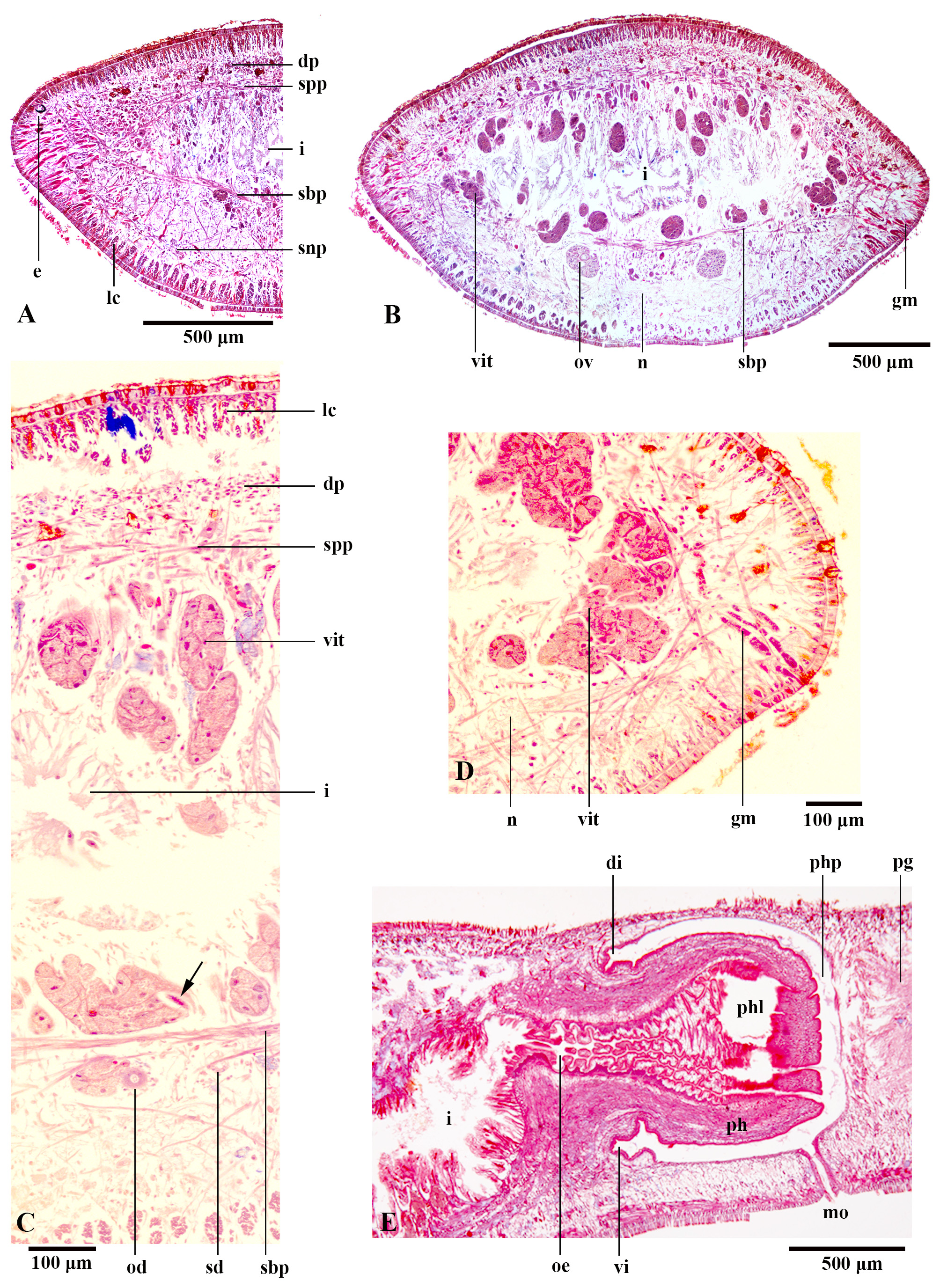
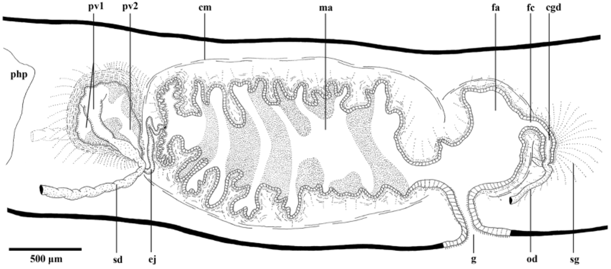
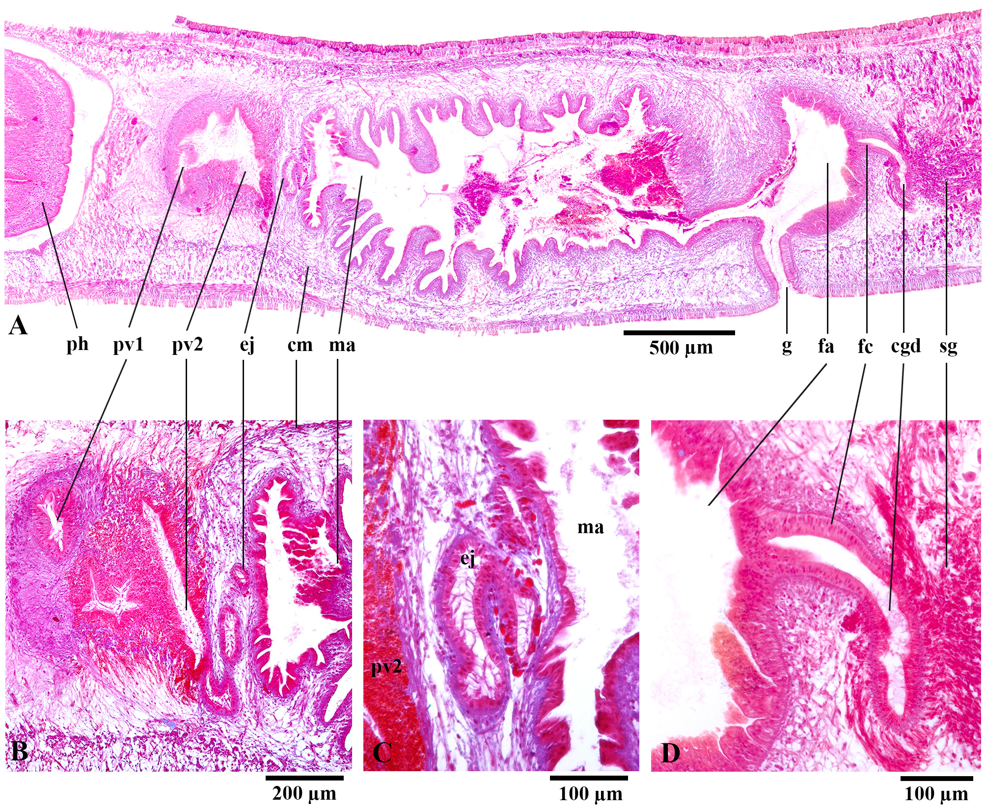
TABLE 4. Measurements (mm) from fixed specimens of Pasipha mbya sp. nov. (DG) distance of gonopore from anterior end, (DM) distance of mouth from anterior end, (DMG) distance between mouth and gonopore. The numbers in parentheses represent the position relative to body length (%).
| MLP-He 6476 | MLP-He 6978 | |
|---|---|---|
| Length | 28 | 46.2 |
| Maximum width | 3 | 3.2 |
| Maximum height | 1.3 | 1.5 |
| DM | 20.2 (72%) | 32.4 (70%) |
| DG | 23.6 (84%) | 38.4 (83%) |
| DMG | 3.4 | 6 |
| Creeping sole (%) | 90% | 90% |
TABLE 5. Thickness of cutaneous (CM) and parenchymatic (PM) musculatures (µm), and CMI and PMI indices at prepharyngeal region of Pasipha mbya sp. nov.
| MLP-He 6476 | MLP-He 6978 | |
|---|---|---|
| CM dorsal | ||
| circular | 5 | 2.5 |
| diagonal | 12.5 | 10 |
| longitudinal | 45 | 75 |
| CM ventral | ||
| circular | 5 | 5 |
| diagonal | 12.5 | 10 |
| longitudinal | 82.5 | 100 |
| CMI (%) | 13% | 14% |
| PM dorsal | 35 | 60 |
| PM supra-intestinal | 10 | 35 |
| PM sub-intestinal | 35 | 35 |
| PMI (%) | 6% | 9% |
TABLE 6. Measurements (mm) of reproductive organs of Pasipha mbya sp. nov. LCGD, length of common glandular ovovitelline duct; LFA, length of female atrium; LFC, length of female canal; LMA, length of male atrium; LPV, length of prostatic vesicle. The numbers in parentheses represent the position relative to body length (%).
| MLP-He 6476 | MLP-He 6978 | |
|---|---|---|
| Anteriormost testes | 6.7 (24%) | 11 (24%) |
| Posteriormost testes | 18.6 (66%) | 29 (63%) |
| LMA | 1.6 | 4 |
| Location of ovaries | 6.2 (22%) | 10 (22%) |
| LCGD | 0.2 | 0.35 |
| LFC | 0.2 | 0.35 |
| LFA | 0.4 | 1.6 |
No known copyright restrictions apply. See Agosti, D., Egloff, W., 2009. Taxonomic information exchange and copyright: the Plazi approach. BMC Research Notes 2009, 2:53 for further explanation.