
Parvalona parva ( Daday, 1905 ) Damme & Kotov & Dumont, 2005
|
publication ID |
https://doi.org/ 10.1080/00222930500060884 |
|
persistent identifier |
https://treatment.plazi.org/id/B762596A-FFD1-0D41-49FA-372CFECB1606 |
|
treatment provided by |
Felipe |
|
scientific name |
Parvalona parva ( Daday, 1905 ) |
| status |
comb. nov. |
Parvalona parva ( Daday, 1905) View in CoL comb. nov.
( Figures 1–3 View Figure 1 View Figure 2 View Figure 3 )
Leydigia parva Daday, 1905, p 186 –187, Plate 11: Figures 20, 21.
Alona parva (Daday) in Smirnov 1971, p 391, 393, Figure 471; Forró and Frey 1982, p 124.
Not Leydigia parva Daday in Goulden 1966, p 101 –103, Plate 3: Figures 3 View Figure 3 –5; Frey 1982, Table 1.
Not Leydigia glabra Smirnov, García Ponce and Silva-Briano 2000, p 1 View in CoL –3, Figures 1 View Figure 1 –10. Not Birgeia travassosi Bergamin 1939 , Plate 2: Figure 5.
Not Alona View in CoL sp. nov. in Brehm 1939, p 184, Plate 36: Figures 20, 21.
Type material
Type locality. From Daday (1905, p 186, 228): ‘‘Curuzu-chica, toter Arm des Paraguayflusses; Estia Postillon, Lagune und deren Ergüsse’’, Paraguay. For remarks, see ‘‘Distribution’’.
Lectotype: parthenogenetic female, 0.47 mm from unknown locality in Paraguay, DAD D III-79 : II/P-721. There are two females on this slide, the lectotype marked by an arrow on the slide; selected by D. G. Frey, July 1965 ( Forró and Frey 1982) . Paralectotype: parthenogenetic female on the same slide with the lectotype.
Other material examined
Three parthenogenetic females, on three slides, kept at UG : two dissected specimens, one complete, from a temporary pool in Mandacaru, coordinates 2 ° 359500S, 42 ° 429460W, Lençóis Maranhenses National Park , Maranhao, Brazil, collected by K. Van Damme and D. Van Damme, 16 August 1996, from samples at UG , Belgium, labelled Brazil 1996.014– 1996.015 ( SIII 1-2 ) .
Amended diagnosis
See diagnosis of the genus.
Redescription
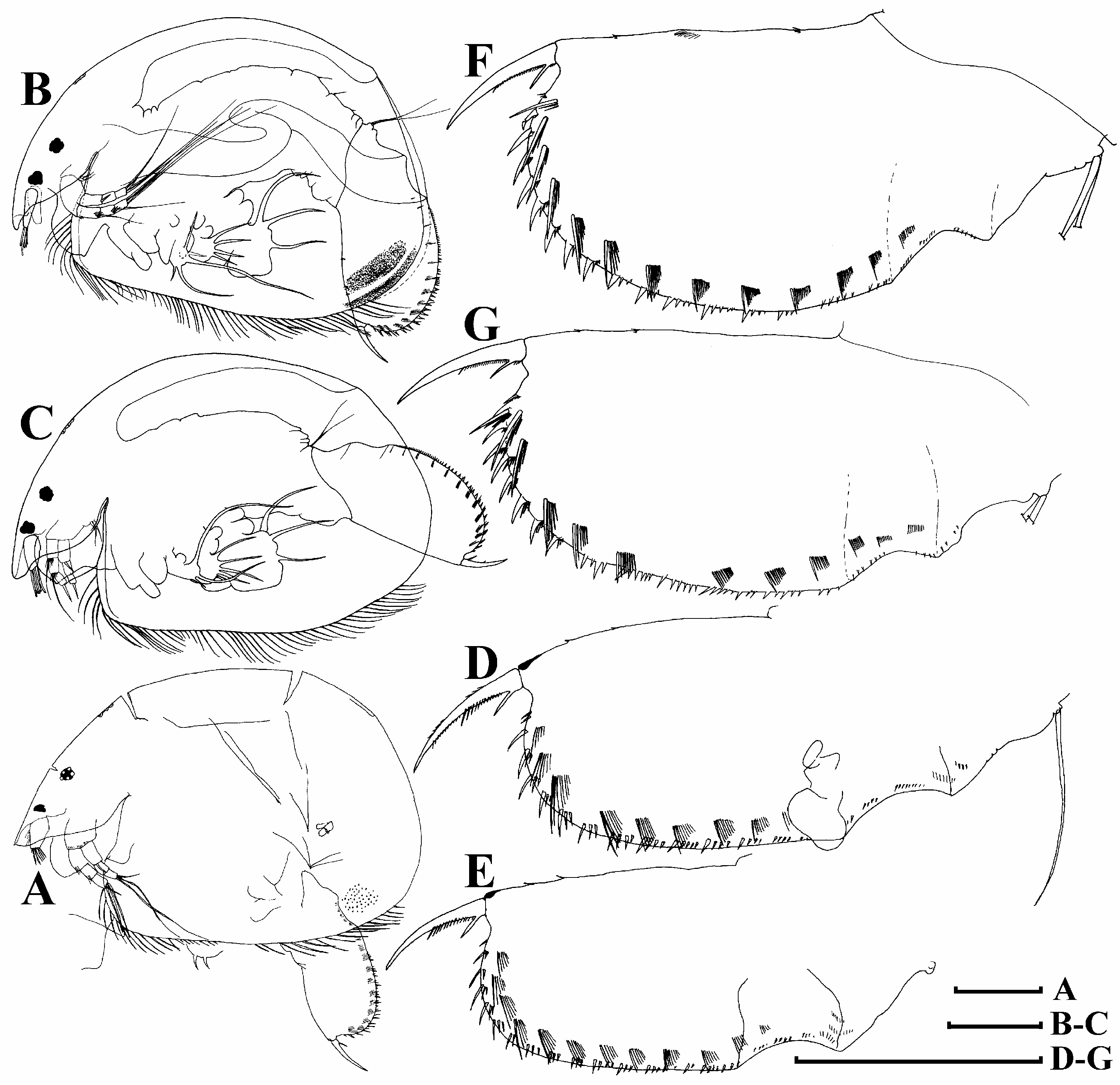
Parthenogenetic female. General: colour (after fixation) colourless to pale yellow ( Daday 1905) to reddish brown. In lateral view body subovoid, high (body height/body length50.6520.73 in adults), with maximum height in middle ( Figure 1A–C View Figure 1 ). In dorsal view, body strongly bilaterally compressed. Dorsal margin regularly arched from tip of rostrum to rounded postero-dorsal angle, posterior margin slightly convex, postero-ventral margin widely rounded, ventral margin convex in posterior half and straight to concave in anterior half. In contrast to Daday’s (1905) description, no wide striation on the carapace was found. Granulate or dotted valves, resulting from internal structures. In the posteroventral portion of the valve, these dots are organized in several bands along the valve margin, as marked by Daday (1905).
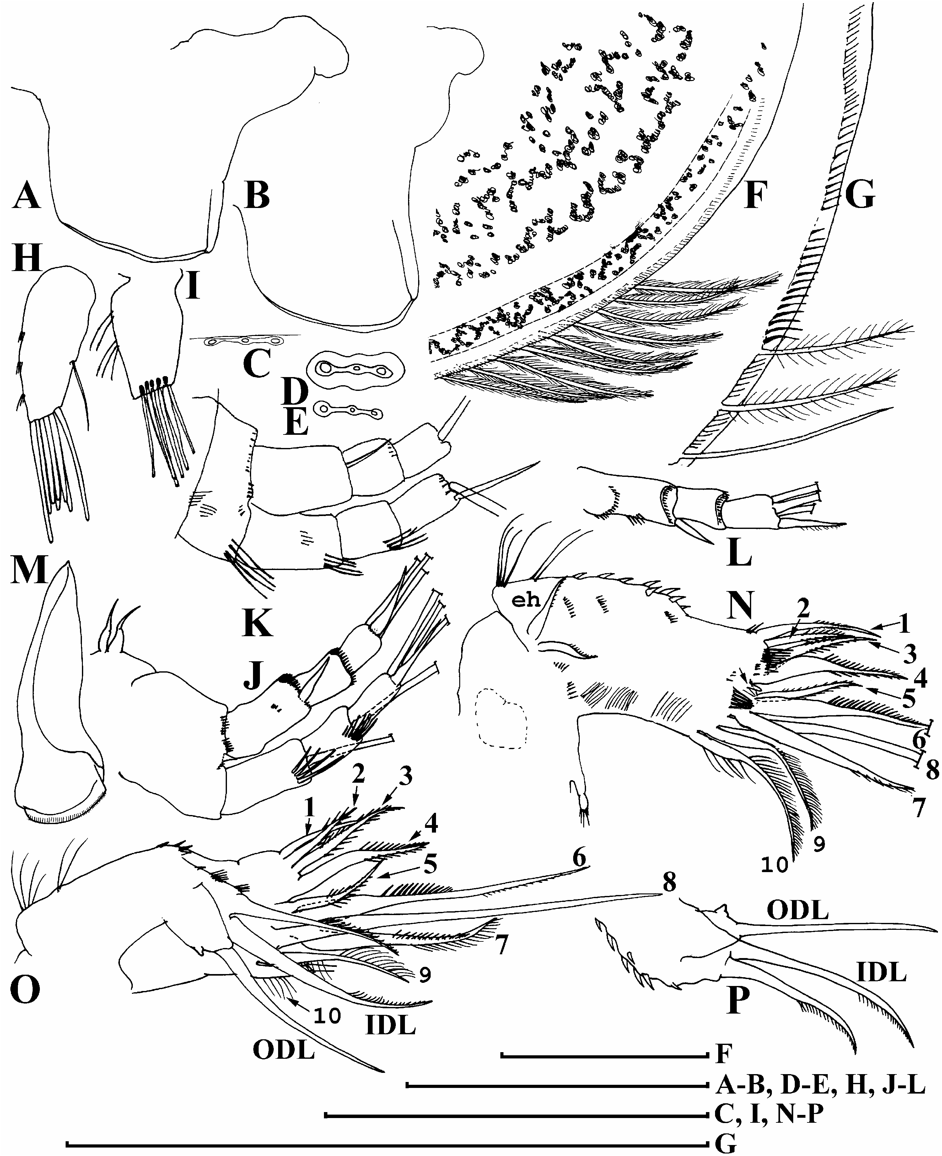
Head: relatively small, rounded-triangular in lateral view, with rostrum short and relatively blunt, in contrast to Daday’s (1905) description. Also, in Daday’s specimens ( Figure 1A View Figure 1 ) the rostrum is truncated, but it seems to be an artefact of excessive compression of the specimens during slide preparation. Eye and ocellus of similar size, ocellus located approximately in middle of distance from eye to tip of rostrum. Head shield with mandibular articulation of alonine-type (see Frey 1967). Three major head pores ( Figure 2C–E View Figure 2 ) with a relatively narrow connection between them, central pore of the same size as anterior or posterior one, or somewhat narrower ( Figure 2D, E View Figure 2 ). Lateral head pores were not seen. In one specimen ( Figure 2D View Figure 2 ), a clear field around the pores was noted, demarcated by a thin line.
Labrum: labrum with a main body, small distal labral plate, and a large medial labral keel ( Figure 2A, B View Figure 2 ). In lateral view, labral keel trapezium-shaped, without setulation. Ventral portion of labral keel with thickened ridge.
Valves: large, subovoid, with numerous (51; average of two specimens, Figure 1B, C View Figure 1 ), relatively long setae, located submarginally in posterior portion of margin bases ( Figure 2F View Figure 2 ). Setae divisible in three groups, of which the most rostral group contains the largest setae, followed by a small group of shorter setae situated ventrally of the region between limbs I and III and a third and final group of larger setae. Posterior margin of valve with a row of numerous setules at some distance from one another, implanted on inner side of carapace ( Figure 2G View Figure 2 ).
Postabdomen ( Figure 1D–G View Figure 1 ): postabdomen with remarkably wide postanal portion, at least four times as long as anal margin. Ventral margin straight to slightly convex, with rows of minute marginal setules. Anal margin relatively short, shifted strongly to base of postabdomen. Preanal margin somewhat longer than anus, straight to depressed (there is a chance that it is deformed in Daday’s specimens), preanal and postanal angle well defined. Whole postanal margin as large arched curve. Inflated basis for postabdominal claws bordered from postanal margin by a distinct depression. Distally, each side of postabdomen provided with a row of 13–16 successive clusters of relatively long marginal denticles, with size increasing distally and each group consisting of mostly three to five denticles; these clusters continue into three to four groups of fine setules on anal margin. Medially to postanal denticles, 9–13 groups of long lateral setules, the distalmost of each group or row being the largest and thicker than the others.
Postabdominal seta: as long as anal plus preanal margin ( Figure 1D View Figure 1 ).
Postabdominal claw: approximately as long as or little longer than preanal margin, slightly and evenly curved. Basal spine large, as marked by Daday (1905), length up to 1.5– 2 times diameter of claw at base; basal spine stout, not pressed to claw. Small basal denticles present, proximally from basal spine ( Figure 2F, G View Figure 2 ).
Antenna I: short (shorter in lectotype, Figure 2I View Figure 2 ), not reaching tip of rostrum, with two to three transverse rows of setules at anterior face ( Figure 2H, I View Figure 2 ). Antennular sensory seta slender, as long as half of antenna I length, arising at distance of one-third of antenna I length from distal end. Nine aesthetascs of different size, longest as long as antenna I, projecting beyond tip of rostrum.
Antenna II: relatively short. Coxal part with two sensory setae ( Figure 2J View Figure 2 ); basal segment robust, with transverse rows of numerous, fine, long setules, rudimentary distal spine and short setules at its distal margin. Antennal branches relatively elongated, exopod shorter than endopod, all segments cylindrical, with rows of four to seven long, stout setules, longest of which reaching up to half of following segment ( Figure 2J, K View Figure 2 ). Antennal formula (exo/endo): setae 1-1-3/0-0-3, spines 0-0-1/1-0-1. Spine on first (basal) segment of endopod long, reaching or nearly reaching tip of second segment. Apical spines of exopod and endopod of similar length, markedly longer than apical segments ( Figure 2J–L View Figure 2 ).
Mandible: elongated, with widened head ( Figure 2M View Figure 2 ) bearing small ridges. Left and right mandibles asymmetrical.
Limb I: epipodite small, globular ( Figure 2N View Figure 2 ). Accessory seta absent, ODL with a long, naked seta, and a rudiment of the second seta ( Figure 2O, P View Figure 2 ). IDL of similar size as ODL, with two relatively large setae of somewhat different size, armed with small setules unilaterally in terminal half. Endite 1 with three posterior soft setae ( Figures 2N, O View Figure 2 , 3A View Figure 3 : 2– 4), with size increasing basally, all armed with short hairs bilaterally, and one anterior seta (1) long, stout, with short setules in second distal half. Endite 2 with three soft setae of unequal size (5–7): seta 5 short, armed bilaterally with short setules, seta 6 longest, asymmetrically armed with long setules proximally, seta 5 armed with short setules; two small elements anterior to seta 5, one of which (arrow) clearly a rudiment of a stiff seta on endite 2. Endite 3 with three soft setae (8–10), of which one (8) naked and significantly larger than the rest, two other setae two-segmented, setulated along one side, seta 10 somewhat longer than seta 9. Two ejector hooks (eh) of equal size anteriorly on outer portion of limb corm. Also here, a series of long setules and groups of short denticles ( Figure 3B View Figure 3 ). Low maxillar process with a single short seta supplied with a bunch of setules distally.
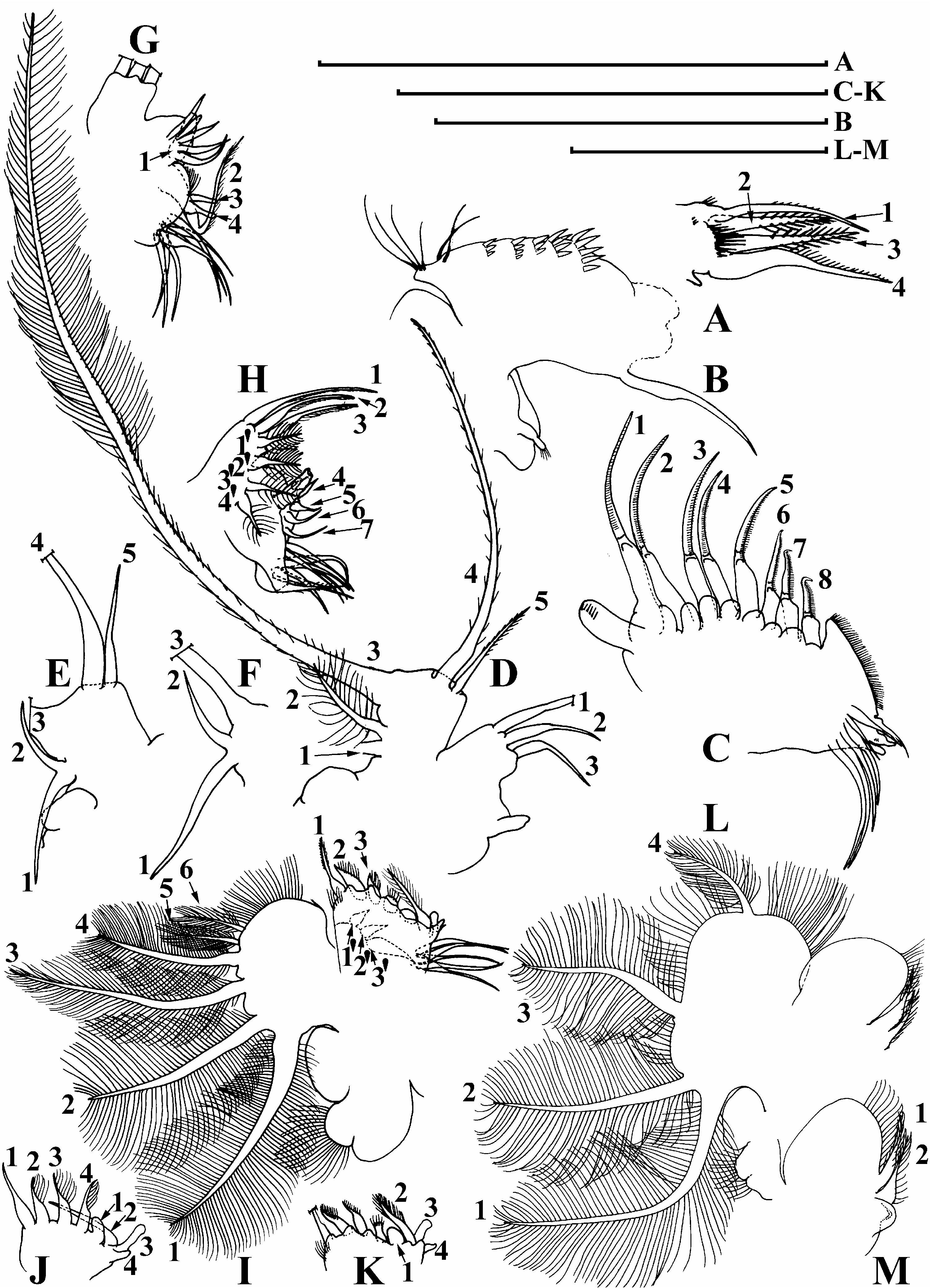
Limb II: exopodite a small, subovoid lobe with a row of setules. Eight scrapers ( Figure 3C View Figure 3 : 1–8), scrapers 1–3 with size slightly decreasing basally, scraper 4 short, scraper 5 longer that 4, scraper 5–8 again with size decreasing basally. A series of hillocks posteriorly to scrapers. Gnathobase with prominent basal-ventral angle, armed with short setules. Distal armature of gnathobase with three elements, filter plate with six setae, two distalmost setae markedly short.
Limb III: epipodite ovoid, exopodite flat, relatively small, with three lateral setae ( Figure 3D–F View Figure 3 : 1–3) and two setae of different size distally ( Figure 3D, E View Figure 3 : 4–5); exopodite setae 4 and 5 widely spaced from one another, their proximal bases (first fourth of seta) perpendicular to each other. Exopodite setae 1 and 2 sparsely setulated with long filter setules, longest exopodite seta 3 armed with long setules in distal half while proximal half bears shorter setules, pressed to the seta; exopodite seta 4, half the length of exopodite seta 3 ( Figure 3D View Figure 3 ), bears short setules over complete length, while exopodite seta 5, one-quarter length of previous seta, is only setulated in distal half (note that setulation of exopodite setae can be described better on SEM photographs because of the three-dimensional nature which is represented less in a two-dimensional drawing). Distal endite armed with three stiff setae ( Figure 3D, H View Figure 3 : 1–3). Basal endite of similar size with distal endite, anteriorly with a sensillum and four, stiff setae ( Figure 3H View Figure 3 : 4–7). Four posterior soft setae (19–49), setulated bilaterally. Distal armature of gnathobase with four setae ( Figure 3G View Figure 3 : 1–4), one of them (1) a thick, bottle-shaped sensillum of middle size. Seven setae of similar size in filter plate III.
Limb IV: pre-epipodite relatively large, setulated; epipodite ovoid. Exopodite large, round, with six setae, not differentiated into lateral and distal group ( Figure 3I View Figure 3 : 1–6); exopodite setae 5 and 6 markedly smaller and more slender than the others. Marginally on inner limb face, a row of four stiff (‘‘torch’’) setae ( Figure 3J View Figure 3 : 1–4), seta 1 longest, stout, naked, each of setae 2–4 armed with fine setules, sometimes seta 4 shortened ( Figure 3K View Figure 3 ). Posteriorly, three soft setae ( Figure 3I View Figure 3 : 19–39). Distal armature of gnathobase with four elements ( Figure 3K View Figure 3 : 1–4): element 1 an ovoid sensillum. Filter plate with five setae of similar size.
Limb V: pre-epipodite small, bipartite and setulated; epipodite ovoid ( Figure 3L View Figure 3 ). Exopodite large, ellipsoid, with a single distal seta (4) and three lateral setae (1–3). Inner limb portion with wide flap-like distal projection, fringed by setules. Two submarginal setae (1–2) on inner face of limb, distal member (1) larger, but not protruding behind distal projection. Gnathobase greatly reduced, as a simple projection, no filter plate was found.
Ephippial female, male. Unknown.
Size. Lectotype, parthenogenetic female 0.47 mm; parthenogenetic females 0.45–0.47 mm (n 52), up to 0.60 mm according to Daday (1905).
Ecology. Daday (1905) did not provide details about the localities from Paraguay. The Brazilian specimens were found in a shallow, temporary waterbody (dimensions: 1.5 m × 100 m × 80 m) between cerrado-fixed dunes in the Lencoís Maranhenses, functioning as a drinking pool for cattle. The waterbody was rich in submerged vegetation, with a mean temperature of 31 ° C, pH 8.4, conductivity of 140 MS cm 21 and oxygen level of 8.4 mg l 21. Regarding faunistic elements, it was rich in aquatic insects (Hemiptera), molluscs ( Ampullaria ), frogs and fish. Most common branchiopods consisted of Cyclestheria hislopi Baird, 1859 , Leydigiopsis curvirostris Sars, 1901 , Alona ossiani Sinev, 1998 ( Alona affinis group), Ephemeroporus hybridus ( Daday, 1905) , and Chydorus ventricosus Daday, 1898 .
Distribution. This is a rare South American species. At present, it is known only from two(?) localities in Paraguay ( Daday 1905) and now a single locality in NE Brazil (first record), both on the Atlantic Coast of the South American continent. It is important to note that although Daday (1905) mentions having studied in his work ‘‘several samples from Paraguay’’, we were not able to pinpoint these two locations. The only current locality of which the name resembles Daday’s ‘‘Curuzu-chica’’ is Curuzú Cuatiá (coordinates 29 ° 489S, 58 ° 029W), a city situated east of the Paraná River in Corrientes Province, Argentina. Situated close to the border of Paraguay, and the Paraná River being a hydrological continuation of the Paraguay River, we believe it possible that this locality might actually be situated in Argentina. Note that all other Central and South American records ( Bergamin, 1939; Goulden 1966; Frey 1982) in reality dealt with other species (see ‘‘Discussion’’).
| UG |
Museo del Departamento de Estratigrafia y Paleontologia |
| V |
Royal British Columbia Museum - Herbarium |
No known copyright restrictions apply. See Agosti, D., Egloff, W., 2009. Taxonomic information exchange and copyright: the Plazi approach. BMC Research Notes 2009, 2:53 for further explanation.
|
Kingdom |
|
|
Phylum |
|
|
Class |
|
|
Order |
|
|
Genus |
Parvalona parva ( Daday, 1905 )
| Damme, Kay Van, Kotov, Alexey A. & Dumont, Henri J. 2005 |
Leydigia glabra Smirnov, García Ponce and Silva-Briano 2000 , p 1
| Smirnov NN & Garcia Ponce H & Silva-Briano M 2000: 1 |
Alona parva (Daday)
| Forro L & Frey DG 1982: 124 |
| Smirnov NN 1971: 391 |
Leydigia parva
| Goulden CE 1966: 101 |
Alona
| Brehm V 1939: 184 |
Leydigia parva
| Daday E 1905: 186 |