
Paralaophonte pacificaemulator, Gómez & Morales-Serna, 2013
|
publication ID |
https://doi.org/ 10.1080/00222933.2012.757657 |
|
DOI |
https://doi.org/10.5281/zenodo.10536624 |
|
persistent identifier |
https://treatment.plazi.org/id/2C4C87C9-DF16-7D31-FE21-2399FD38F9FE |
|
treatment provided by |
Felipe |
|
scientific name |
Paralaophonte pacificaemulator |
| status |
sp. nov. |
Paralaophonte pacificaemulator sp. nov.
( Figures 21 View Figure 21 –29)
Material examined
One female holotype (EMUCOP-080205-03), one male allotype (EMUCOP- 080205-04) preserved in alcohol. Paratypes preserved in alcohol: one CIV
(EMUCOP-080205-19), five adult females, 21 adult males, four CV, and four CIV (EMUCOP-080205-18), two adult females (EMUCOP-080205-17), two adult females and 2 CV (EMUCOP-230691-13), one adult female (EMUCOP-240691-01), four adult females, one adult male, one CIV and four CV (EMUCOP-230691-14), and four adult females, one adult male, one CV and one CIV (EMUCOP-230691-39). Dissected paratypes: seven females (EMUCOP-010591-01, EMUCOP-010591-02, EMUCOP- 010591-03, EMUCOP-010591-06, EMUCOP-050205-01, EMUCOP-050205-02, EMUCOP-090301-30), and 12 males (EMUCOP-010591-05, EMUCOP-010591-07, EMUCOP-010591-08, EMUCOP-010591-09, EMUCOP-010591-10, EMUCOP- 010591-11, EMUCOP-020591-04, EMUCOP-040591-04, EMUCOP-050205-05, EMUCOP-050205-06, EMUCOP-090301-29, EMUCOP-090301-35), and one male CV (EMUCOP-010591-04). Collected from Ensenada del Pabellón lagoon (Sinaloa
State, north-western Mexico) (24 ◦ 19 ′ – 24 ◦ 35 ′ N, 107 ◦ 28 ′ – 107 ◦ 45 ′ W), stn. 2, 3, 4, and 14 (see Gómez-Noguera and Hendrickx, 1997, for more information regarding nitrogen and carbon content and sediment type), brackish, less than 2 m depth, 1, 2 and 4 May 1991, 23 and 24 June 1991; from Urías System (Sinaloa State, north-western Mexico) (23 ◦ 11 ′ 06 ′′ N, 106 ◦ 25 ′ 06 ′′ W), stn. 1, 2, 7, and 9 (see Morales-Serna, Gómez, and Bustos-Hernández 2006, for more information regarding organic matter content, chlorophyll a content and sediment type), brackish, less than 2 m depth, 9 March 2001, 5 and 8 February 2005, coll. S. Gómez (Ensenada del Pabellón lagoon) and F. N. Morales-Serna, F. E. Vargas-Arriaga and S. Gómez (Urías System).
Type locality
Urías System (Sinaloa State, north-western Mexico) (23 ◦ 11 ′ 06 ′′ N, 106 ◦ 25 ′ 06 ′′ W) GoogleMaps .
Other localities
Ensenada del Pabellón lagoon (Sinaloa State, north-western Mexico) (24 ◦ 19 ′ – 24 ◦ 35 ′ N, 107 ◦ 28 ′ – 107 ◦ 45 ′ W) GoogleMaps .
Etymology
The specific epithet pacificaemulator ( pacifica after P. pacifica ; Latin, aemulor, to emulate) makes reference to the strong resemblance to P. pacifica .
Description
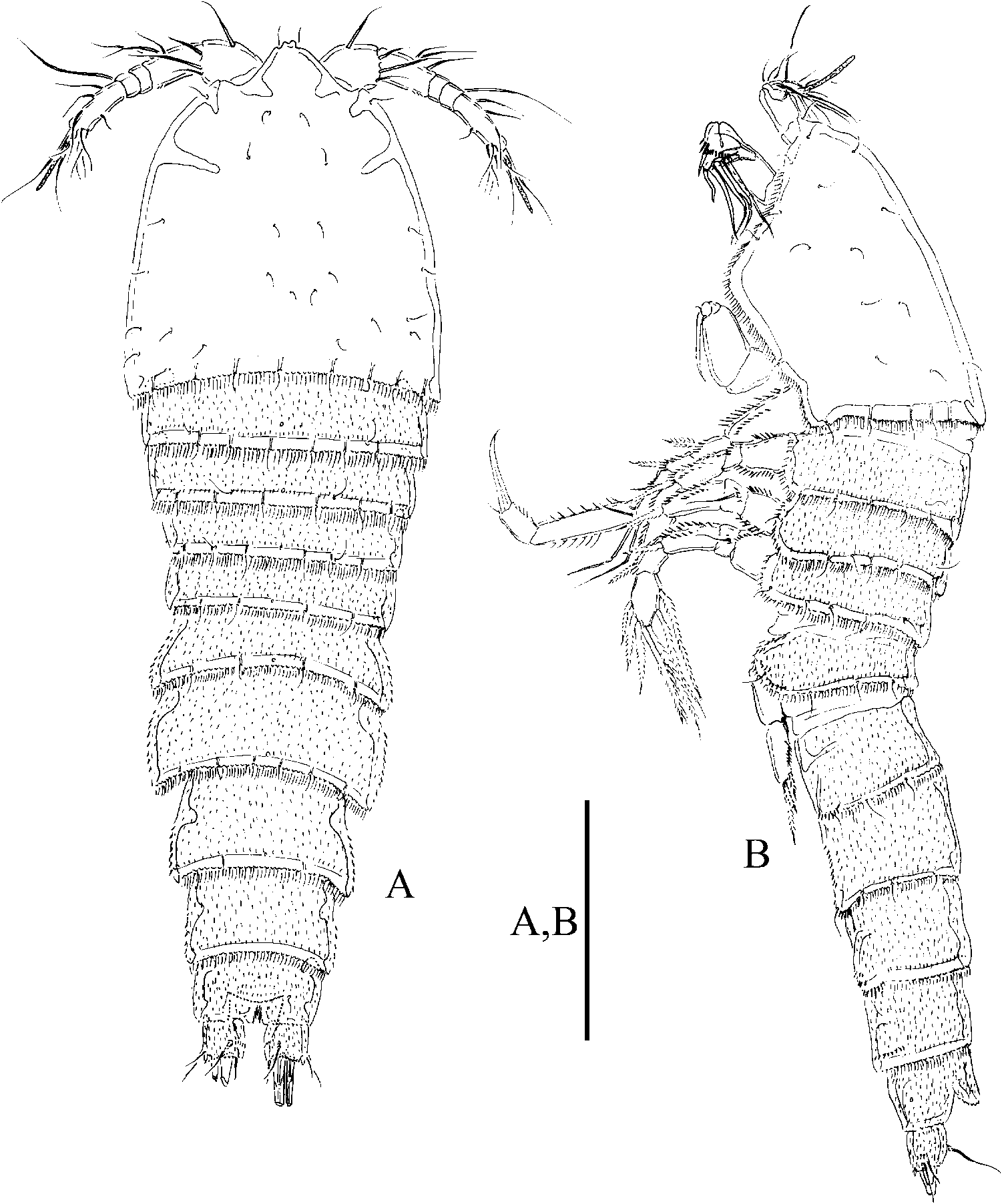
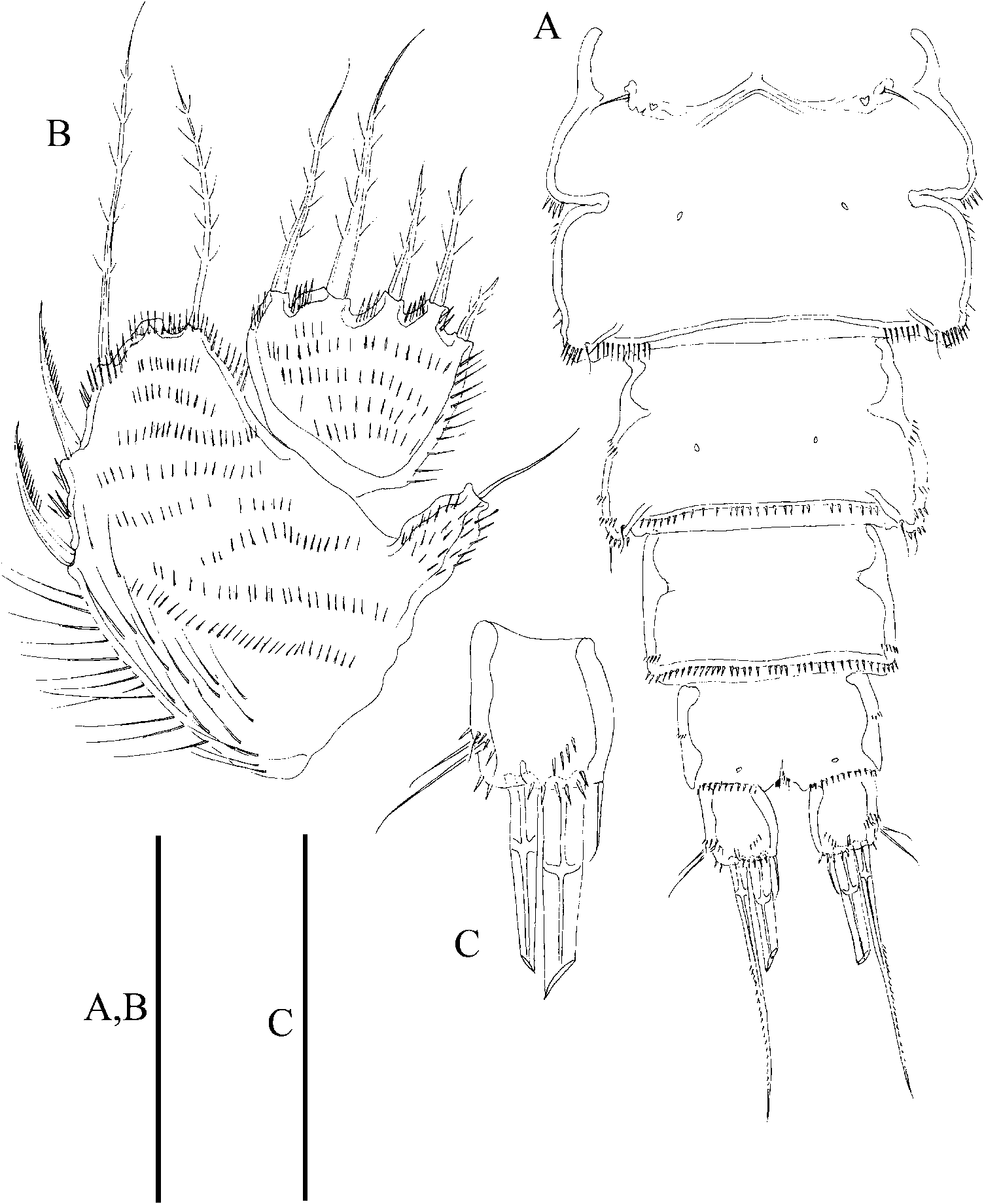
Female. Habitus fusiform ( Figure 21A, B View Figure 21 ). Total body length measured from tip of rostrum to posterior margin of caudal rami ranging from 405 µm to 480 µm (mean = 447 µm; n = 9). Rostrum fused to cephalic shield, triangular, with bilobed tip flanked by pair of sensilla. Cephalothorax dorsally and laterally as shown ( Figure 21A, B View Figure 21 ), with posterior margin minutely serrate dorsally, with setules along posterior margin dorsally and laterally. P2–P5-bearing somites covered by tiny spinules, with posterior margin minutely serrate, with setules along posterior margin dorsally and laterally. Genital double-somite distinct dorsally and laterally ( Figure 21A, B View Figure 21 ); fused ventrally ( Figure 22A View Figure 22 ); both somites of the genital double-somite with posterior margins minutely serrate and ornamented as previous somites dorsally and laterally, with lateral expansions moderately developed ( Figures 21A View Figure 21 , 22A View Figure 22 ), and with sets of spinules; posterior half of genital double-somite with set of spinules close to posterior corner ventrally. Fourth and fifth urosomites as previous somites dorsally; fourth urosomite with, fifth urosomite without lateral expansions; both urosomites with setules (very fine spinules?) along posterior margin. Anal somite ( Figures 21A, B View Figure 21 , 22A View Figure 22 , 26C, D View Figure 26 ) covered with tiny spinules dorsally and laterally, with spinules along posterior margins dorsally and ventrally; rounded anal operculum with minutely serrate posterior margin and flanked by pair of sensilla. Caudal rami ( Figures 21A, B View Figure 21 , 22A, C View Figure 22 , 26C, D View Figure 26 ) about 1.2 times as long as wide; covered with tiny spinules dorsally and laterally and with rows of stronger spinules ventrally; with seven elements; seta I very small, inserted ventrally to seta II, the latter about three times longer, both inserted laterally on distal fourth; seta III longer than seta II and situated almost at the same level, ventral to seta II and posterior to seta I; seta IV pinnate; seta V longest; seta VI arising from inner distal corner, nearly as long as seta II; seta VII situated dorsally, on the distal third of the inner margin, triarticulated.
Antennule ( Figure 23A View Figure 23 ) six-segmented; first segment with medial and distal inner rows of spinules, without process; second segment with some inner spinules proximally, with blunt conical outer process; third segment with inner spinules proximally and with some transverse rows of minute spinules along outer margin medially and distally; fourth and fifth segments small, each with outer spinular row; sixth segment elongate, about three times as long as wide, with outer spinular row proximally. Armature formula, I-(1); II-(8); III-(7); IV-(2 + ae); V-(1); VI-(9 + acrothek) (acrothek consisting of two setae and one aesthetasc fused basally).
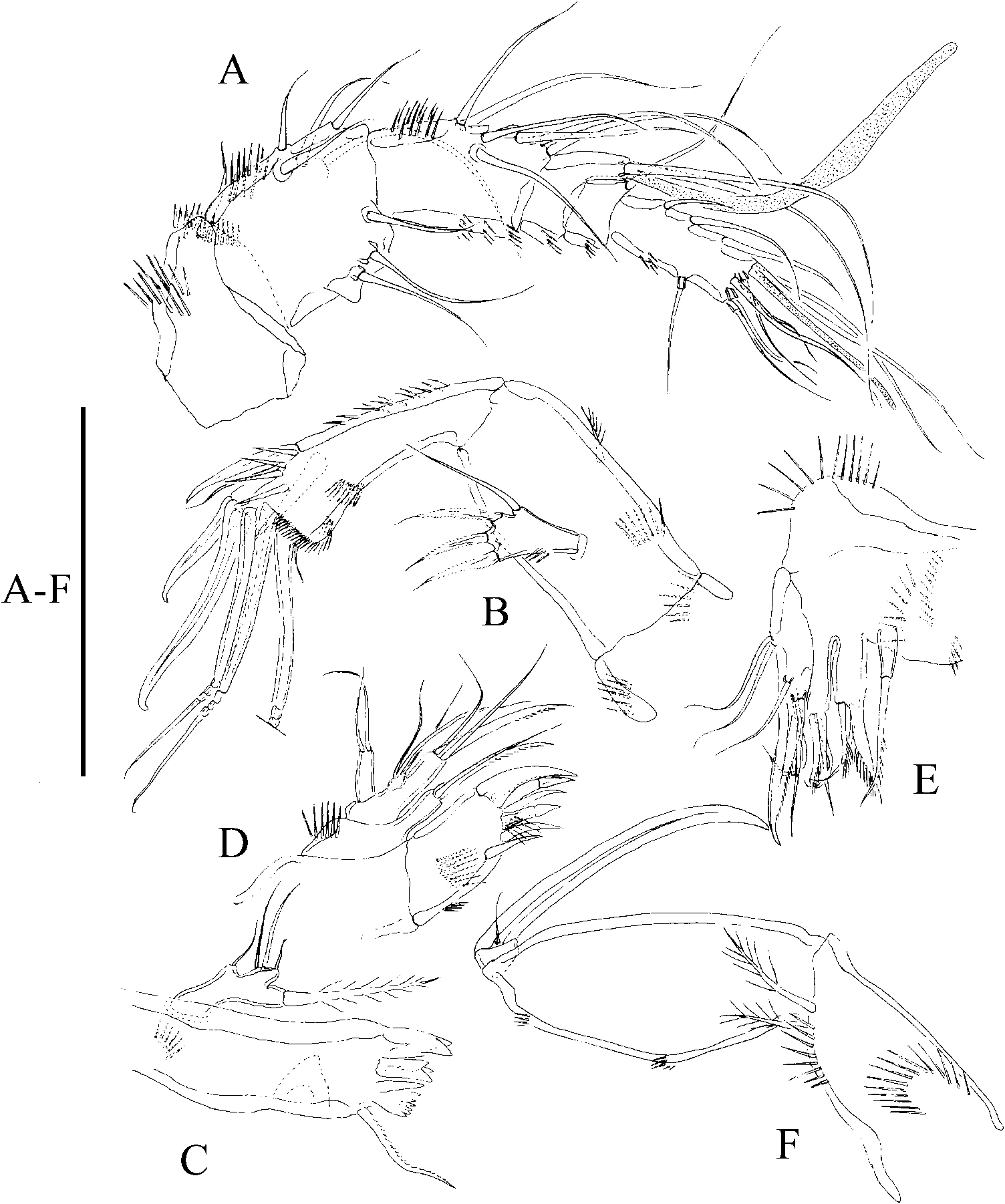
Antenna ( Figure 23B View Figure 23 ). Coxa with some spinules as depicted. Allobasis with one small, unipinnate abexopodal seta, and with inner row of spinules proximally. Exopod one-segmented, with two lateral (proximal one longer, bare, slender) and two distal elements. Free endopodal segment with inner row of spinules, with two outer frills, laterally with two spines and a slender seta, and apically with two strong spines, two geniculate single setae and one geniculate seta fused to tiny element basally.
Mandible ( Figure 23C View Figure 23 ). Strong gnathobase with bi- and multicuspidate teeth distally and one pinnate seta laterally. Palp one-segment, with five seta (one basal, one exopodal, three endopodal).
Maxillule ( Figure 23D View Figure 23 ). Arthrite with some spinules as depicted, with five strong apical spines and some spinules, and one lateral element. Coxa with some proximal spinules, with two setae. Basis with three apical elements (one of them stronger). Exopod one-segmented, elongate, with two setae. Endopod small, one-segmented, with three setae.
Maxilla ( Figure 23E View Figure 23 ). Syncoxa with outer and inner spinules as shown; with three endites; proximal endite with one seta, middle and distal endites each with three elements as figured (one of them fused to endite basally). Allobasis drawn into strong claw with three accessory setae. Endopod represented by two long setae.
Maxilliped ( Figure 23F View Figure 23 ). Syncoxa with spinular rows as depicted, with two distal setae. Basis with small outer spinules. Endopod drawn out into claw with one accompanying seta.
P1 ( Figure 24A View Figure 24 ). Coxa with several spinule rows as figured. Basis with longitudinal rows of spinules, with inner and outer spine-like element. Exopod three-segmented, reaching to slightly below the middle of ENP1. Endopod two-segmented, elongate; ENP1 long, about 6.7 times as long as wide, with inner setules and outer spinules; ENP2 about twice as long as wide, with outer and apical spinules, with one small apical seta and one strong claw, the latter about 1.7 times as long as supporting segment.
P2 ( Figure 24B View Figure 24 ). Praecoxa with transverse row of outer spinules. Coxa with spinular rows as figured. Basis with spinules at base of outer, spine-like seta. Exopod three-segmented; EXP1 without, EXP2 with inner seta; EXP3 with three outer spines, two apical and one inner element. Endopod two-segmented, reaching slightly beyond insertion site of inner seta of EXP2, ENP1 as long as EXP1; ENP1 without armature; ENP2 with two inner (proximalmost smaller) and two apical setae.
P3 ( Figure 25A View Figure 25 ). Praecoxa as in P2. Coxa and basis as in P2, except for slender and bare basal seta of P3. Exopod three-segmented; EXP1 without, EXP2 with inner seta; EXP3 with three outer spines, two apical and two inner elements. Endopod twosegmented, reaching insertion site of inner seta of EXP2; ENP1 without armature; ENP2 with three inner setae, two apical and one outer element.
P4 ( Figure 25B View Figure 25 ). Praecoxa, coxa, basis and exopod as in P3 (though P4 EXP3 somewhat different in general shape). Endopod two-segmented, barely reaching distal margin of EXP1; first segment without armature; second segment with one inner seta, two apical and one outer element.
P5 ( Figure 22B View Figure 22 ) large, with separate rami. Baseoendopodal lobe well developed, not reaching tip of EXP, with spinules as shown; with four setae; outer basal seta arising from short setophore. Exopod broad, covered with spinules, with five setae, outermost smallest (about half length of adjacent seta).
P6 ( Figure 22A View Figure 22 ) represented by one seta.
Armature formula of P1–P5 as in Table 2.
sp. nov.
Male. Habitus ( Figures 26A, B View Figure 26 , 27A, B View Figure 27 ) as in female except for separate second and third urosomites ventrally, and for coarser and more abundant spinules on third, fourth and fifth urosomites ventrally ( Figure 28A View Figure 28 ). Urosome more slender than in female. Anal somite and caudal rami ( Figure 26C, D View Figure 26 ) as in female. Total body length ranging from 420 µm to 470 µm measured from tip of rostrum to posterior margin of caudal rami (mean = 447 µm; n = 13).
Antennule ( Figure 28A View Figure 28 ) eight-segmented, subchirocer; second segment with conical outer process; sixth segment with two acute projections. Armature formula difficult to define: I-(1); II-(9);III-(6);IV-(1);V-(10 + ae);VI-(0);VII-(1);VIII-(8 + acrothek). Acrothek consisting of two setae and one aesthetasc fused basally.
Antenna, mandible, maxillule, maxilla and maxilliped (not shown) as in female.
P1 ( Figure 28B View Figure 28 ) as in female.
P2 EXP as in female. P2 ENP ( Figure 28C View Figure 28 ) dimorphic, two-segmented, distalmost inner seta modified as depicted.
P3 (Figure 29A) dimorphic. Exopod three-segmented, segments more robust than in female; first segment without, second segment with one, third segment with two inner spines; all setae and spines very strong and without ornamentation except for pinnate inner element of EXP2; relative length of spines on EXP3 as depicted. Endopod three-segmented; ENP1 without armature; ENP2 with outer apophysis reaching beyond ENP3, the latter with two inner and two apical setae.
P4 (Figure 29B) as in female except for exopodal segments more strongly developed, and for comparatively stronger and more spinulose outer spines, outer apical element of EXP3, and shorter inner seta of EXP 2 in male. Endopodal segments comparatively much larger than in female.
Left and right P5 fused (Figure 29C); baseoendopodal lobe with one seta plus outer seta of basis; exopod with five setae (innermost considerably stronger).
P6 (Figure 29D) represented by two plates, each bearing one outer slender seta and one inner strong spine-like element.
Remarks
See Remarks for P. pacificavicimum sp. nov.


| CV |
Municipal Museum of Chungking |
No known copyright restrictions apply. See Agosti, D., Egloff, W., 2009. Taxonomic information exchange and copyright: the Plazi approach. BMC Research Notes 2009, 2:53 for further explanation.