
Paraheligmonelloides ennisae, Smales & Heinrich, 2010
|
publication ID |
https://doi.org/ 10.11646/zootaxa.2672.1.1 |
|
DOI |
https://doi.org/10.5281/zenodo.5308630 |
|
persistent identifier |
https://treatment.plazi.org/id/FF7CEC67-FD63-FF9A-FF3A-FAA3FD23751F |
|
treatment provided by |
Felipe |
|
scientific name |
Paraheligmonelloides ennisae |
| status |
sp. nov. |
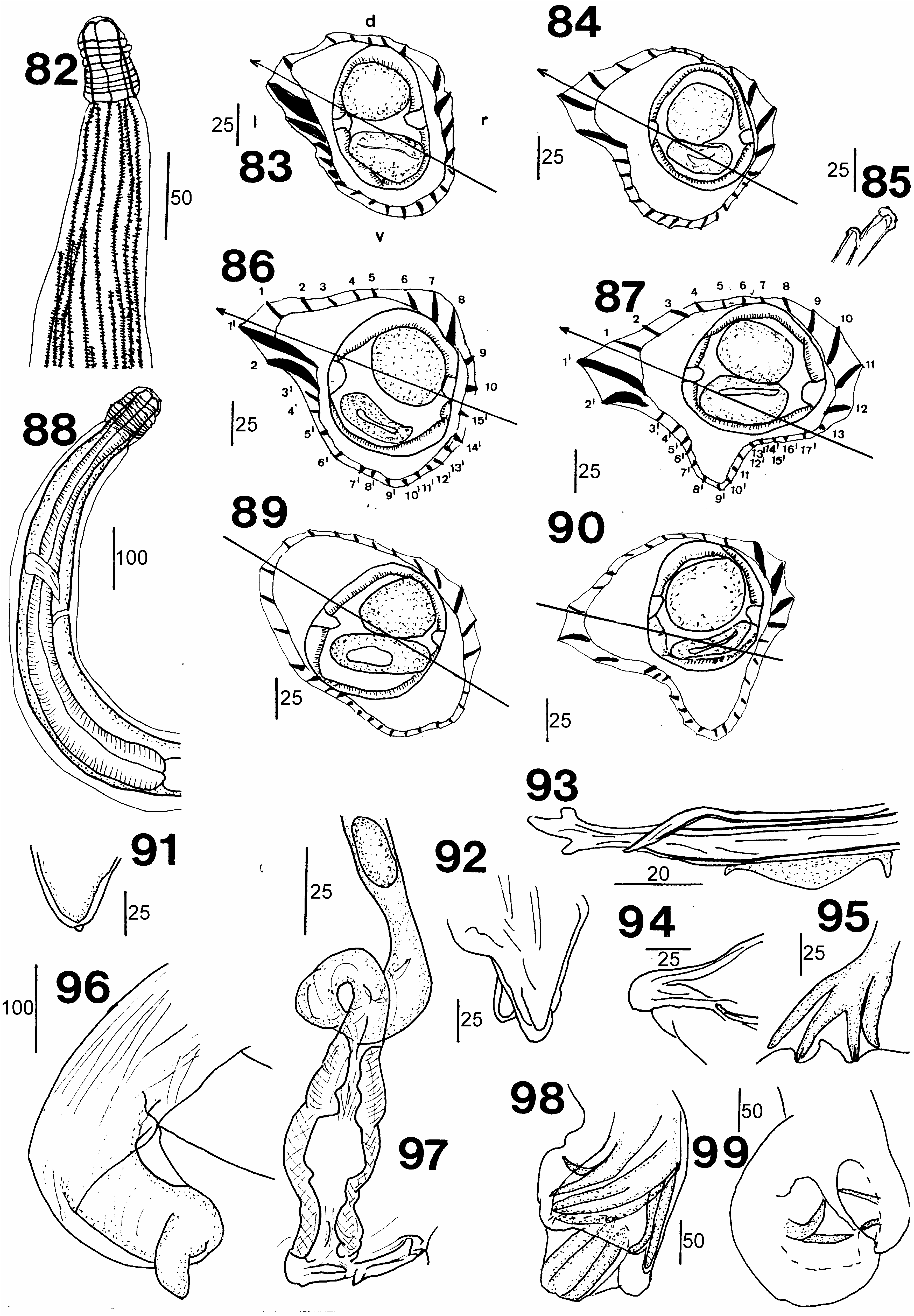
Paraheligmonelloides ennisae sp. nov.
( Figs 82–99 View FIGURES 82–99 )
Type host. Paramelomys rubex (Thomas)
Site in host. Small intestine.
Material examined. Holotype male, allotype female from Paramelomys rubex from hill south of Tifalmin (5° 7´S; 141° 25´E), West Sepik District, Sanduan Province, Papua New Guinea, coll. T. Flannery 15. iv. 1986, AM W36799 View Materials , W.36800; paratypes 9 males, 9 females, same data AM W.36801. GoogleMaps
Other material examined. From Paramelomys rubex Papua New Guinea, Sanduan Province; 1 male, 3 females Somoro Summit, Torricelli Mts (3° 24´S; 142° 8´E) GoogleMaps , 4 males, 10 females Fiminterr above Hindenburg Wall (5° 7´S; 141° 25´E) GoogleMaps , 1 female Ofektaman, Telefomin area : Indonesia, Papua ; 8 females Mokwan area, Arfak Mts (1° 6´S; 133° 56´E), AM W.36793, W.36794, W.36795, W.36796, W.36797, W.36798 GoogleMaps .
Etymology. The species is named after T. Ennis who helped collect many of the hosts.
Description. General: Nematodes slightly or tightly coiled; prominent cephalic vesicle present with about 9 annulations; buccal capsule vestigial. Mouth opening triangular with rudimentary lips; labial and cephalic papillae not observed. Oesophagus claviform. Nerve ring in mid oesophageal region; deirids and excretory pore immediately posterior to nerve ring. Synlophe (based on sections from 15 worms) of pointed longitudinal cuticular ridges in both sexes extends from posterior margin of cephalic vesicle to immediately anterior to bursa or vulva; 24–26 ridges in anterior, 26–29 in midbody, 28–31 in posterior. Axis of orientation of ridges passing from ventral right to dorsal left sides, inclined about 65° from sagittal axis in mid body; 9– 13 ridges dorsal side, 14–18 ridges ventral side. In anterior and mid body ridges 1´, 2´more developed than ridges 1, 2; ridges 1–4 decreasing, 5–8 increasing in size; ridges 1´–3´decreasing, 4´–17´about the same size. In posterior body ridges reduce in size.
Male: (Measurements of 10 males) Length 2800–3900 (3200), maximum width 102–142 (117). Cephalic vesicle 40–53 (47) long. Oesophagus 380–440 (415) long; nerve ring 190, deirids, excretory pore 220, 300 from anterior end. Bursa (based on 12 worms) with left lobe larger than right, left rays more robust; pattern of rays 2–3 for both lobes; rays 2, 3 diverge distally, recurved ventrally; rays 4, 5, 6, recurved dorsally. Dorsal lobe with median notch, shorter than laterals; dorsal trunk bifurcates at about two thirds its length, each branch dividing again at distal tip; terminal divisions, rays 9, 10, symmetrical, rays 8 arising at slightly different levels from dorsal trunk proximally to division of dorsal ray. Genital cone short, conical, dorsal lip more robust than ventral lip. Spicules filiform, right spicule with trifid tip, left spicule with curved simple tip 330–460 (386) long. Gubernaculum 37.5 long.
Female: (Measurements of 10 females) Length 3200–4200 (3750), maximum width 100–155. (130) Cephalic vesicle 43–53 (49.5) long. Oesophagus 410-450 (435) long; excretory pore, deirids 190, 240 from anterior end. Monodelphic ovejector, vulva near posterior end, 130, 190 from tail tip; vestibule and sphincter about same length, 100, infundibulum longer, looped in some specimens. Tail usually twisted dorsally then ventrally, coiled in some specimens, held in place by cuticle, 43, 46 long; tail tip conical, ending in small knob. Eggs thin shelled, ellipsoidal, up to 8 in utero 55–75 (70.5) by 34–49.5 (38.8).
Remarks. The new species belongs to the genus Paraheligmonelloides as it has a synlophe with a cuticular dilatation supported by a much more developed ridge 1’ than ridge 1. The orientation and relative sizes of the ridges is as described for the south east Asian members of the genus ( Ow Yang et al. 1983; Hasegawa et al. 1999). Of these P. ennisae with 26–29 ridges in the synlophe of the mid body is closest to P. singauwaensis Smales, 2009 with 20–23 ridges and P. amplicaudae with 21–24 ridges, all other species having fewer than 22 ridges. Paraheligmonelloides ennisae n. sp. can be distinguished from both P. amplicaudae and P. singauwaensis in having longer spicules 330–460, the right tip trilobed, compared with shorter spicules, 280–365 and 230–385 respectively, both with simple tips. Females of P. ennisae with a twisted posterior end and the tail tip ending in a knob also differ from females of P. amplicaudae which has an enlarged posteror body and P. singauwaensis which has the posterior end of the female neither twisted nor enlarged and the tail tip ending in a spike.
| T |
Tavera, Department of Geology and Geophysics |
| AM |
Australian Museum |
No known copyright restrictions apply. See Agosti, D., Egloff, W., 2009. Taxonomic information exchange and copyright: the Plazi approach. BMC Research Notes 2009, 2:53 for further explanation.