
Neurostigma xanthopterum New, 1980
|
publication ID |
https://doi.org/ 10.11606/1807-0205/2022.62.052 |
|
persistent identifier |
https://treatment.plazi.org/id/FE7D8790-5B7D-744E-FCF2-0D41B81BFE7F |
|
treatment provided by |
Felipe |
|
scientific name |
Neurostigma xanthopterum New, 1980 |
| status |
|
Neurostigma xanthopterum New, 1980 View in CoL ( Figs. 1-11 View Figure 1 View Figure 2 View Figure 3 View Figure 4 View Figure 5 View Figure 6 View Figure 7 View Figure 8 View Figure 9 View Figure 10 View Figure 11 )
Neurostigma xanthoptera New, 1980: 194 View in CoL ; Rafael et al., 1983:916 (Deposition of type); New,1984:3 (Ecology).
Neurostigma xanthopterum Lienhard & Smithers, 2002: 122 (Catalogue) View in CoL ; García-Aldrete & Mockford, 2009:667 (species list); Mendivil-Nieto et al., 2020:132 (new species and new record); Silva-Neto & GarcíaAldrete, 2020: 3 (species list); González-Obando et al., 2021: 83 (new species).
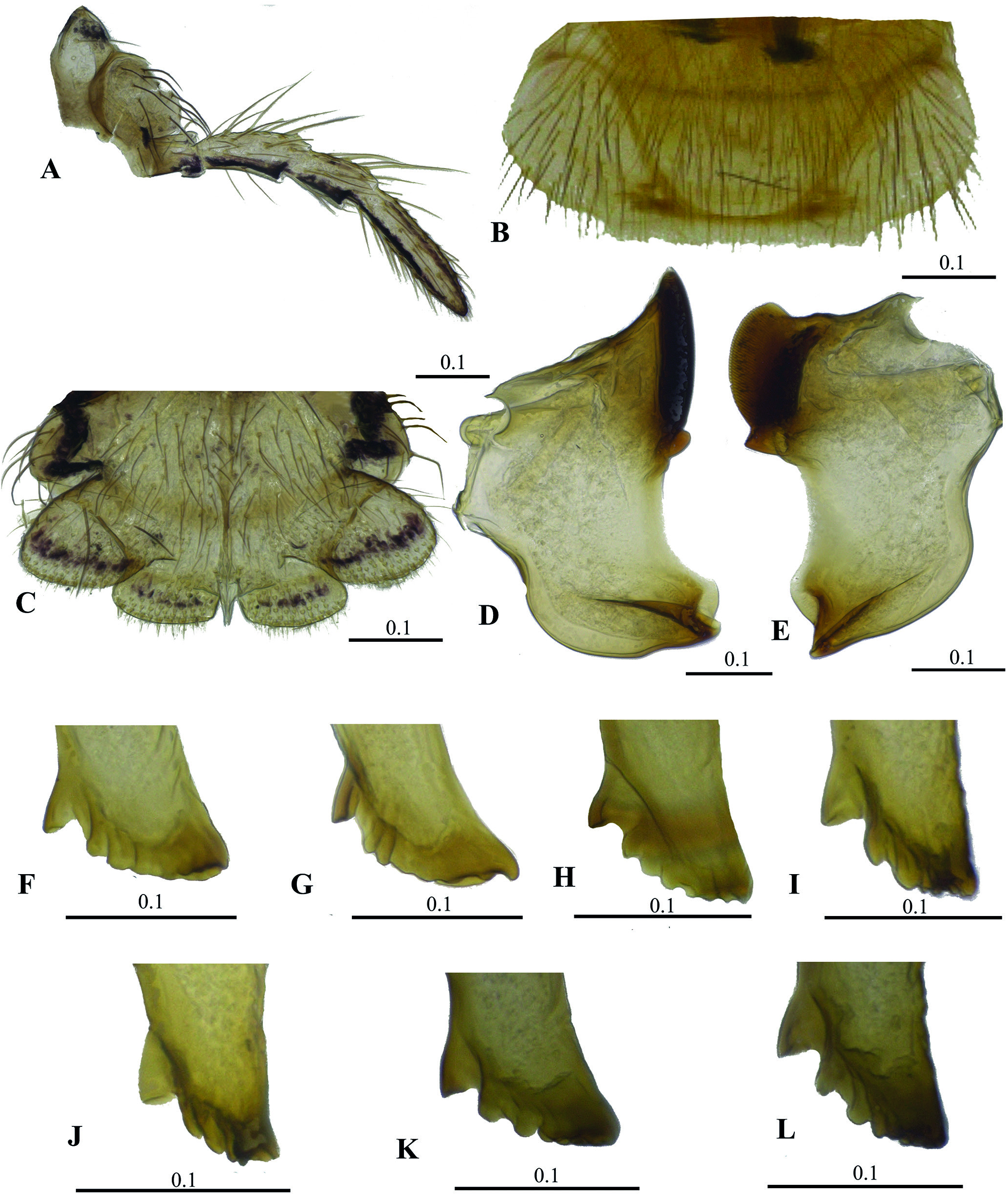
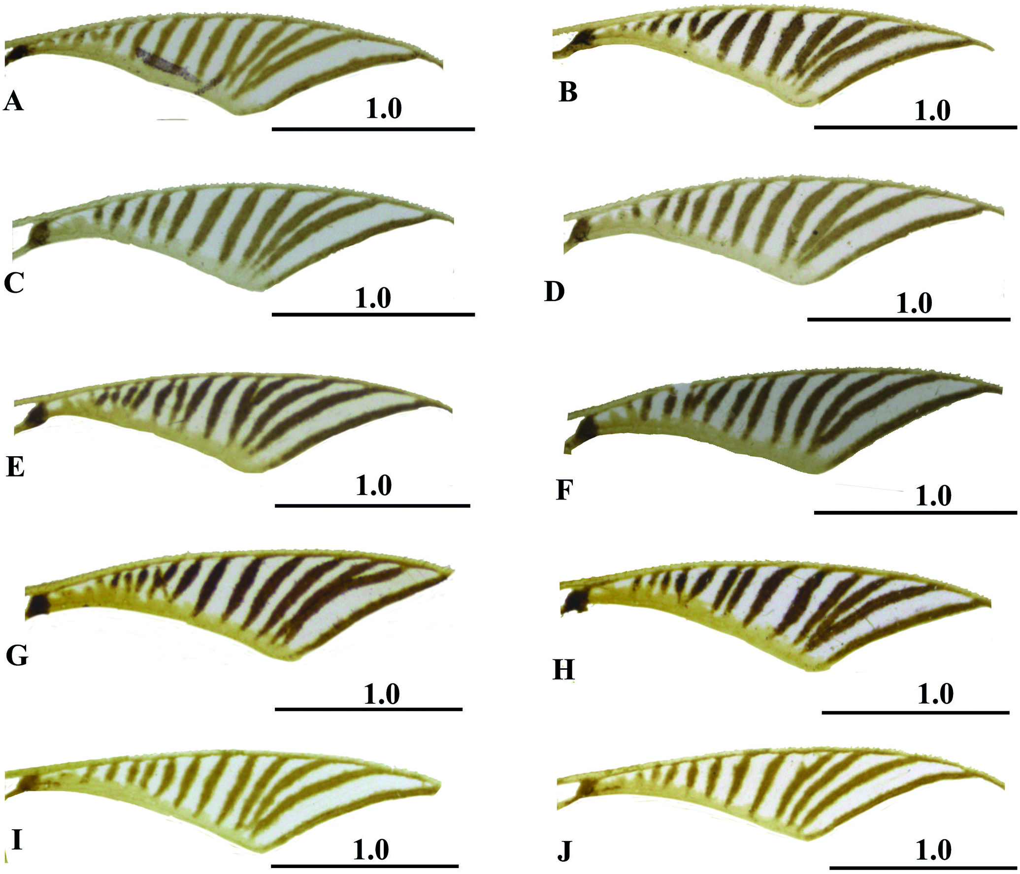
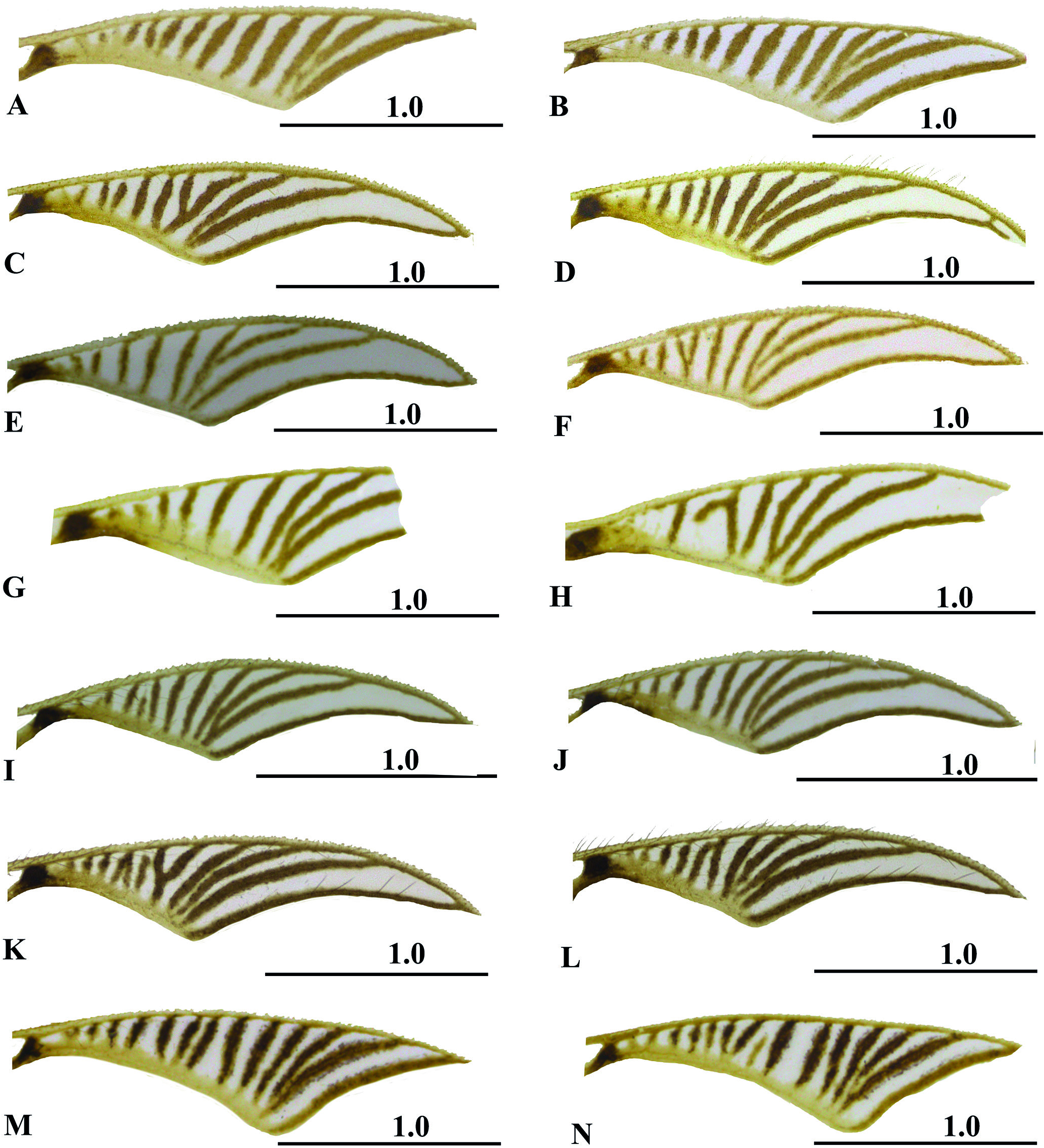
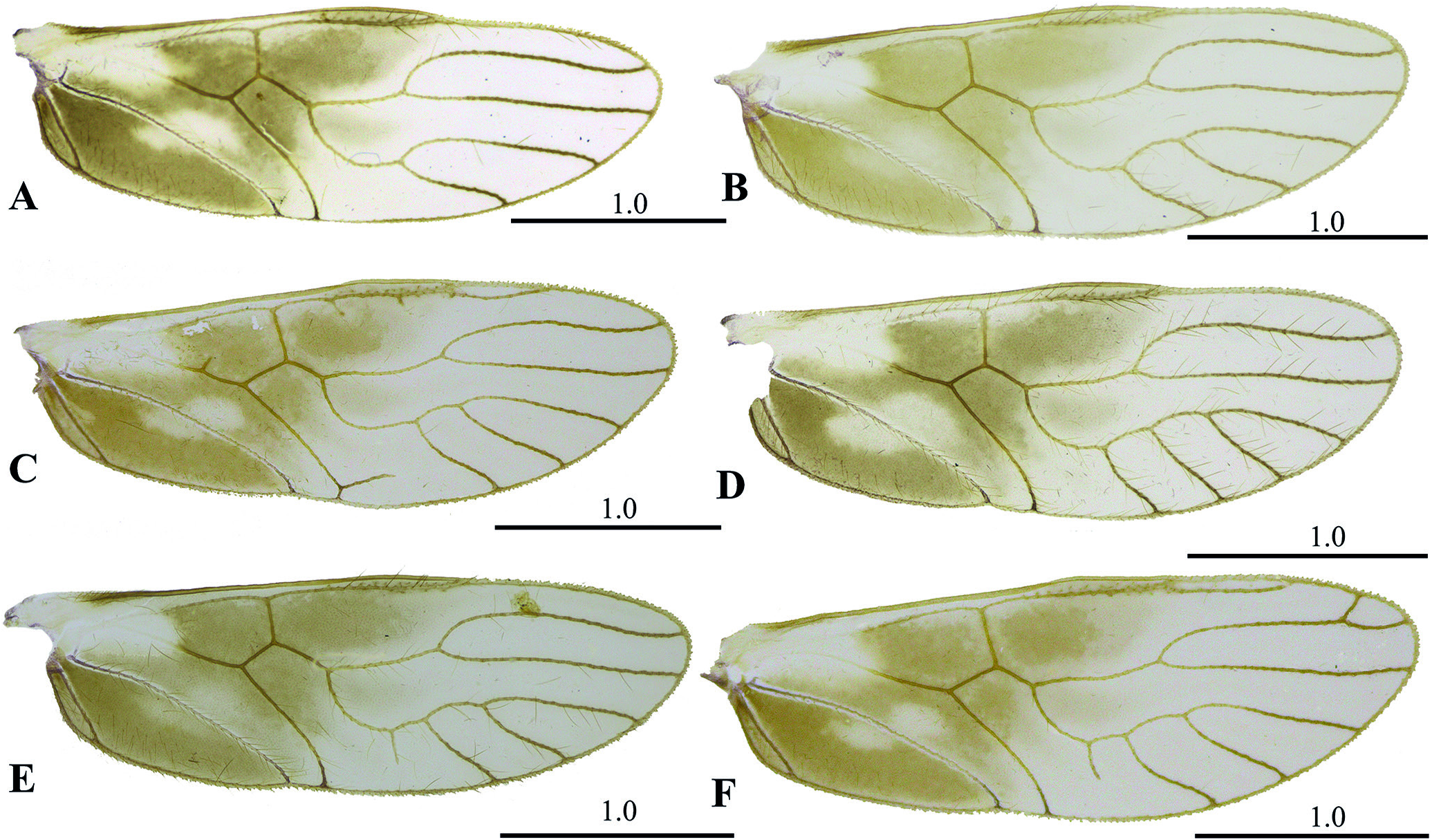
Update diagnosis: Head with vertex narrowed, bilobed, raised and distinctly emarginate medially ( Figs.1 View Figure 1 , 2A View Figure 2 , 3B View Figure 3 , 4A View Figure 4 ).Forewings with their proximal part half yellowish to brown, basally with a large club-shaped hyaline area between vein R and vein M+CuA; with large irregular dark brown spots from base of Pterostigma to CuP ( Figs. 10 View Figure 10 A-B, 10D-G) or present only from M+CuA to CuP ( Figs.2 View Figure 2 B-C,4B,10C).A small dark brown spot at the base of the Pterostigma and this one with its transverse veins pigmented along its entire length ( Figs.2 View Figure 2 B-C,7-9).Hindwings with their proximal part half yellowish to brown, with two hyaline areas almost elliptic ( Figs.2G View Figure 2 , 4C View Figure 4 , 11 View Figure 11 ). Phallosomal arch narrow and widening at the ends, anteriorly with a U-shaped area, then with a small transverse part, almost straight, posteriorly elongated vertically, ending in an almost elliptical area, with convex inner margin and blunt outer margin ( Fig. 2J View Figure 2 ). Endophallus heart-shaped, radular sclerites with small denticles, more pronounced in midline ( Fig. 2J View Figure 2 ). Hypandrium U-shaped, more sclerotized on the sides ( Fig. 2I View Figure 2 ). Ninth sternum with a v-shaped recess in the middle of the posterior margin and a deep recess in the anterior region, giving rise to two similar areas, almost elliptical and anteriorly open ending in a membranous area ( Fig. 4E View Figure 4 ). Gonapophyses with short, blunt end- ed heel, distally culminated, with v2 heavily sclerotized, with three setae on outer lobe; V3 weakly sclerotized, swollen and elongated ( Fig. 4G View Figure 4 ).
Male
As described by New (1980).
Female
Color: Body dark brown, with yellowish spots with tergal lobes of meso and metathorax light brown; thoracic pleura brown with clear spots ( Figs. 3 View Figure 3 A-C).Head with light reddish spots between front and postclypeus, and on the inner inferior margin of compound eyes and the antennal orbit; vertex yellowish;black compound eyes;ocelli blackened ( Figs. 3 View Figure 3 B-C, 4A). Escape and pedicel with dark spots, light brown flagelomeres. Maxillary palps with blackened contours ( Fig. 5A View Figure 5 ). Labrum basally yellow, distally pale yellow ( Fig. 5B View Figure 5 ). Labium anteriorly with black spots near the lateral margins of the prementum, with transverse linear spots on the labial palps and paraglosas ( Fig. 5C View Figure 5 ). Coxae and trochanters brown;femurs brown,darker basally,with four dark brown spots in the distal region;light brown tibiae;tarsomeres 1 and tarsomeres 2 light brown.Forewings with their proximal part half yellowish to brown, basally with a large club-shaped hyaline area between vein R and vein M+CuA; with large irregular dark brown spots from M+CuA to CuP; brown veins ( Figs. 3A View Figure 3 , 4B View Figure 4 ). A small dark brown spot at the base of the Pterostigma and this one with its transverse veins pigmented along its entire length ( Figs. 3A View Figure 3 , 4B View Figure 4 , 7 View Figure 7 ). Hindwings with their proximal part half yellowish to brown, with two hyaline areas almost elliptic; brown veins ( Figs. 3A View Figure 3 , 4C View Figure 4 ).Brown abdomen.Clunium hyaline. Subgenital plaque, gonapophysis and ninth sternum pale brown. Epiproct and paraproct almost hyaline. Eggs almost entirely yellow, with whitish margins ( Figs. 3 View Figure 3 D-E).
Morphology: Head with abundant macrosetae,mainly on vertex,it is narrowed,bilobed,raised and distinctly emarginate medially;small compound eyes ( Figs.3 View Figure 3 A-C,4A).Outer cusp of lacinial tip broad, with 5-7 denticles ( Figs. 5 View Figure 5 F-L; see also variation of males with 4-7 lacinia denticles ( Figs. 6 View Figure 6 F-W)). Labrum with abundant setae with labral sclerites curved outwards at bases ( Fig. 5B View Figure 5 ). Labial palps almost elliptical ( Fig. 5C View Figure 5 ). Mandibles asymmetrical, elongate and with outer margin angled ( Figs. 5 View Figure 5 D-E). Thorax with many setae and with four large sets of macrosetae, one on each side of the anterior scutal lobe and one on each side of the anterior margins of the lateral scutal lobes ( Figs.3 View Figure 3 B-C).Forewings with narrow pterostigma extending distally, with number and shape of transverse veins varying ( Fig. 7 View Figure 7 , see also variation of males ( Figs. 8-9 View Figure 8 View Figure 9 )); Rs, proximal to transverse vein r-m, strongly convex; R₂₊₃ and R₄₊₅ almost touching basally and separating distally; M with five branches ( Fig. 4B View Figure 4 ). Hindwings elongate; M simple, unbranched,R₂₊₃ vein slightly curved at the base and R₄₊₅ vein almost straight ( Fig.4C View Figure 4 ).Subgenital plate broad,U-shaped, no pigmented area,setae as illustrated ( Fig. 4D View Figure 4 ).Ninth sternum with a v-shaped recess in the middle of the posterior margin and a deep recess in the anterior region,giving rise to two similar areas, almost elliptical and anteriorly open ending in a membranous area ( Fig. 4E View Figure 4 ). Gonapophyses with short, blunt ended heel, distally culminated, with v2 heavily sclerotized, with three setae on outer lobe; V3 weakly sclerotized, swollen and elongated ( Fig. 4G View Figure 4 ). Epiproct trapezoidal, with invaginations in the center and with setae as illustrated ( Fig. 4F View Figure 4 ). Paraprocto broad, almost elliptical, sensory fields with 28-29 tricobothria on basal rosettes;setae as illustrated ( Fig. 4F View Figure 4 ). Eggs almost elliptical, with an almost triangular tip like a peduncle ( Figs. 3 View Figure 3 D-E).
Measurements (in microns): FW:4666,HW: 3146,F: 910, T: 1694, t1: 523, t2: 158, f1: 577, f2: 406, f3: 521, Mx2: 126, Mx4: 264, IO: 626, D: 351, d: 279.


No known copyright restrictions apply. See Agosti, D., Egloff, W., 2009. Taxonomic information exchange and copyright: the Plazi approach. BMC Research Notes 2009, 2:53 for further explanation.
|
Kingdom |
|
|
Phylum |
|
|
Class |
|
|
Order |
|
|
Family |
|
|
Genus |
Neurostigma xanthopterum New, 1980
| Reategui, Natália Soares, Rafael, José Albertino & Silva-Neto, Alberto Moreira da 2022 |
Neurostigma xanthopterum
| Gonzalez-Obando, R. & Carrero-Gironza, N. & Mendivil-Nieto, J. & GarciaAldrete, A. N. 2021: 83 |
| Mendivil-Nieto, J. A. & Gonzalez Obando, R. & Carrero Gironza, N. S. 2020: 132 |
| Garcia-Aldrete, A. N. & Mockford, E. L. 2009: 667 |
| Lienhard, C. & Smithers, C. N. 2002: 122 |
Neurostigma xanthoptera
| New, T. R. 1984: 3 |
| Rafael, J. A. & Penny, N. D. & Harada, A. Y. 1983: 916 |
| New, T. R. 1980: 194 |