
Mitrapsylla aeschynomenis, Rendón-Mera & Burckhardt & Cavichioli & Queiroz, 2020
|
publication ID |
https://doi.org/ 10.11646/zootaxa.4887.1.1 |
|
publication LSID |
lsid:zoobank.org:pub:B9A17D69-EBE7-49F4-AB01-54CA617FED02 |
|
DOI |
https://doi.org/10.5281/zenodo.4338379 |
|
persistent identifier |
https://treatment.plazi.org/id/03A687A2-8745-FF81-58C7-C701F836F8BF |
|
treatment provided by |
Plazi |
|
scientific name |
Mitrapsylla aeschynomenis |
| status |
sp. nov. |
Mitrapsylla aeschynomenis sp. nov.
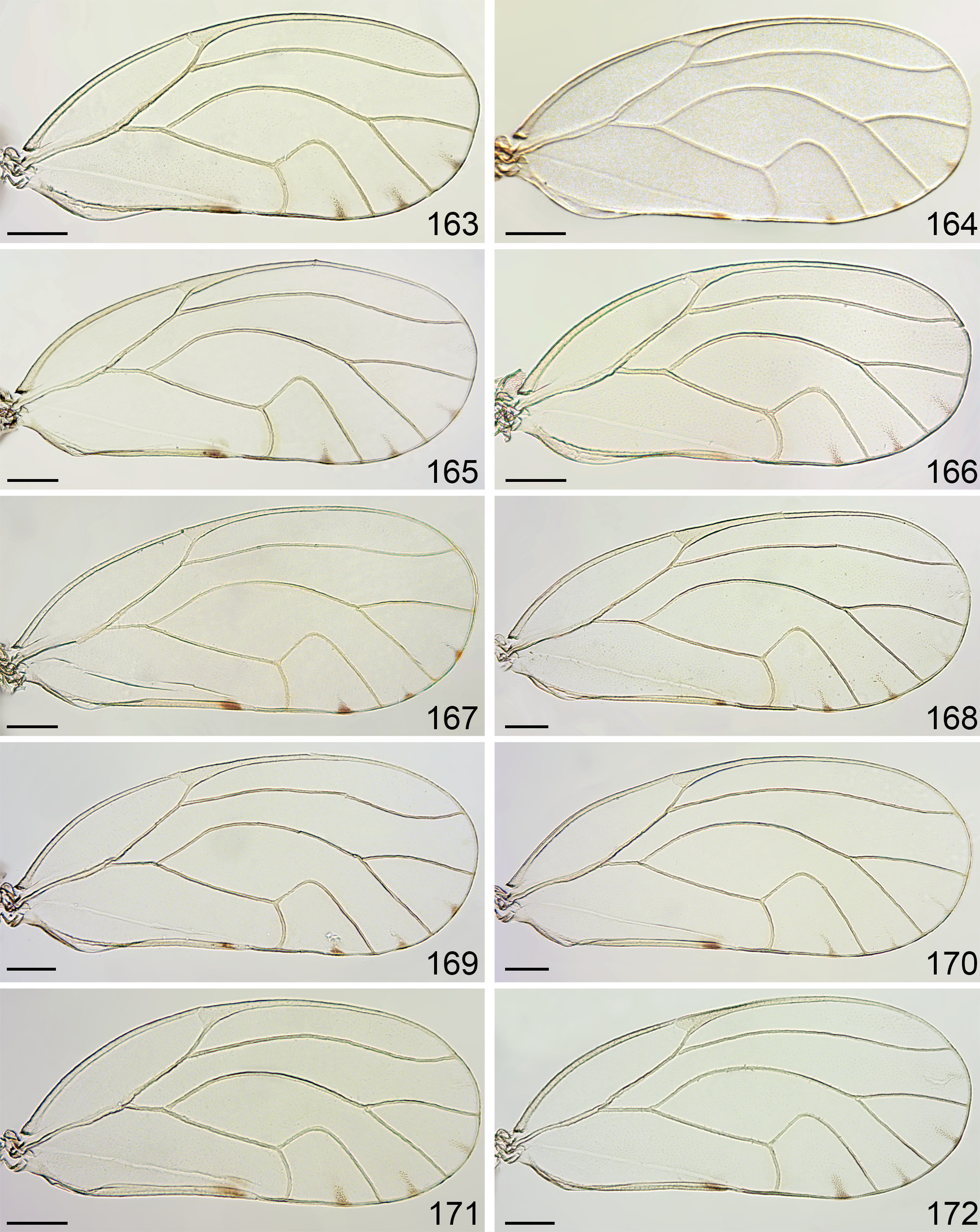
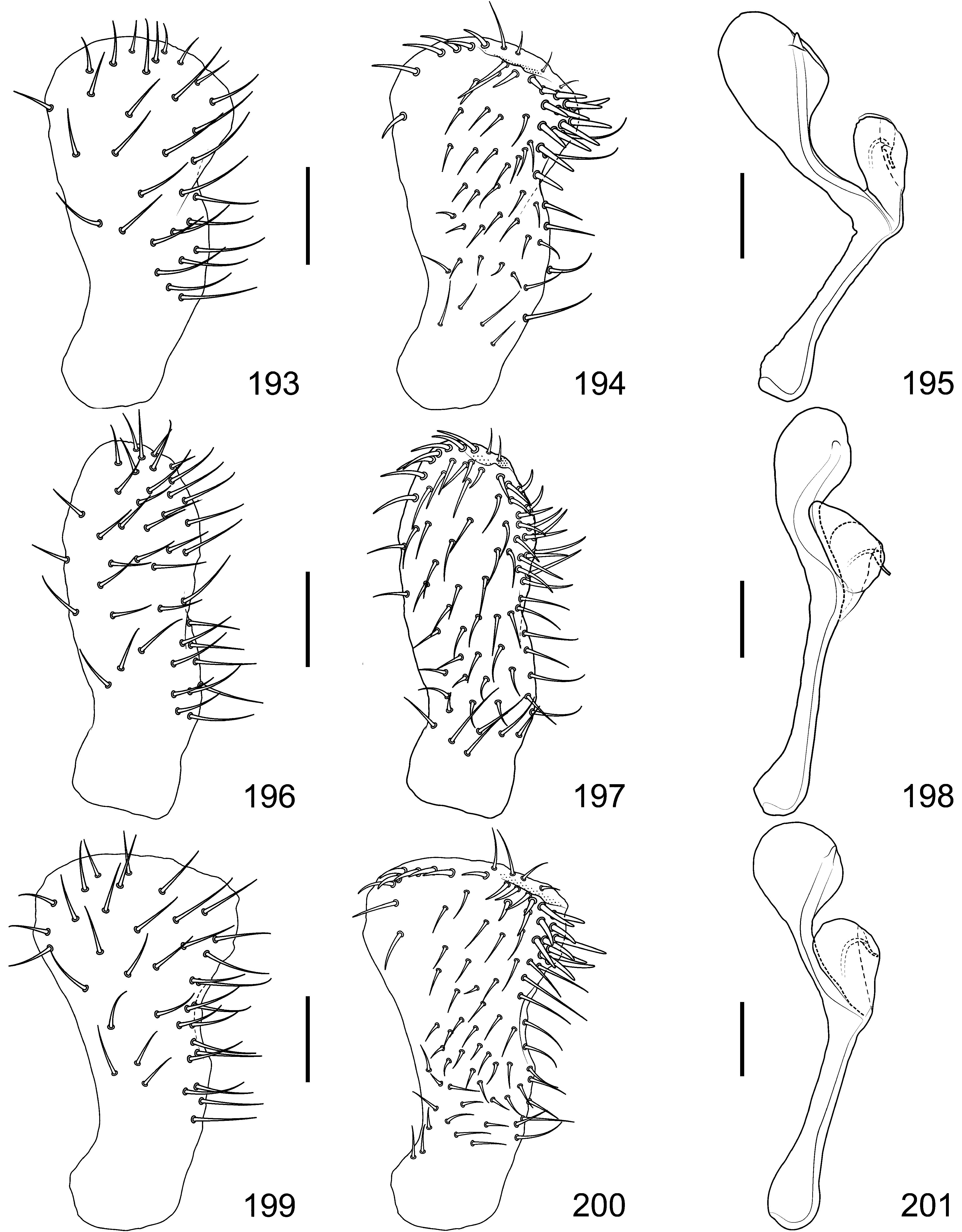
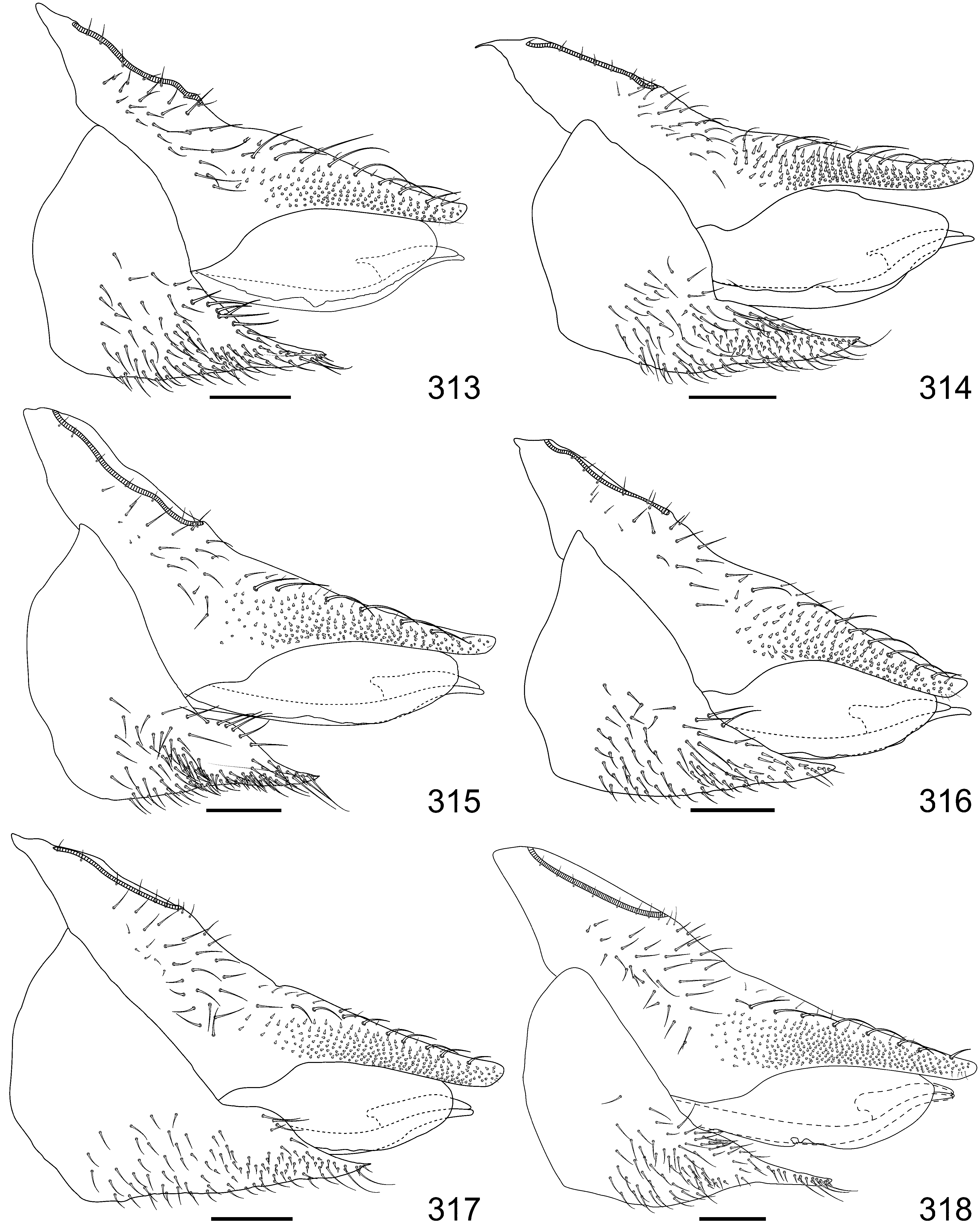
( Figs 103 View FIGURES 103–112 , 133 View FIGURES 133–147 , 163 View FIGURES 163–172 , 193 View FIGURES 193–201 ‾195, 283, 313, 343, 373)
LSID: urn:lsid:zoobank.org:act:5C0B006E-F20D-43E0-A8A9-5C57EABEA4D5
Material examined. Holotype ♁, Brazil: Maranhão, Araioses, Povoado Parangi , BR-402 9 km from MA-PI border, -3.0633, -41.9983, 30 m, 29.vi.2016, Aeschynomene paniculata (D. Burckhardt & D.L. Queiroz) , #210(2) ( DZUP 215391 View Materials , dry). GoogleMaps
Paratypes. Maranhão: 50 ♁, 52 ♀, 4 immatures, same data as holotype (D. Burckhardt & D.L. Queiroz), #210(2) ( DZUP, NHMB, dry, slide mounted, 70% ethanol) GoogleMaps .
Diagnosis. Genal process, in dorsal view, expanded; with broadly or narrowly rounded apex. Paramere, in lateral view, clavate; anterior margin almost straight in median two quarters, somewhat abruptly curving towards apex; apex directed posteriorly; sclerotized ridge medially; in dorsal view, sclerotised ridge irregularly inward directed. Aedeagus complex unipartite; in lateral view, ventral process with apical expansion larger than dorsal lobe.
Description. Colouration. Body with whitish striped-pattern; variation: somewhat faint; vertex with stripe along posterior margin weak or absent; mesoscutellum sometimes with additional stripe along anterior margin; older specimens with markings with dark outline. Head and thorax light yellowish-brown, light orange to light orange-brown. Gena sometimes light brownish anteriorly and ventrally; genal process lighter than rest of gena. Eye grey to dark red; ocelli colourless to orange. Antenna light yellow, segments 1–2 sometimes darker. Clypeus yellow to orange, lighter medially and darker along edges; rostrum light to dark yellow. Thorax usually with margins of sclerites darker. Pronotum usually slightly lighter than rest of thorax. Mesopraescutum rarely with posterior half irregularly coloured. Forewing colourless to slightly yellowish; veins dark yellow to light brown, sometimes slightly darker towards apex; pterostigma concolorous or slightly lighter than veins. Hindwing colourless. Fore- and midleg dark yellow with tarsi brown to dark brown, hindleg light yellow with apical tarsomere sometimes brownish. Abdomen lighter to concolorous with rest of body, sometimes darker ventrally; intersegmental membranes light straw-coloured; spiracular sclerites concolorous with tergites. Male terminalia irregularly light yellow to yellowishbrown, proctiger usually darker than rest of terminalia. Female terminalia irregularly light yellow, proctiger darker than subgenital plate and brownish apically.
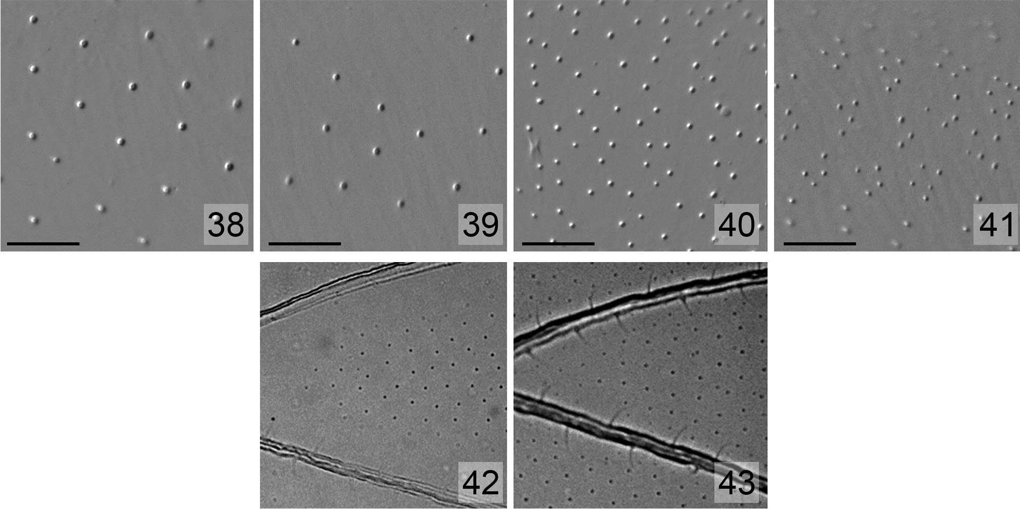
Structure. Body length ♁ 2.1–2.2 mm (2.14± 0.05 mm), ♀ 2.2–2.4 mm (2.26± 0.07 mm) (5 ♁, 5 ♀). Genal process ( Fig. 133 View FIGURES 133–147 ) expanded, evenly or irregularly narrowing towards broadly or narrowly rounded apex, 0.4–0.5 times as long as vertex along midline. Antenna 1.9–2.1 times as long as head width; longest terminal seta shorter than segment 10. Apical labium segment 0.1–0.2 times longer than head width and 0.6–0.7 times longer than median segment. Forewing ( Fig. 163 View FIGURES 163–172 ) 2.7–2.8 times as long as head width, 2.2–2.3 times as long as wide, obovoid or suboval, narrowly or broadly rounded apically; vein M+Cu 1 0.3–0.4 times as long as Cu 1; ratio a/b 1.4–1.6; ratio c/d 0.8–1.0; ratio e/f 0.6–0.9. Surface spinules usually moderately spaced, forming rhomboids ( Fig. 39 View FIGURES 38–43 ), denser towards apex of cells; absent, much reduced or covering apical half of cell c+sc, covering apical half, third or all cell r 1, apical half of cell r 2, around radular areas of cells m 1, m 2 and cu 1 (seldom much reduced), m 2 basally, and most of cell cu 2; leaving spinule-free spaces along veins ( Fig. 42 View FIGURES 38–43 ). Metatibia 0.6–0.7 times as long as head width.
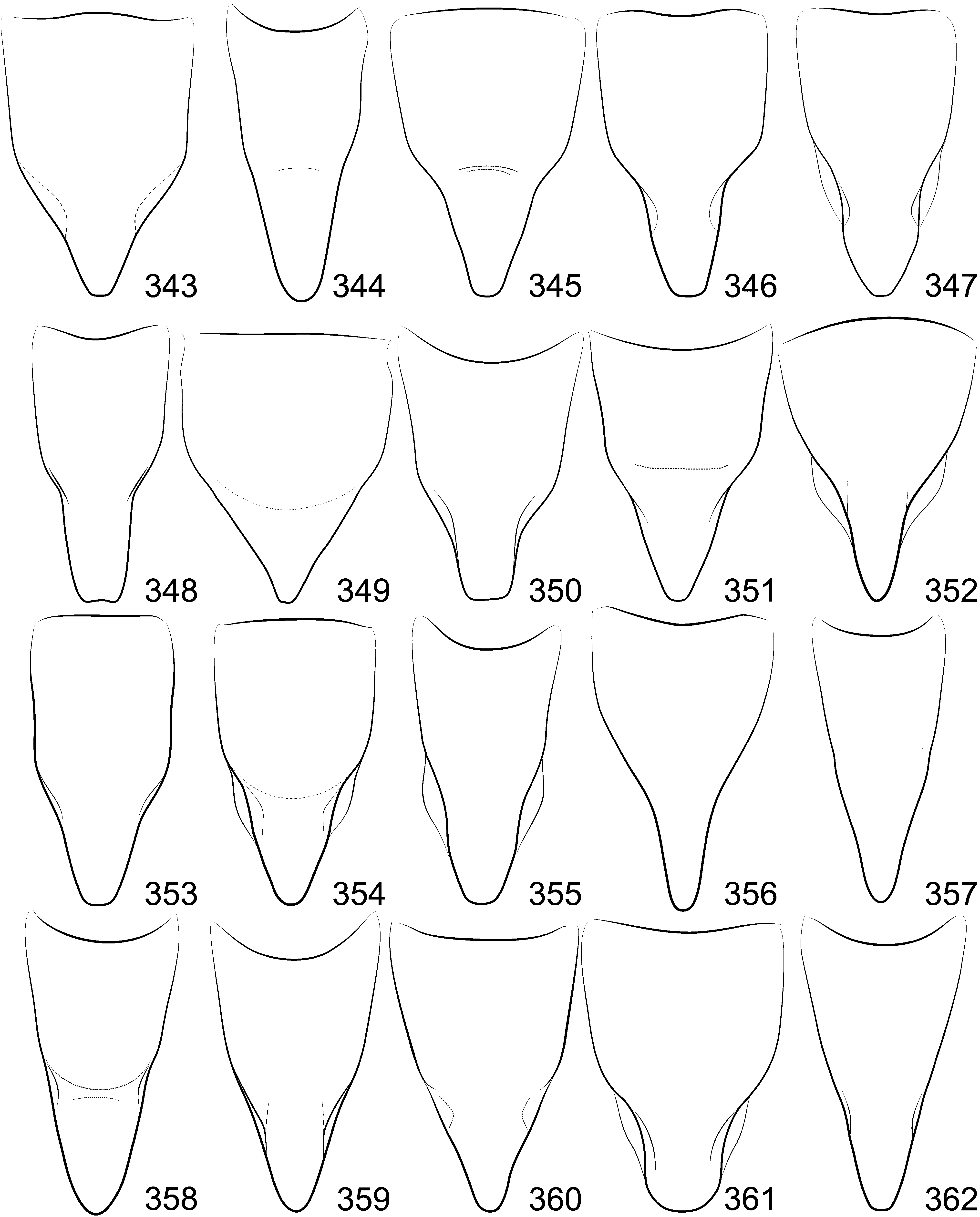
Terminalia. Male. Proctiger, in lateral view, 0.4 as long as head width; with long, blunt or weakly tapered, weakly to strongly down-curved posterior lobe. Paramere, in lateral view ( Figs 193 View FIGURES 193–201 ‾195) 0.8 times as long as proctiger; clavate, moderately expanded apically; anterior margin almost straight in median two quarters, somewhat abruptly curving towards apex; posterior margin angulate and expanded in about apical third, weakly convex in basal two thirds; apex irregularly rounded, slightly directed posteriorly, with sclerotised ridge medially ( Fig. 194 View FIGURES 193–201 ); inner surface ( Fig. 194 View FIGURES 193–201 ) covered with short setae, longer basally and along posterior margin, with row of thick setae along apical anterior margin, several thick setae below sclerotised ridge, and group of stout setae on apical posterior margin; in dorsal view ( Fig. 283 View FIGURES 283–312 ), sclerotised ridge irregularly inward directed, bearing posterior tooth. Aedeagus ( Fig. 195 View FIGURES 193–201 ) complex unipartite; in lateral view, dorsal lobe obovoid; ventral process weakly upturned, with apical expansion larger than dorsal lobe, irregularly globular, bearing short, conical tubercle.—Female ( Fig. 313 View FIGURES 313–318 ). Proctiger, in lateral view, 0.9–1.0 times as long as head width; dorsal outline weakly to strongly concave distal to circumanal ring, apical extension slightly to strongly sinuous, apex slightly to moderately upturned, smoothly obliquely truncate; circumanal ring 0.3 times as long as proctiger. Subgenital plate, in lateral view, 0.5–0.6 times as long as proctiger; apex well-developed; ventral outline almost straight to slightly sinuous, sometimes slightly notched submedially; covered with medium long setae in median third and ventrally throughout, shorter setae in apical third, long setae at apex, and group of long setae on dorsum subapically, with seta-free patch subapically; in ventral view ( Fig. 343 View FIGURES 343–362 ), lateral margins somewhat unevenly, strongly narrowing at half towards narrow, subtruncate apex.
Measurements (in mm) (3 ♁, 3 ♀). HW ♁ 0.58–0.59 (0.59±0.01), ♀ 0.58–0.60 (0.59±0.01); AL ♁ 1.16–1.26 (1.21±0.05), ♀ 1.15–1.23 (1.18±0.04); LAB2 ♁ 0.13–0.15 (0.14±0.01), ♀ 0.13–0.14 (0.14±0.01); LAB3 ♁ 0.09, ♀ 0.08–0.09 (0.09±0.01); FL ♁ 1.56–1.65 (1.61±0.05), ♀ 1.58–1.66 (1.62±0.04); TL ♁ 0.38–0.41 (0.40±0.02), ♀ 0.38– 0.41 (0.39±0.02); MP 0.24–0.25 (0.25±0.01); PL 0.20; DL 0.24– 0.27 (0.25±0.01); FP 0.52–0.56 (0.54±0.02).
Etymology. Named after its host-plant genus, Aeschynomene .
Distribution. Brazil: Maranh„o.
Host-plant. Aeschynomene paniculata Vogel (Leguminosae, Papilionoideae , Aeschynomeneae).
Habitat. Unknown.
Comments. Mitrapsylla aeschynomenis sp. nov. resembles M. aurantia sp. nov., M. cubana Crawford and M. didyma sp. nov. in the body dimensions and the paramere weakly expanded apically in lateral view, with sclerotised ridge medially; but differs in the expanded genal process with rounded apex (rather than subconical with acute apex), the anterior margin of the paramere almost straight in median two quarters and somewhat abruptly curving towards apex, and the sclerotised ridge irregularly inward directed in dorsal view.


No known copyright restrictions apply. See Agosti, D., Egloff, W., 2009. Taxonomic information exchange and copyright: the Plazi approach. BMC Research Notes 2009, 2:53 for further explanation.
|
Kingdom |
|
|
Phylum |
|
|
Class |
|
|
Order |
|
|
Family |
|
|
Genus |