
Metopiasoides jefersoni Asenjo
|
publication ID |
https://doi.org/ 10.11646/zootaxa.4097.4.11 |
|
publication LSID |
lsid:zoobank.org:pub:81880E86-F327-4794-92F0-043D4578786D |
|
DOI |
https://doi.org/10.5281/zenodo.6063584 |
|
persistent identifier |
https://treatment.plazi.org/id/03AD2A5F-FFB9-CF18-ACB7-F933FC6CF90E |
|
treatment provided by |
Plazi |
|
scientific name |
Metopiasoides jefersoni Asenjo |
| status |
sp. nov. |
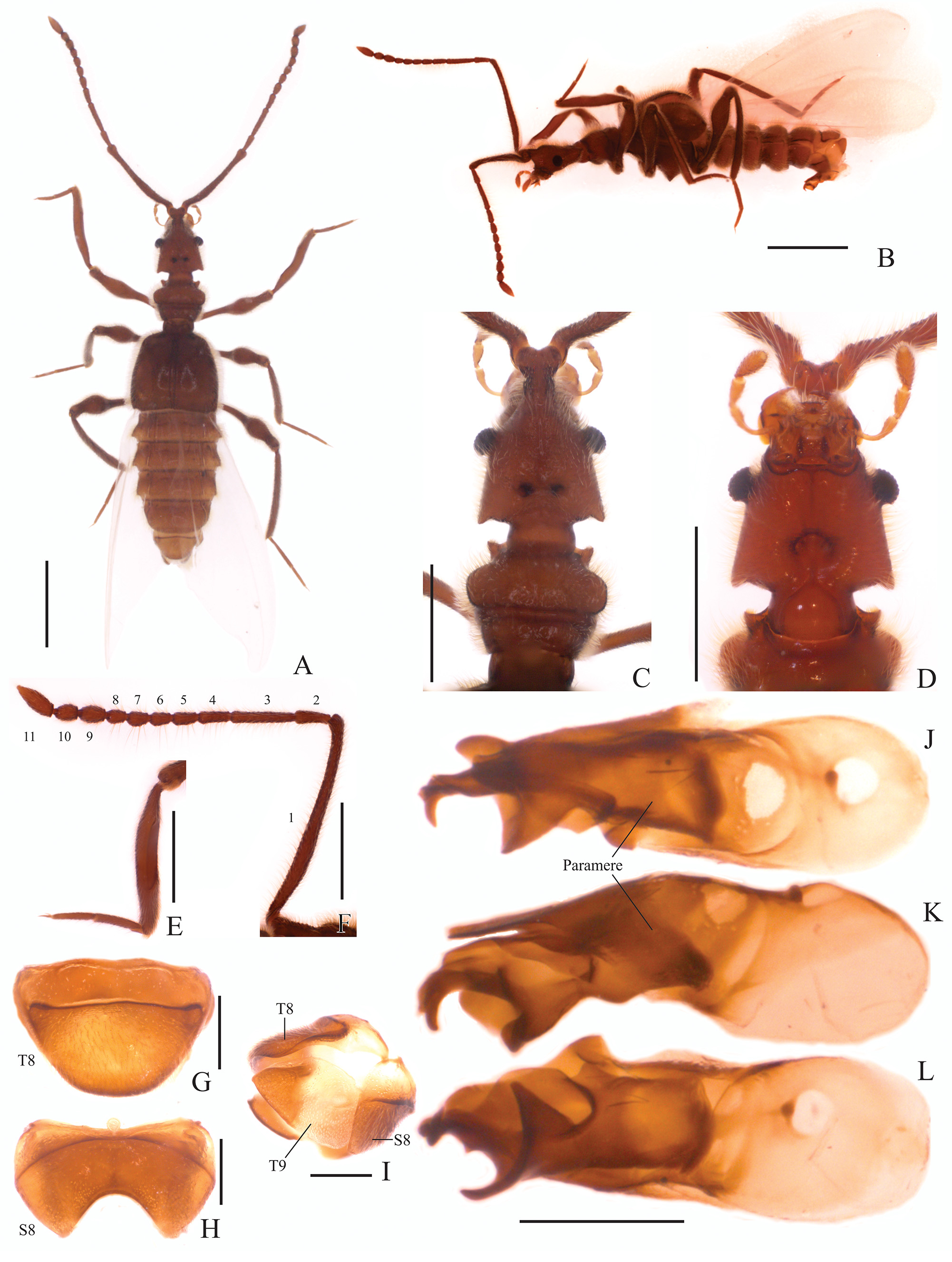
Metopiasoides jefersoni Asenjo View in CoL , new species ( Figs 1 View FIGURE 1 A–L)
Type material (1 ♂). Holotype: PERU: ♂, labeled “ PERU: CU[Cusco state], Santa Teresa / distrito, 13°02'42.9"S [South], / 72°38'39.5"W [West], 11.xii [December].2014, / 2035m [altitude in relation to sea level], Pan trap (yellow), / leg. J. Suarez De la Cruz”; “ HOLOTYPE [red label]/ Metopiasoides / jefersoni sp. nov. Asenjo / Desig. Asenjo, 2016 ” ( MUSM).
Diagnosis. Among Metopiasoides species, M. jefersoni is similar to M. grandior Comellini from Panama (Cerro Colorado), in having the posterolateral margin of the head with a cone-like prolongation ( Fig. 1 View FIGURE 1 A,C). Metopiasoides jefersoni is distinguished by the medioapical emargination of sternite VIII ( Fig. 1 View FIGURE 1 H) and the shape of the aedeagus ( Fig. 1 View FIGURE 1 J–L).
Description. Holotype male, BL: 5.19. Body, mouthparts, antennae and tarsi reddish dark brown ( Fig. 1 View FIGURE 1 A–D).
Head: pyriform ( Fig. 1 View FIGURE 1 C–D), longer (HL: 0.86) than wide (HW: 0.68), anterior region distinctly narrower ending and slightly raised apically at the antennal tubercle. Posterior margin of head abruptly narrowing and with a small conelike projection at the posteriolateral angles. Neck almost half width of head and with margins rounded. Head with two vertexal foveae [vf] ( Fig. 1 View FIGURE 1 C) near posterior margin, connected by transverse sulcus. Vertex longitudinally impressed with a shallow sulcus running from anterior margin of antennal tubercle to neck. Ventral surface of head ( Fig. 1 View FIGURE 1 D) with long gular sulcus widening towards neck, interrupted in the posterior third by two gular foveae [gf] connected by a curved sulcus. Eyes ( Fig. 1 View FIGURE 1 C–D) prominent, situated at middle of head length and directed anteriorly. Antennae ( Fig. 1 View FIGURE 1 F) 2/3 of length of body, scape almost half antenna length, last three antennomeres gradually broadening. Scape length 1.55 mm, width 0.11 mm, pedicel shorter than scape (0.22: 0.09), antennomere 3 (0.38: 0.09) longer than wide, antennomere 4 (0.18: 0.09) longer than wide, antennomere 5 (0.14: 0.10) longer than wide, antennomere 6 (0.11: 0.10) subquadrate, antennomere 7 (0.11: 0.10) subquadrate, antennomere 8 (0.10: 0.10) quadrate, antennomere 9 (0.15: 0.11) longer than wide, antennomere 10 (0.14: 0.11) longer than wide, antennomere 11 (0.27: 0.09) longer than wide; all antennomeres covered by long microsetae.
Thorax: pronotum ( Fig. 1 View FIGURE 1 C) wider than long (PL: 0.60; PW: 0.72) with a weak median longitudinal sulcus. Pronotum convex with apices produced into acute projections, lateral margin to a deep transverse sulcus at half pronotum length. Pronotum with a slight transverse impression at base. Pronotal basal margin concave. Prosternum with lateral procoxal fovea [lpcf]. Mesoventrite with prepectal fovea [ppf] and lateral mesosternal fovea [lmsf]. External margin of mesocoxal cavities with lateral mesocoxal foveae [lmcf]. Metaventrite with two pointed tubercles in the middle.
Elytra: wider than long (EL: 1.08; EW: 1.20), sides gradually broadening apically ( Fig. 1 View FIGURE 1 A). Posterior margins concave, humeri with an obtuse tubercular swelling and sutural stria [ss] present. Elytron with two basal elytral foveae [bef] at anterior margin, one close to the elytral suture, while the second is smaller and located near the middle of elytral width at base. Apical-lateral margin of elytra distinctly notched. Flight wings ( Fig. 1 View FIGURE 1 A–B) well developed, three times as long as elytra when fully extended and covered by minute setae.
Legs: Legs long and slender. Femora thickened in apical half. Tibiae curved and similar in length to femora, all tibiae thickened at apex. Protibiae lacking microsetae on the concave, mesial face, which is carinate. Tarsi 3-segmented, first tarsomeres very short and last 2 tarsomeres longer, tarsomere 2 longer that segment 3; all tarsi with a single claw and a minute accessory seta.Procoxae, mesocoxae conical and prominent, metacoxae transverse, region that articulates with the trochanter conical shaped. Procoxae with small prosternal process apically pointed, mesocoxae widely separated, metacoxae contiguous.
Abdomen: strongly marginated, with five visible tergites (morphological tergites IV–VIII), tergite III reduced to small plate under elytra, tergite VIII with rounded apex. Tergites IV–VII bordered by distinct paratergites. Sternite III with transversal depressed plate completely nude and placed under the metacoxae, this transversal plate have a longitudinal projected carina in the middle. Sternite VIII ( Fig. 1 View FIGURE 1 H) widely emarginated at apex.
Aedeagus: asymmetric with the parameres fused, median lobe slightly bulbous at base ( Fig. 1 View FIGURE 1 J–L).
Female. Unknown.
Habitat. The specimen was collected in the Peruvian Amazon forest at Urubamba Valley (at an altitude of 2035 meters).
Distribution. Only known from the type locality.
Etymology. This species is named in honor to my friend and colleague Jeferson Suarez De la Cruz, who collected the specimen and work at the MUSM.
Comments. The new species belongs to the genus Metopiasoides based on the relative size of the third antennomere ( Fig. 1 View FIGURE 1 F), which is longer than the second and fourth antennomeres. The second and fourth antennal segments are similar in length and width. The mesial face of the protibia ( Fig. 1 View FIGURE 1 E) is carinate.
No known copyright restrictions apply. See Agosti, D., Egloff, W., 2009. Taxonomic information exchange and copyright: the Plazi approach. BMC Research Notes 2009, 2:53 for further explanation.