
Metaleptobasis turbinata, Ellenrieder, 2013
|
publication ID |
https://doi.org/ 10.11646/zootaxa.3738.1.1 |
|
publication LSID |
lsid:zoobank.org:pub:77D1A6F6-C320-442B-AF31-83324E5EAF3B |
|
persistent identifier |
https://treatment.plazi.org/id/03E187ED-667A-FFF7-D7A8-FD76E16CF86F |
|
treatment provided by |
Felipe |
|
scientific name |
Metaleptobasis turbinata |
| status |
sp. nov. |
Metaleptobasis turbinata View in CoL new species
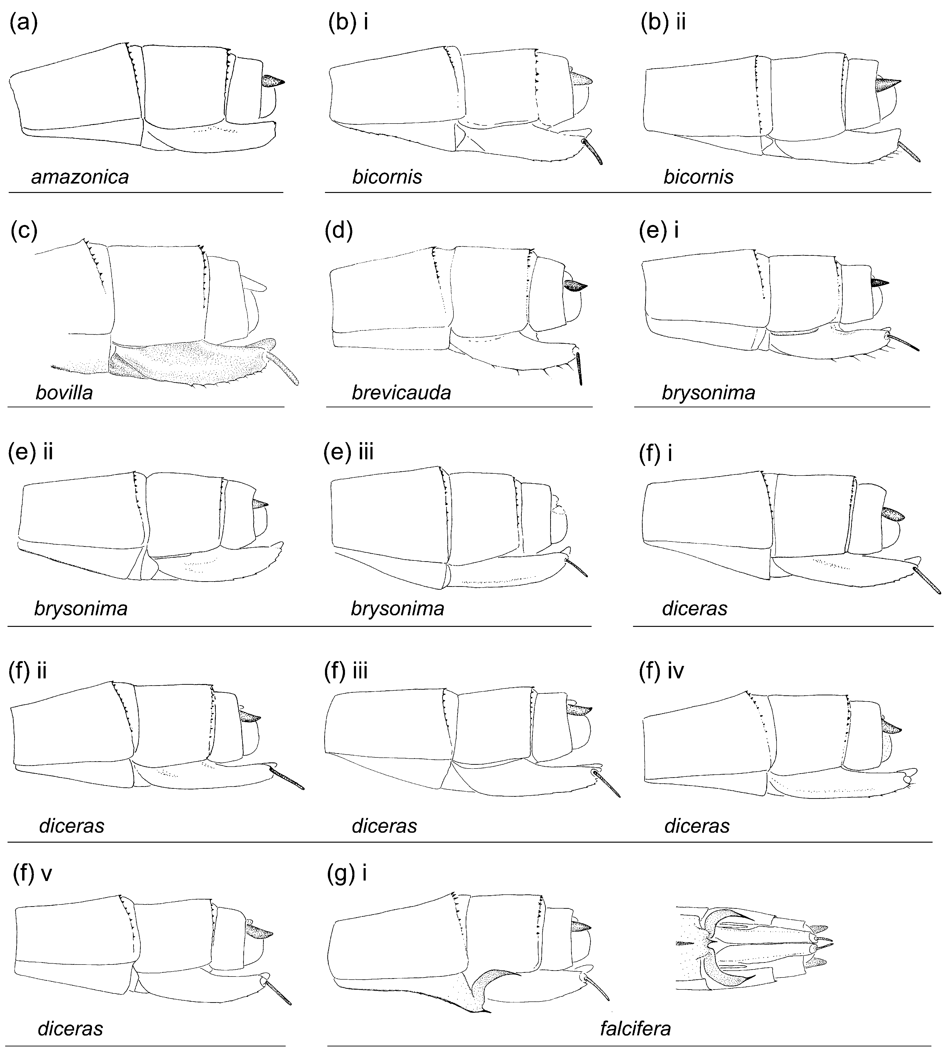
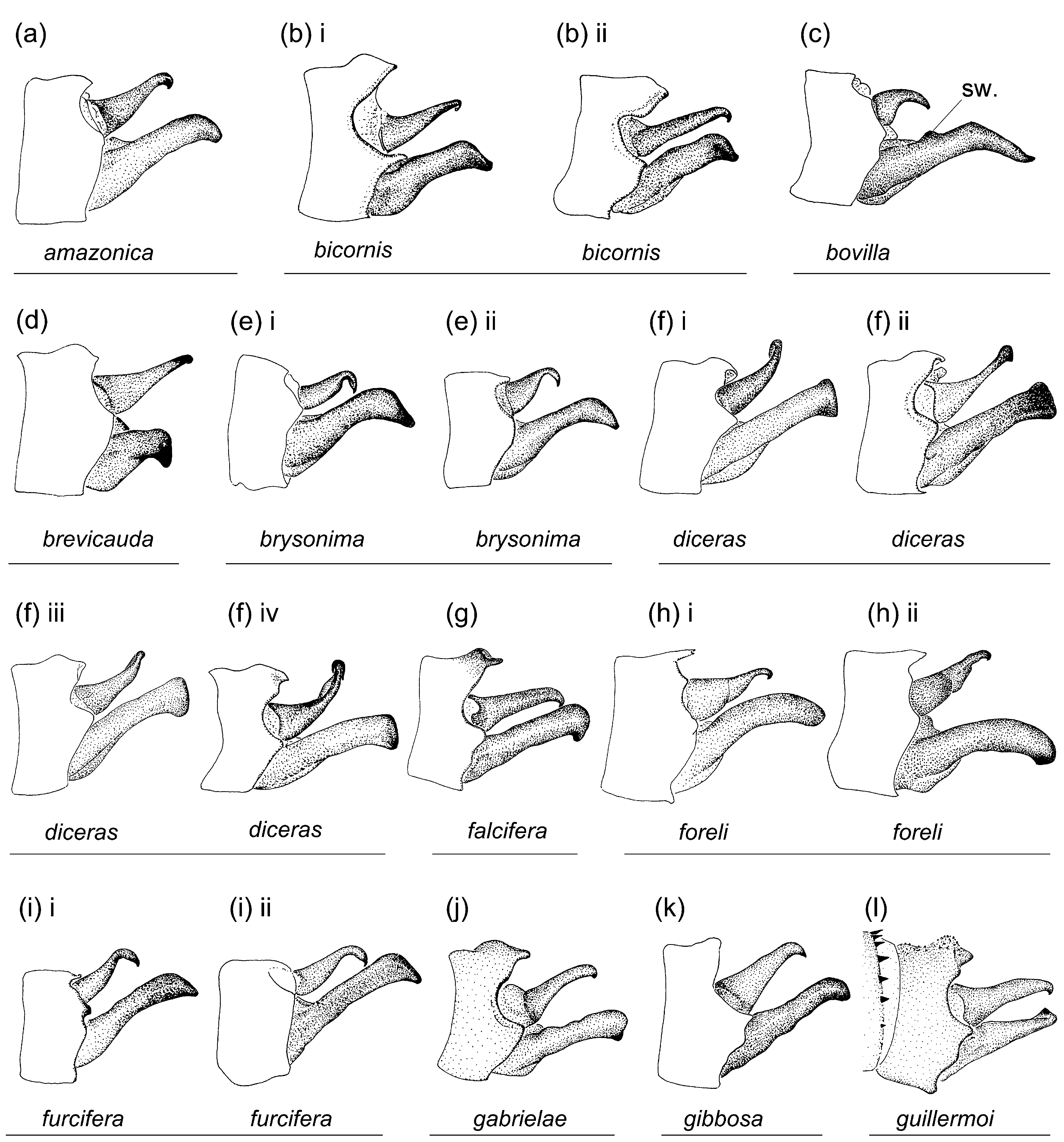
Figs. 1 View FIGURE 1 ae; 3ae; 4ae; 5ae; 8ae; 9ad; 10ae; 11ae; 12ae; 13m; 14a
Etymology. From Latin turbo (noun), meaning ' that which whirls', referring to its unique genital ligula with coiled flagella.
Types. ( all *) Holotype ♂: PERU, Loreto Dep., Tamshiyacu-Tahuayo Reserve , forest swamp (4°24'18''S, 73°14'38''W), 25 ii 2010, TF leg. [ RMNH]; 1 ♀ paratype: same data as holotype [ TF]. GoogleMaps
Specimens examined. Total: 1 ♂, 1 ♀.
Description of male holotype. Labium, base of mandible, and rear of head ivory; labrum pale brown with black line along latero-basal margins and a medio-basal black spot; eyes brown, in life bright green with orange dorsal spot ( Fig. 13m i View FIGURE 13 ); gena pale yellowish green with a black spot along frons; anteclypeus pale yellowish brown; postclypeus brown with black stripe along anterior margin; antefrons brown; postfrons and epicranium brown with black areas as depicted in Fig. 1 View FIGURE 1 ae i; postocular lobes rounded. Thorax. Color as described for genus, with middorsal dark stripe on pterothorax black with metallic green reflections, wider than inter-laminal sinus, slightly wider than 0.33 of mesanepisterna width, parallel sided, extending along sides of antealar sinus ( Fig. 3 View FIGURE 3 ae i). Pronotum anterior lobe smooth; anterior area of propleuron with a prominent triangular tubercle, opposite ventral margin of anterior lobe of pronotum; anterior and middle lobes of pronotum separated dorso-laterally by a groove; anterior margin of middle lobe of pronotum forming a bilobed crest; pronotum posterior lobe slightly trilobed, with medial lobe smoothly convex, and lateral lobes entire and smoothly convex, almost as long as medial lobe, with lateral corners rounded ( Fig. 4 View FIGURE 4 ae i). Mesanepisternal horns with bases separated, as long as 1.5 times mesostigmal plate width, of medium thickness, compressed antero-posteriorly, directed dorso-laterally, parallel to each other at base and diverging at tip, forming an angle of 30° with dorsum in lateral view ( Fig. 5 View FIGURE 5 ae i). Wings hyaline, veins dark brown; Pt sub-rectangular, with anterior and posterior sides longer than distal side, and membrane dark brown margined by pale brown; 12 pnx in Fw, 11 in Hw. Abdomen. Postmortem background color pale yellow on S1–10, in life pale greenish blue on S1–7, yellow on S8–10; S1 with a dark reddish brown dorsoposterior spot; dorsum of S2 dark reddish-brown, of S3–10 black, with a basal pale incomplete ring at anterior edge on S2–8 interrupted by dorso-longitudinal dark line, and with a small diffuse pale transverse spot near posterior margin on each side on S2–6, on S3–6 dark dorsal color posterior to pale transverse spot darker and extended ventrally along sides of lateral terga; sterna medio-longitudinal carina brown on S3, 7–8; cercus pale brown with apex black; paraproct pale yellow with distal 0.33 pale brown and apex black. Genital lobe short, less than 0.50 of anterior hamule height, smoothly curved; posterior hamule digit-like and small, shortly surpassing ventral margin of genital fossa in lateral view; curvature of basal segment of genital ligula marked by a slight concave depression; genital ligula distal segment sub-rectangular, with ratio maximum width/length of 0.60, with apex bearing a pair of long coiled flagella, lacking an ectal fold ( Fig. 8 View FIGURE 8 ae). Medial portion of S10 postero-dorsal margin ( Figs. 10 View FIGURE 10 ae; 11ae; 12ae) not projected posteriorly, lacking a medial incision, with a dorsal prominence. Cercus sub-cylindrical at base, with distal 0.66 compressed with medial concavity, gradually narrowing distally, about parallel sided in dorsal view ( Fig. 10 View FIGURE 10 ae); tip rounded in dorsal view, ending in a point directed ventrally ( Fig. 11 View FIGURE 11 ae); ratio of cercus length to S10 maximum length in lateral view 1.85; ratio of cercus length to paraproct length in lateral view 1.1; paraproct with base sub-cylindrical, distal 0.66 compressed with an inner concavity, narrowed at distal half ( Fig. 12 View FIGURE 12 ae); tip curved medially at about 90°, bluntly pointed.
Dimensions. Hw 20.2; abdomen 33.2; total length 39.7.
Female paratype (collected with the type). Head. As in holotype ( Fig. 1 View FIGURE 1 ae ii; 13m ii).— Thorax. As in holotype but lateral lobes of posterior lobe of pronotum slightly longer than medial lobe ( Fig. 4 View FIGURE 4 ae ii); mesanepisternal horns narrowing to tip, directed dorso-laterally at an angle of 40° with dorsum in lateral view ( Fig. 5 View FIGURE 5 ae ii); Pt pale brown margined with pale yellow; 12 (right) and 13 (left) pnx in Fw, 12 (right) and 11 (left) in Hw.— Abdomen. Color pattern as in holotype but medio-longitudinal carina brown on sterna S2–8; posterior margin of S8 sternum smooth, lacking any denticles, spines, or processes ( Fig. 9 View FIGURE 9 ad); external valve of ovipositor and stylus dark reddish brown; ovipositor surpassing slightly level of tip of cercus; cercus dark reddish brown, epiproct and paraproct brown.
Dimensions. Hw 20.6; abdomen 32.2; total length 38.7.
Diagnosis. Metaleptobasis turbinata shares antero-dorsal margin of middle lobe of pronotum forming a distinct crest (cr.) only with M. foreli and M. quadricornis . It differs from both of them, and from all other species in the genus, by the crest being bilobate ( Figs. 4 View FIGURE 4 ae; 5ae; vs. entire, Figs. 4h View FIGURE 4 ; 5h, y View FIGURE 5 ) and by its distal segment of genital ligula bearing a pair of long coiled latero-apical flagella ( Fig. 8 View FIGURE 8 ae; vs. lacking flagella, Figs. 8a View FIGURE 8 – ad).
The only known female differs from male in shape of pronotum posterior lobe and shape and orientation of mesanepisternal horns.
Habitat. Forest swamps.
Distribution. Loreto Dep. in N Peru ( Fig. 14a).
Phylogenetic analysis
Characters. The phylogenetic analysis was based on the 33 characters listed below (23 binary, 10 multistate). The plesiomorphic state (0) is given for each character (based on results of the analysis) followed by the apomorphic states (1, 2, 3); consistency index followed by retention index for each character are indicated in square brackets. Illustrations of characters states of outgroop taxa can be found in Garrison et al. (2010).
1 Labium medio-distal cleft: (0) shallower than 0.50 of labium length; (1) as deep as 0.50 or more of labium length [1, 1].
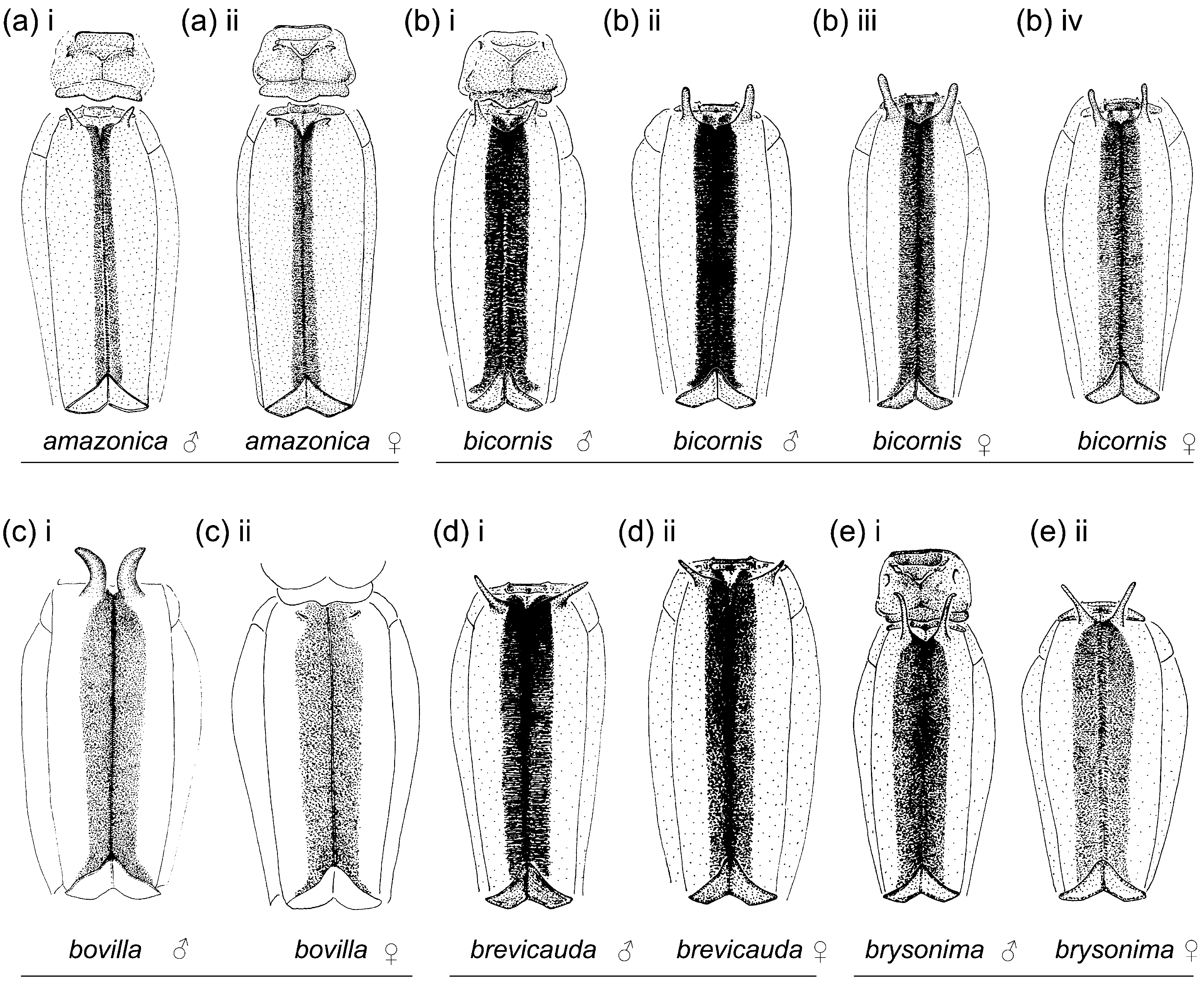
2 Black on dorsum of head: (0) extensive, covering about 0.50 to most of dorsum ( Figs. 1c, e, g–l, r, t–w, y View FIGURE 1 , ab– ac, ae); (1) covering entire dorsum; (2) limited to isolated spots and stripes covering less than 0.50 of dorsum ( Figs. 1a–b, d, f, m–q, s, x, z View FIGURE 1 –aa, ad) [0.4, 0.83].
3 Frons: (0) rounded; (1) angulate [0.5, 0.0].
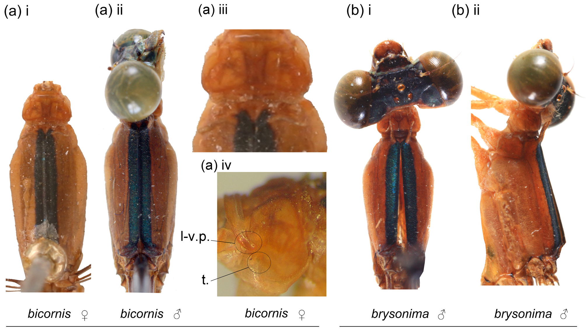
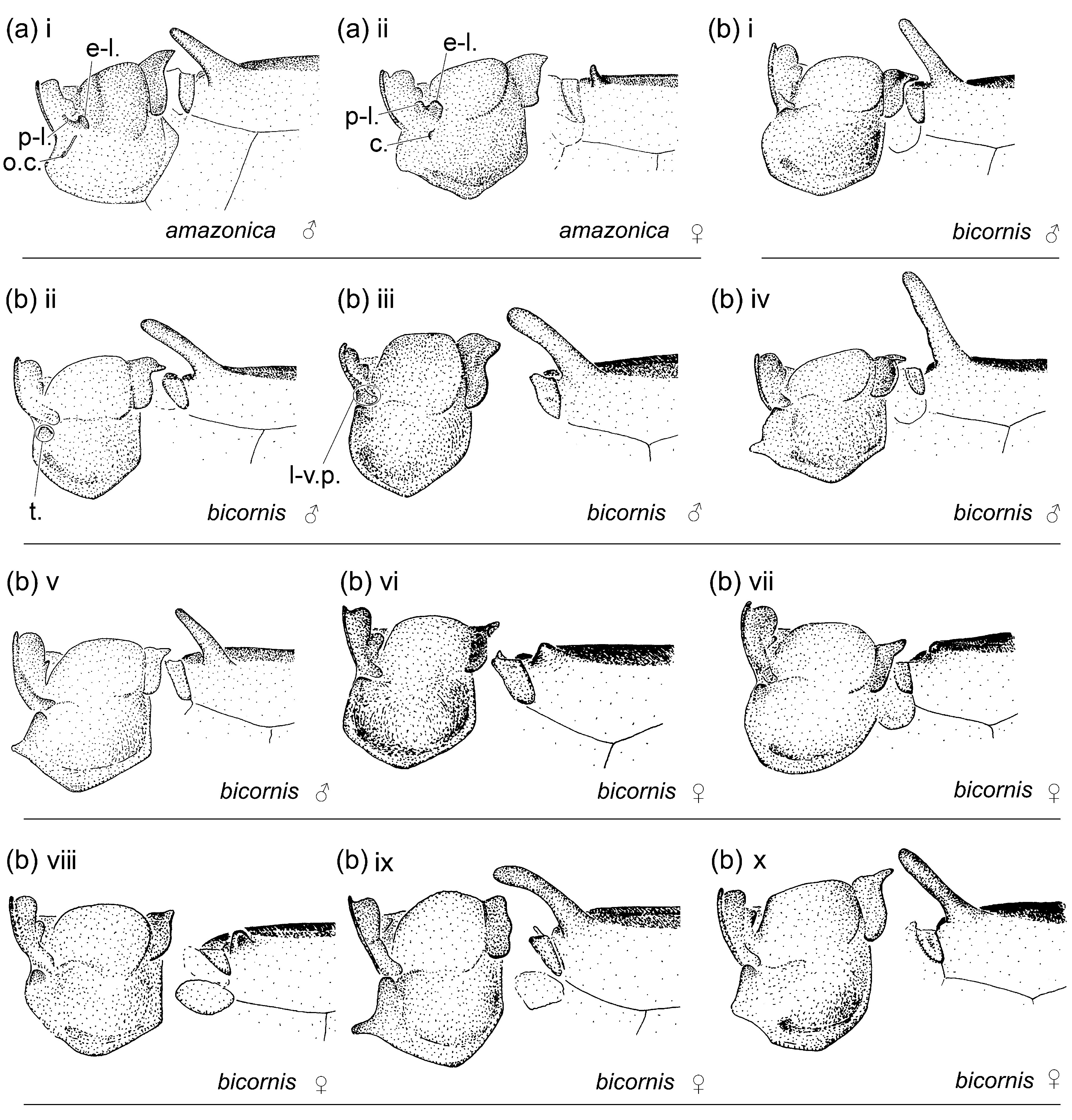
4 Latero-ventral margin of anterior lobe of pronotum on each side: (0) not projected ventro-posteriorly ( Figs. 5a, c–w, y View FIGURE 5 –ae); (1) projected ventro-posteriorly (l-v.p., Figs. 2a View FIGURE 2 iv; 5b, x) [1, 1].
5 Anterior portion of propleuron on each side with a rounded or plate-like tubercle: (0) absent ( Figs. 5a, c, f–g, m, o–q, s–t, w View FIGURE 5 , aa, ac–ad); (1) low (t., Figs. 5e, i, n, u, z View FIGURE 5 ); (2) prominent (t., Figs. 2a View FIGURE 2 iv; 5b, d, h, j–l, r, v, x–y, ab, ae) [0.33, 0.73].
6 Anterior portion of propleuron on each side with two sub-vertical crests: (0) absent ( Figs. 5a–e, g–o, r–z View FIGURE 5 , ab– ac, ae); (1) present (l.c., Figs. 5f, p–q View FIGURE 5 , aa, ad) [1, 1].
7 Anterior and middle lobes of pronotum dorso-laterally: (0) separated by an evident groove (g., Figs. 5a–o, r–z View FIGURE 5 , ab–ac, ae); (1) almost touching, separated by a narrow fissure (f., Figs. 5q View FIGURE 5 , aa, ad) [0.5, 0.66].
8 Anterior margin of middle lobe of pronotum: (0) smooth ( Figs. 4a–g; i–x; z View FIGURE 4 –ad); (1) forming a crest (c., Figs. 4h, y View FIGURE 4 , ae) [0.5, 0.5].
9 Paired ear-lobe projections on anterior portion of female middle lobe of pronotum: (0) absent ( Figs. 4b–f, h– k, m–q, s, u, x View FIGURE 4 –ab, ad–ae); (1) present, digit like or tongue-shaped, oriented laterally or postero-laterally ( Figs. 4a View FIGURE 4 iii, g, t, v–w, ac), flap like with a dorsal concavity, oriented dorso-ventrally ( Fig. 4l View FIGURE 4 ii), or upright ( Fig. 4r View FIGURE 4 vi–vii) [0.33, 0.71].
10 Posterior margin of female pronotum posterior lobe: (0) entire, bilobed, trilobed, or sinuous ( Figs. 4a–d, f–h, j View FIGURE 4 –ae); (1) with a deep medial u- or v-shaped incision ( Figs. 4 e, i View FIGURE 4 ) [1, 1].
11 Dorsal surface of female lateral portions of posterior lobe of pronotum: (0) smoothly convex or flat ( Figs. 4a– f, h–v View FIGURE 4 , ac, ae); (1) globose (gl., Figs. 4g, w View FIGURE 4 , ac) [1, 1].
12 Male mesanepisternal horns: (0) absent; (1) present, forming an angle of 0°–15° with dorsum in lateral view ( Figs. 5d, p, s, x View FIGURE 5 ); (2) present, forming an angle of 30°–90° with dorsum in lateral view ( Figs. 5a–c, e–o, q–r, t–w, y View FIGURE 5 –ae) [0.4, 0.77].
13 Vein descending from quadrangle forming: (0) a zigzag; (1) an unbroken straight line to wing margin (v.q., Fig. 6a View FIGURE 6 ) [1, 1].
14 Pterothoracic black color: (0) absent or including dark stripes or areas additional to a mid-dorsal stripe; (1) limited to a mid-dorsal stripe ( Figs. 3a–e View FIGURE 3 ) [1, 1].
15 Supplementary tooth of pretarsal claw: (0) well developed; (1) vestigial [1, 1].
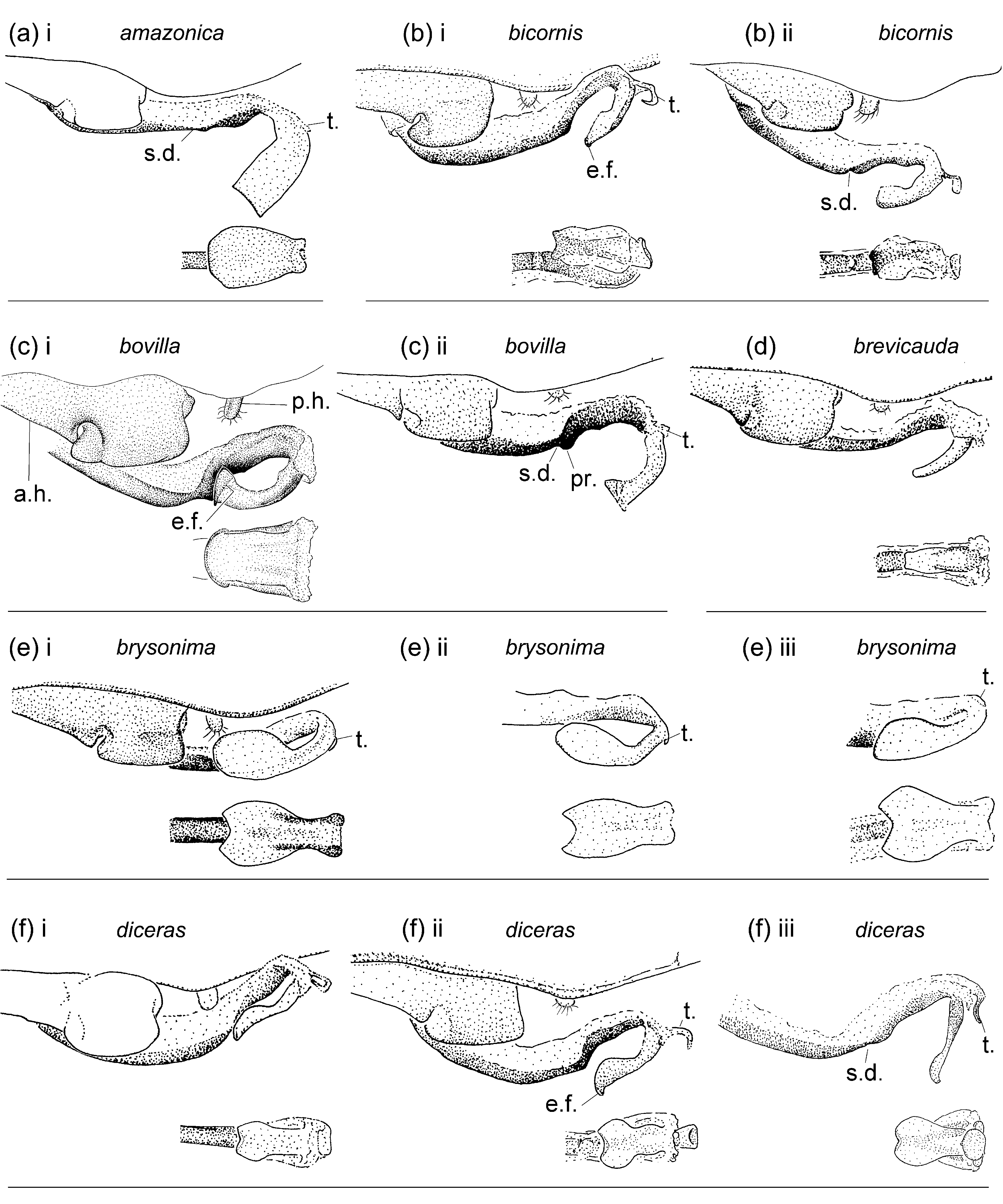
16 Male genital lobe (g.l.) height: (0) distinctly shorter than 0.50 of anterior hamule height ( Figs. 8a–f, h–v, x View FIGURE 8 –ab, ad–ae); (1) as high as or higher than 0.50 of anterior hamule height ( Figs. 8g, w View FIGURE 8 , ac) [1, 1].
17 Male posterior hamule (p.h.): (0) digit-like and small, with at most only tip surpassing ventral margin of genital fossa in lateral view ( Figs. 8a–f, h–i, m–q, s, u, x View FIGURE 8 –ab, ad–ae); (1) laminar and large, clearly surpassing ventral margin of genital fossa in lateral view ( Figs. 8g, j–l, r, t, v–w View FIGURE 8 , ac) [1, 1].
18 Deep concave depression (d.d.) at curvature of basal segment of genital ligula: (0) absent ( Figs. 8a–f, h–i, m–q, s, u, x View FIGURE 8 –ab, ad–ae); (1) present ( Figs. 8g, j–l, r, t, v–w View FIGURE 8 , ac) [1, 1].
19 Inner fold of genital ligula: (0) present; (1) absent ( Figs. 8a View FIGURE 8 –ae) [1, 1].
20 Shape of genital ligula distal segment in ventral view: (0) sub-rectangular ( Figs. 8b, d, k–l, n, p–s, x, z View FIGURE 8 –aa, ad– ae); (1) sub-quadrate ( Figs. 8a, c, m, o View FIGURE 8 ); (2) pear-shaped, distinctly widened sub-apically ( Figs. 8e–f, g–j, t–w, y View FIGURE 8 , ab–ac) [0.33, 0.73].
21 Apex of distal segment of genital ligula in ventral view: (0) transverse or with a shallow concavity ( Figs. 8a–b, d–g, i–r, t View FIGURE 8 –aa, ac–ae); (1) with a deep concavity or cleft, as deep as wide or deeper ( Figs. 8s View FIGURE 8 , ab); (2) convex ( Figs. 8c, h View FIGURE 8 ); (3) pointed [0.43, 0.2].
22 Ectal fold on apex of genital ligula distal segment: (0) absent ( Figs. 8a, i, m–o, q, s, x View FIGURE 8 –ae); (1) narrow ( Figs. 8b, f–g, j–l, p, r, t–w View FIGURE 8 ); (2) wide ( Figs. 8c, h View FIGURE 8 ) [0.4, 0.77].
23 Posterior margin of female S8 sternum: (0) smooth ( Figs. 9a–f, h–i, m–p, r–s, w–z View FIGURE 9 , ab–ac); (1) with 1 to 6 denticles or small spines ( Figs. 9k–l, q View FIGURE 9 ); (2) with a pair of strong spines ( Figs. 9j, u View FIGURE 9 ); (3) with a two ( Figs. 9g, t, v View FIGURE 9 ) or three-pronged curved or arcuate process ( Fig. 9 View FIGURE 9 ab) [1, 1].
24 Medial u-shaped incision or longitudinal slit on female S10 postero-dorsal margin: (0) present; (1) absent [0.5, 0.83].
25 Medial u-shaped incision or longitudinal slit on male S10 postero-dorsal margin: (0) absent ( Figs. 10c, g–h, j– l, r, t, v–w View FIGURE 10 , ab–ac, ae); (1) present ( Figs. 10a–b, d–f, i, m–q, s, u, x View FIGURE 10 –aa, ad) [0.14, 0.66].
26 Postero-medial projection on male S10 postero-dorsal margin: (0) absent ( Figs. 10c, k, s View FIGURE 10 , ae; 11c, k, s, ae); (1) present ( Figs. 10a–b, d–j, l–r, t View FIGURE 10 –ad; 11a–b, d–j, l–r, t–ad) [0.25, 0.73].
27 Sub-apical prominence on male S10 dorsal surface: (0) absent, dorsum flat ( Figs. 12e, h–i, s, y View FIGURE 12 ); (1) present ( Figs. 12a–d, f–g, j–r, t–x, z View FIGURE 12 –ae) [0.2, 0.69].
28 Articulated spur on ventral base of male cercus: (0) absent ( Figs. 11a View FIGURE 11 –ae); (1) present [0.33, 0.5].
29 Male cercus in dorsal view: (0) curved medially ( Figs. 10a–d, f–g, j–o, q–x, z View FIGURE 10 –ae); (1) about straight ( Figs. 10e, h–i, p, y View FIGURE 10 ) [0.25, 0.77].
30 Width of male cercus in dorsal view: (0) uniform ( Figs. 10q, r, t, w, z View FIGURE 10 , ad–ae); (1) narrowing to tip ( Figs. 10a, c– e, h–i, m–p, s, u–v, x–y View FIGURE 10 , aa–ac); (2) widening sub-apically ( Figs. 10b, f–g, j–l View FIGURE 10 ) [0.18, 0.4].
31 Tip of male cercus in dorsal view: (0) pointed ( Figs. 10a–e, g–n, p, r–y View FIGURE 10 , aa–ac); (1) rounded or blunt ( Figs. 10f, o, q, z View FIGURE 10 , ad–ae) [0.166, 0.28].
32 Male paraproct width of distal third in lateral view: (0) narrower than medial third ( Figs. 12a, c, m–n, q–r, z View FIGURE 12 ); (1) about as wide as medial third ( Figs. 12b, h, k–l, o, s, x–y View FIGURE 12 , ab, ae); (2) wider than medial third ( Figs. 12d– g, i–j, p, t–w View FIGURE 12 , aa, ac–ad) [0.2, 0.65].
33 Male paraproct tip: (0) with smooth medial surface and no apical tooth ( Figs. 11 View FIGURE 11 ab, ae); (1) with smooth medial surface and ending on an apical tooth ( Figs. 11a, c, g–h, j–k, m, o, q–w, y View FIGURE 11 , aa, ac–ad); (2) with a ridge (r.) on medial surface ending on an apical tooth (s.t., Figs. 11b, d–f, n, x, z View FIGURE 11 ); (3) with a ridge on medial surface with apical and basal teeth (te., Figs. 11i, l, p View FIGURE 11 ) [0.25, 0.4].
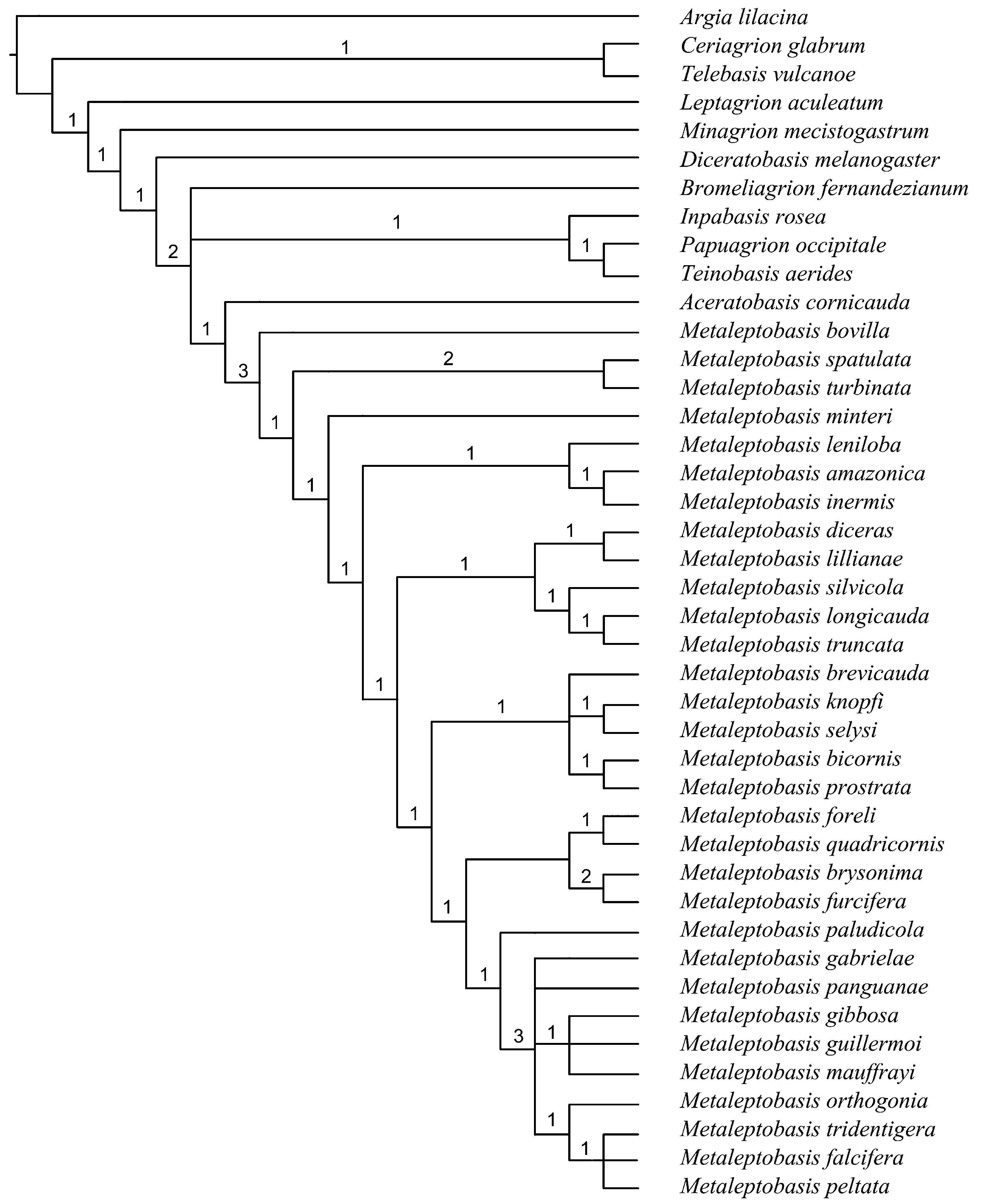
The phylogenetic analysis resulted in four equally parsimonious trees (length = 122, C.I. = 0.37, R.I. = 0.73). Bremer support values are shown on the branches of the consensus tree ( Fig. 15 View FIGURE 15 ). The four trees varied only in the arrangement of species within the outgroup and within one clade of Metaleptobasis . Within the outgroup, the two different arrangements obtained differ in the position of Bromeliagrion ; in one ( Fig. 16 View FIGURE 16 ) it is the sister group of (( Inpabasis ( Teinobasis + Papuagrion )) + ( Aceratobasis + Metaleptobasis )), and in the other one it is the sister group of ( Inpabasis ( Teinobasis + Papuagrion ), as part of the sister clade to ( Aceratobasis + Metaleptobasis ). The different arrangements within Metaleptobasis vary only in the position of M. panguanae , which is placed as sister to ( M. orthogonia ( M. tridentigera ( M. falcifera + M. peltata ))) or as sister to ((( M. gibbosa + M. guillermoi ) M. mauffrayi ) + ( M. orthogonia ( M. tridentigera ( M. falcifera + M. peltata ))). I show the tree with the latter arrangement as the basis for discussion ( Fig. 16 View FIGURE 16 ).
Metaleptobasis was recovered as part of the clade including taxa with an angulate frons and an articulated spur on ventral base of male cercus. Frons could have reverted to round in Bromeliagrion , and articulated spur could have being secondarily lost in Metaleptobasis . Within this clade, Metaleptobasis constitutes the sister group of Aceratobasis ( Fig. 16 View FIGURE 16 ), with which it shares the vein descending from quadrangle forming an unbroken straight line to wing margin (v.q., Fig. 6a View FIGURE 6 ).
The monophyly of Metaleptobasis is supported by two apomorphies: presence of male mesanepisternal horns ( Figs. 4–5 View FIGURE 4 View FIGURE 5 ), and pterothoracic dark color limited to mid-dorsal stripe ( Figs. 3 View FIGURE 3 ; 13 View FIGURE 13 ).
Within the genus, M. bovilla appears as sister to all other species. The similarities between its genital ligula and that of M. foreli —both presenting a convex apex with a wide ectal fold—are thus to be interpreted as homoplasies.
Metaleptobasis spatulata and M. turbinata are placed in turn as sister group of all remaining species ( Fig. 16 View FIGURE 16 ), forming a clade characterized by a prominent triangular plate-like tubercle on propleuron (t., Figs. 4 View FIGURE 4 ab, ae; 5ab, ae) and male paraproct tip with smooth medial surface and no apical tooth ( Figs. 10 View FIGURE 10 ab, ae; 11ab, ae). Both species also share mesanepisternal horns slightly compressed antero-posteriorly ( Figs. 4 View FIGURE 4 ab, ae; 5ab, ae), which was not coded for this analysis. The bilobed crest on anterior margin of middle lobe of pronotum of M. turbinata is thus interpreted as a convergence with the entire crest on anterior margin of middle lobe of pronotum of M. foreli and M. quadricornis .
Metaleptobasis minteri appears as sister to the remainder species, with which it shares the presence of a medial incision on postero-dorsal margin of male S10 ( Fig. 10s View FIGURE 10 ), interpreted as secondarily lost in the clade including M. mauffrayi . Several clades then branch off sequentially ( Figs. 15–16 View FIGURE 15 View FIGURE 16 ).
The first one, including M. amazonica , M. leniloba , and M. inermis , is defined by the squarish distal segment of genital ligula ( Figs. 8a, o, m View FIGURE 8 ).
A clade defined by the presence of two sub-vertical crests on each side on anterior portion of propleuron (l.c., Figs. 5f, p–q View FIGURE 5 , aa, ac) follows, including M. diceras , M. lillianae , M. longicauda , M. silvicola , and M. truncata .
Then M. bicornis , M. brevicauda , M. knopfi , M. prostrata , and M. selysi integrate a clade characterized by male paraproct tip with a sub-apical ridge on medial surface ending on an apical tooth ( Figs. 10b, d, n, x View FIGURE 10 ; 11b, d, n, x View FIGURE 11 ), representing the sister clade to all remainder species, which share head dorsum with extensive black color ( Figs. 1e, g–l, r, t–w, y View FIGURE 1 , ac).
Two clades are defined within this group, one including M. brysonima , M. foreli , M. furcifera , and M. quadricornis , which share absence of a sub-apical prominence on dorsum of male S10 ( Figs. 11e, h–i, y View FIGURE 11 ; 12e, h–i, y View FIGURE 12 ) and male cercus in dorsal view about straight ( Figs. 10e, h–i, y View FIGURE 10 ), and the other one comprising M. paludicola as sister group to M. gabrielae , M. panguanae , M. gibbosa , M. guillermoi , M. mauffrayi , M. tridentigera , M. orthogonia , M. falcifera , and M. peltata .
This last clade is defined by the presence of a narrow ectal fold (e.f., Figs. g, j–l, r, t–w, ac) on distal segment of genital ligula, which also occurs convergently in M. bicornis , M. diceras , and M. lillianae . The clade excluding M. paludicola has strong support ( Fig. 15 View FIGURE 15 ), and is characterized by the absence of the medial incision on dorsum of male S10 ( Figs. 10g, j–l, r, t, v–w View FIGURE 10 , ac) and by three apomorphies: male posterior hamule laminar and large, clearly surpassing ventral margin of genital fossa in lateral view; presence of a deep concave depression at curvature of basal segment of genital ligula ( Figs. 8g, j–l, r, t, v–w View FIGURE 8 , ac); and posterior margin of female S8 sternum with two strong spines ( Figs. 9j, u View FIGURE 9 ), which in some species are replaced by denticles or small spines ( Figs. 9k–l, r View FIGURE 9 ), or by a two- ( Fig. 9t View FIGURE 9 ) or three-pronged process ( Figs. 9g, v View FIGURE 9 , ab).


| RMNH |
National Museum of Natural History, Naturalis |
| TF |
Department of Mineral Resources |
No known copyright restrictions apply. See Agosti, D., Egloff, W., 2009. Taxonomic information exchange and copyright: the Plazi approach. BMC Research Notes 2009, 2:53 for further explanation.
|
Kingdom |
|
|
Phylum |
|
|
Class |
|
|
Order |
|
|
Family |
|
|
Genus |