
Meridialaris spina Pescador & Peters 1987
|
publication ID |
https://doi.org/ 10.5281/zenodo.199177 |
|
DOI |
https://doi.org/10.5281/zenodo.6209733 |
|
persistent identifier |
https://treatment.plazi.org/id/746187D4-405B-A21E-D1D9-FA46FCB9F92D |
|
treatment provided by |
Plazi |
|
scientific name |
Meridialaris spina Pescador & Peters 1987 |
| status |
|
Meridialaris spina Pescador & Peters 1987 View in CoL
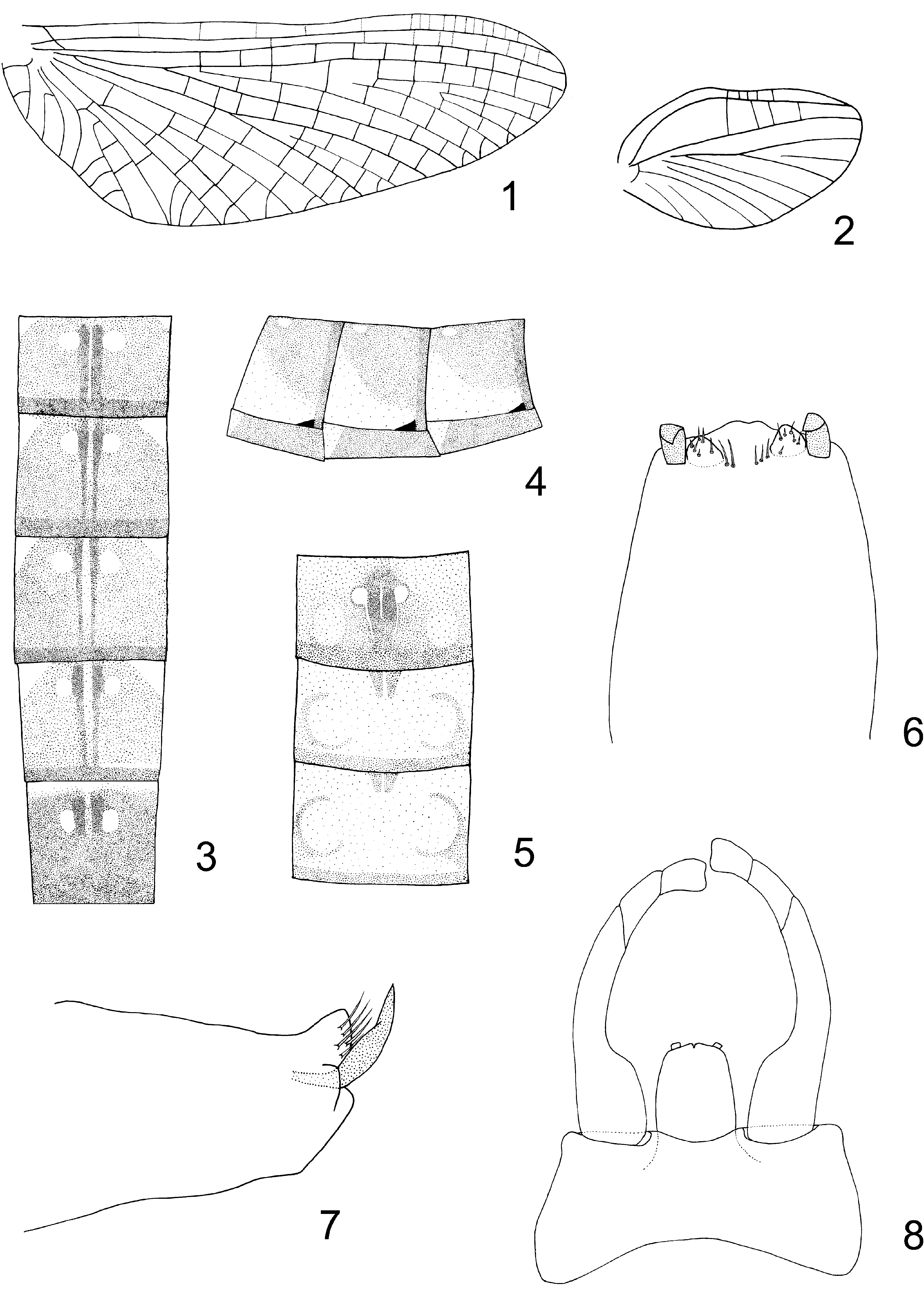
( Figs 1–8 View FIGURES 1 – 8 )
Meridialaris spina Pescador & Peters 1987: 174 View in CoL ; Hubbard et al. 1992; Domínguez et al.1994: 56; Domínguez et al. 2006; Pessacq 2009: 156.
Specimens examined. Nine nymphs, Argentina, Chubut province, unnamed stream, 500 m before Epuyen Lake, access road to Puerto Patriada, 42°08’17” S 71°31’56” W, 5/3/05, Di Prinzio leg. Three nymphs, same data except 12/5/05. Four nymphs, same data except 3/20/06. 32 nymphs, same data except 11/16/08, Pessacq leg. Three 3 and two Ƥ imagos, same data except, Malaise trap 12/2–16/08. One 3 imago + exuvia, same data except, 12/9/08, emerged at laboratory 12/ 13/08. Two 3 imagos + exuvia, same data except, 12/16/08, emerged at laboratory 12/16/08.
Male imago: measurements (mm, n=5): total length: 8.4–9.2 (8.8 ± 0.4), fore wing: 8.9–9.8 (9.5 ± 0.42).
Head: dark brown; antennae brown; ocelli whitish; upper portion of eyes light brown and lower portion black.
Thorax: nota dark brown; pleura light brown with irregular darker areas; sterna dark brown. Forewing ( Fig. 1 View FIGURES 1 – 8 ) membrane hyaline; longitudinal veins light brown, transversal cross veins lighter than longitudinal ones; costal cross veins and veins between Sc and R1 barely visible; pterostigma cloudy white; ICu1 slightly divergent from CuA; fourteen to eighteen costal cross veins. Hind wing ( Fig. 2 View FIGURES 1 – 8 ) oval, wing membrane hyaline; longitudinal veins barely visible, except for Sc light brown; reduced number of transversal cross veins, represented by four to six light brown costal cross veins and four barely visible transversal veins between Sc and R1. Legs yellow; apex of femur brown; basal fore tibia brown.
Abdomen ( Figs 3, 4 View FIGURES 1 – 8 ): all terga brown; terga II–VI with two dark brown longitudinal dorsal bands and two light spots at anterior sides of bands, sides with a light brown pattern basally wider; tergum VII with an anterior lighter stripe and two elongated black dorsal maculae with two light spots. Sternum I brown; sterna II–VIII with a medial brown spot, anterior half yellow, posterior half light brown; sternum IX white. Genitalia ( Fig. 6–8 View FIGURES 1 – 8 ): forceps whitish, angular bend on inner margin of segment one well developed, at about 1/3 distance from base; segment three slightly shorter than segment two. Styliger plate with a shallow U shape posteromedian emargination ( Fig. 8 View FIGURES 1 – 8 ). Penes ( Fig. 6, 7 View FIGURES 1 – 8 ) whitish; approximately rectangular, with distal margins slightly convergent, apex with a shallow U shaped cleft and with two apical curved blade-like processes. Inner to this last structures, two conical lobes covered with hair-like setae. Caudal filaments and cerci whitish, segments covered with short spine-like setae and with an apical ring of brown long spinelike setae.
Female imago: measurements (mm, n=1): total length: 9.1, fore wing: 10.
Head: anterior dorsal region half gray; posterior dorsal half light brown, antennae light brown, ocelli whitish with gray base, eyes black.
Thorax: pronotum light brown, meso and metathoracic nota brown, pleura light brown and sterna brown; wings same as in male except veins darker; 16–18 subcostal cross veins in forewing; legs same as in the male.
Abdomen ( Fig. 5 View FIGURES 1 – 8 ): tergum I brown, terga II–III light brown, with a medial dark brown area with two lateral yellow spots and a medial yellow narrow stripe, two distolateral big light brown spots and a dark brown stripe on segment distal margin; terga IV–VI light brown, with two small brown triangular areas on middle of segment basal margin and with two distolateral brown semicircles and a dark brown stripe on segment distal margin; terga VII–VIII same as previous but slightly darker; terga IX and X brown. Sterna I–VII brown, VIII–IX whitish. Ninth sternum with rounded apex.
Comments on the nymph. The nymphs show an abdominal coloration pattern characterized by terga I–VII and X mostly brown (with two big middle light yellow spots in segments IV and V in some specimens), segment VIII with a big triangular light yellow area with its base occupying most of the segment distal margin, and segment IX almost entirely light yellow. Femora show a wide variation in coloration pattern and in thoracic pro and mesonota there are two lateral small yellow spots.
In the original description ( Pescador & Peters 1987), a wide variation in the color pattern is addressed, most nymphs have “a reduced tergal maculae that are confined near the posterolateral corners”, but a few specimens have “slender and elongated sublateral maculae that extend almost the entire length of the terga” or “abdominal terga faintly washed with brown which obfuscates the pattern of maculae”. Additionally, “pro and mesonota lack spots” and femora shows “basal and apical yellow spots”. Structural characters agree with the original description.
Diagnose and comments on related species. The imago of Meridialaris spina can be easily separated from other species of the genus by the presence of two conical lobes covered with hair-like setae on penes apex ( Figs. 6, 7 View FIGURES 1 – 8 ).
This species can be separated from all other congeners by combination of the following characters: 1) ICu1 slightly divergent from CuA; 2) reduced number of transversal cross veins in hind wing, represented by four to six costal cross veins and four transversal veins between Sc and R1; 3) femora lack median band; 4) abdominal coloration pattern as in Figs. 3–4 View FIGURES 1 – 8 ; 5) angular bend on inner margin of segment one of forceps well developed.
Based on the overall similarity of the penes, particularly the presence of hair-like setae, and the well developed bend on inner margin of segment one of the genital forceps, M. spina appears closely related to M. chiloeense and M. biobionica . In M. biobionica , ICu1 is strongly divergent from CuA, while in M. chiloeense this vein is parallel to slightly divergent from CuA (slightly divergent in M. spina ). Additionally, the margins of penes are slightly apically attenuated in these two species, but almost parallel in M. spina .
Meridialaris spina was formerly known only from Chile and the provinces of Neuquén and Río Negro in Argentina ( Pessacq 2009), this is the first record from the Chubut province.
No known copyright restrictions apply. See Agosti, D., Egloff, W., 2009. Taxonomic information exchange and copyright: the Plazi approach. BMC Research Notes 2009, 2:53 for further explanation.
|
Kingdom |
|
|
Phylum |
|
|
Class |
|
|
Order |
|
|
Family |
|
|
Genus |
Meridialaris spina Pescador & Peters 1987
| Pessacq, Pablo 2010 |
Meridialaris spina
| Pessacq 2009: 156 |
| Dominguez 1994: 56 |
| Pescador 1987: 174 |