
Melchisedec thevenot, Fannes, 2010
|
publication ID |
https://doi.org/ 10.1206/3702.2 |
|
DOI |
https://doi.org/10.5281/zenodo.4565621 |
|
persistent identifier |
https://treatment.plazi.org/id/C60887CE-1E6F-FFA9-21AD-2FF68152F929 |
|
treatment provided by |
Felipe |
|
scientific name |
Melchisedec thevenot |
| status |
sp. nov. |
Melchisedec thevenot View in CoL , new species
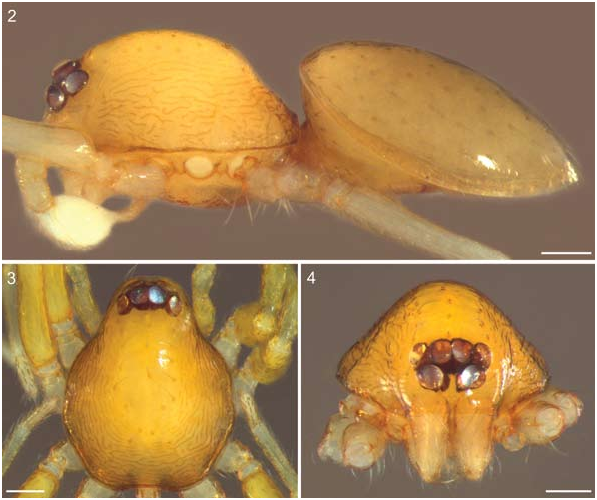
Figures 2–41 View FIGS View FIG View FIGS View FIGS View FIGS View FIGS View FIGS View FIGS View FIGS , 43–55 View FIGS View FIGS View FIGS View FIGS View FIGS , 57 View FIGS , 60, 61 View FIGS , 68–82 View FIGS View FIGS View FIGS
TYPES: Female holotype: Cameroon, Faro Game Reserve , 8°23ʹ09.5˝N 12°49ʹ59.7˝E, Apr. 27, 2007, wooded savannah, canopy fogging GoogleMaps , R. Jocqué, K. Loosveldt, L. Baert, and M. Alderweireldt (PBI_ OON 9209 and MRAC 228.972 View Materials ). Paratype: Cameroon, Atlantic Mountains , 8°31ʹN 12°36ʹE, Apr. 24, 2007, litter among rocks, sieving GoogleMaps , R. Jocqué, K. Loosveldt, L. Baert, and M. Alderweireldt (PBI_ OON 9208 and MRAC 221.480 View Materials ), 1 male . Paratype: Cameroon, Faro Game Reserve , 8°24ʹN 12°49ʹE, Apr. 25, 2007, gallery forest, sieving, same collectors (PBI_ OON 9210 and MRAC 221.441 View Materials ), 1 female GoogleMaps .
ETYMOLOGY: The specific name is a noun in apposition and refers to Melchisédec Thévenot.
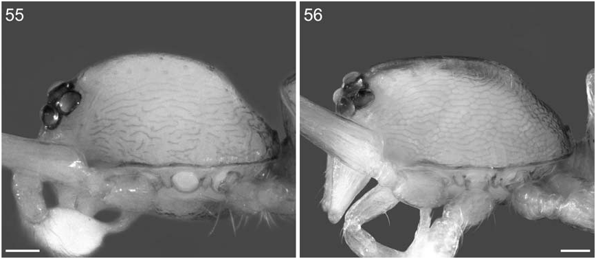
DIAGNOSIS: M. thevenot can be distinguished from M. birni by its more elevated pars cephalica (fig. 55), and by its carapace microsculpture, which consists of widely spaced ridges (figs. 6, 55).
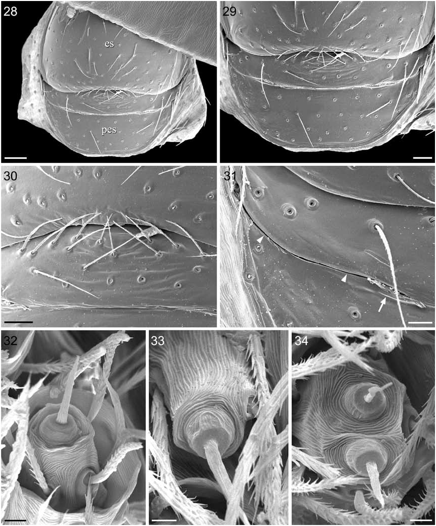
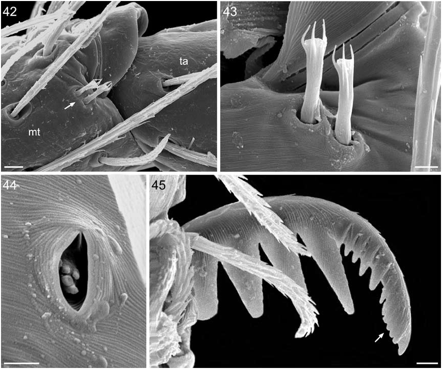
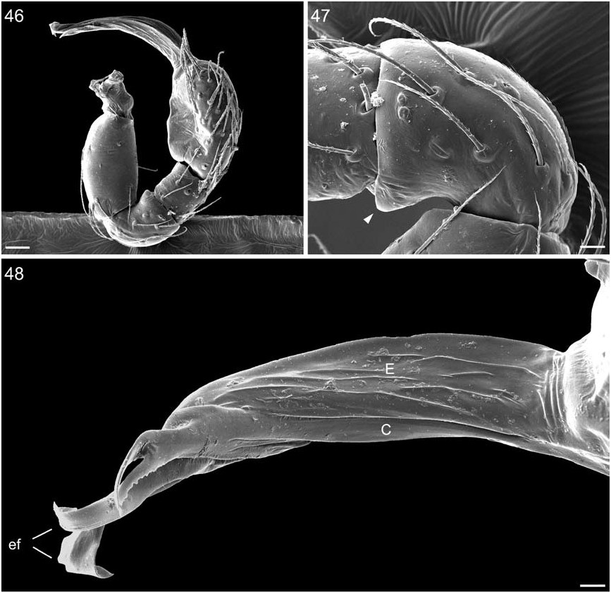
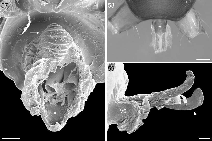
MALE: TL 1.17, CL 0.53, CW 0.43, DSL 0.63, DSW 0.41, Ti I l 0.26, Ti I l/d 5.68. Habitus as in fig. 2. Carapace yellow. Pars cephalica elevated, reaching its highest point at the level of coxal insertion I, thereafter slowly declining (fig. 2). Carapace microsculpture consisting of dark, widely spaced ridges; ridges varying from short and straight to long, meandering, and repeatedly branching (figs. 2, 6, 55). Sternum pale yellow, without hair tufts, with a single pouch situated behind labium (fig. 9); no sclerotized triangles in front of anterolateral corners. Anterior face of paturon light grey. DS light grey, covering entire dorsum. ES and PES pale yellow, fused into single ventral scutum that covers full length of abdomen (fig. 27). Lateral apodemes rather short; a faint brown band connecting anterior ends of apodemes (fig. 27). Left leg II examined with SEM; morphology as in female (see below). Sperm pore small, circular, situated anterior of brown band (fig. 27). Right and left pedipalps symmetrical, not strongly sclerotized, proximal segments yellow, cymbium and bulb white, ECC brown with black tip. Palpal trochanter without ventral projection; femur more than two times as long as trochanter, attaching to patella basally; patella much shorter than femur, not enlarged (fig. 47); tibia approximately as long as patella, with three dorsal trichobothria (structure as on legs). Cymbium approximately as long as bulb (fig. 46); fused to bulb but with clearly defined seam between them; without distal patch of setae; tarsal organ situated on distal third of cymbium, structure as on legs. Bulb distally giving rise to long ECC (figs. 46, 48); bulbal apex not conical. ECC longer than bulb, inward curved (fig. 46), consisting of two elements, a dorsal embolus and a ventral conductor (fig. 48). Dorsal surface of embolus densely grooved (fig. 49). Embolus distally splitting into two embolar flanges (figs. 48–50); one flange is distally broadened, the other is somewhat ribbon shaped (broadened flange, bf, and ribbonlike flange, rf; figs. 51–54). Ventral surface of broadened flange showing fold (fig. 53). Sperm duct opening could not be located with certainty; possibly, the opening is situated at the fold (fig. 53). Conductor distally splitting into a dorsal and ventral branch (figs. 50–52, 54); dorsal branch flat and relatively broad (fig. 51); ventral branch slender, somewhat threadlike (fig. 52), not visible from dorsal point of view. Both branches have a crenulate retrolateral edge (figs. 50–52). Tip of ECC held in sternal pouch (fig. 9).
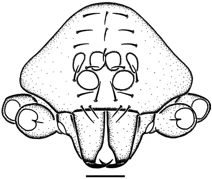
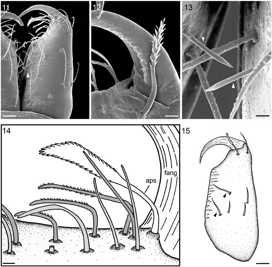
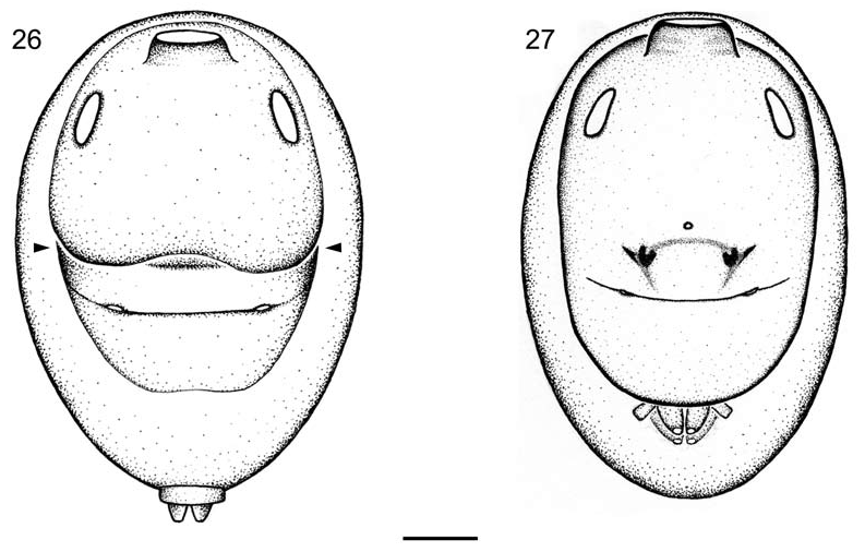
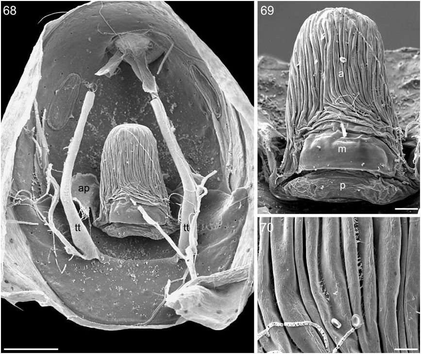
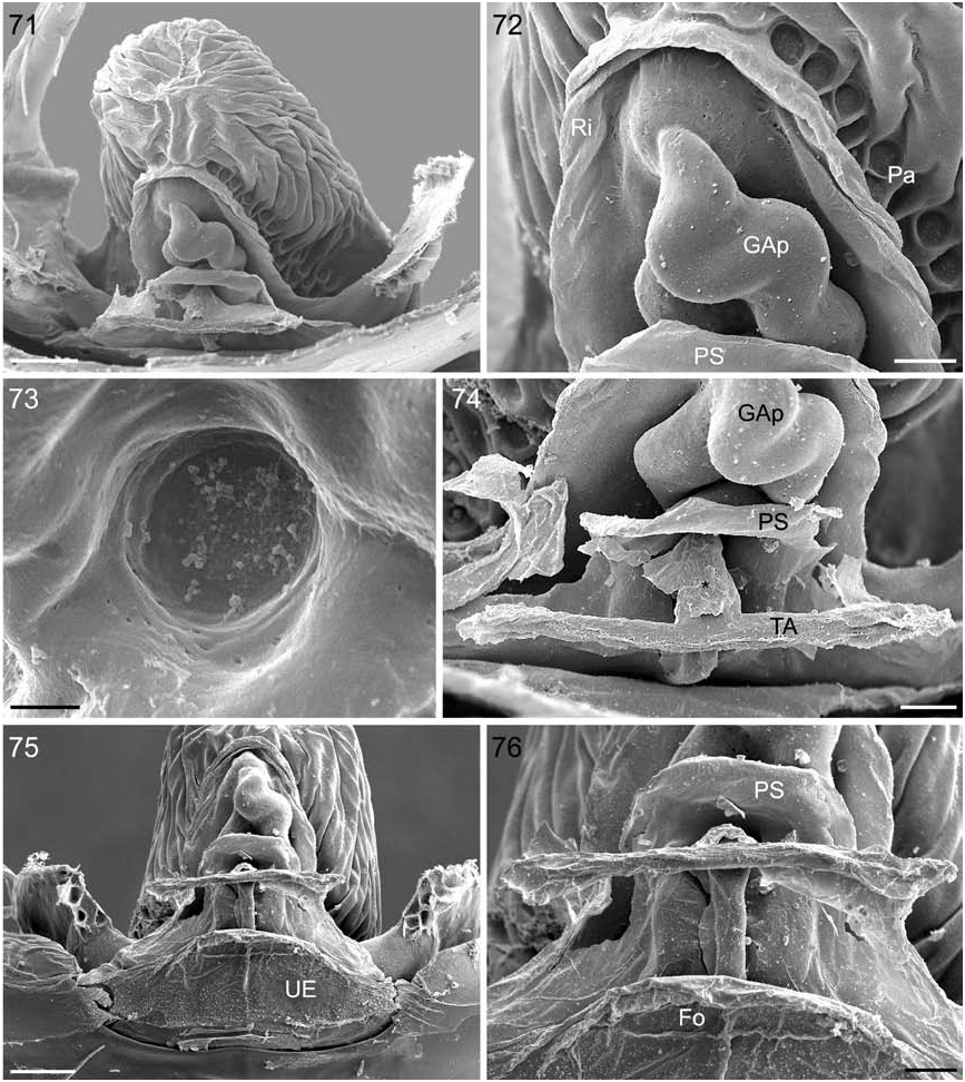
FEMALE: TL 1.30, CL 0.56, CW 0.46, DSL 0.74, DSW 0.57, Ti I l 0.28, Ti I l/d 4.78. Carapace as in male. Clypeus rebordered. Sternum pale orange. Infracoxal grooves without pores; a slit sensillum on posterior part of each groove. Anterior face of paturon pale yellow, with sparse setae (fig. 11). Cheliceral teeth absent. Cheliceral fang without basal process (fig. 11), tip unmodified; anterior edge of fang smooth, posterior edge serrated near base (fig. 12). A short, apically pointed seta arising from inner surface of each chelicera (figs. 11, 13). Promargin flanked by double row of setae (inner and outer row, fig. 14); each row with about six setae; most setae of inner row bent and with small, triangular teeth on shaft (fig. 14). Between double row and fang base a plumose hair (anterior plumose seta, aps; fig. 14). Posterior surface of paturon with two medially directed setae and three shorter spines (fig. 15). Fang base flanked posteriorly by two hairs (fig. 15), innermost one plumose (posterior plumose seta). At least 17 setae on labium: eight on distal margin, four situated subdistally, five situated medially (fig. 16). Labrum as in figure 17. Serrula with about 24 teeth in single row. Female palp without claw; structure of trichobothria as on legs (fig. 18). Soft portions of dorsum white, without color pattern. Two groups of small, rounded tubercles above pedicel tube (fig. 22). Outer surface of pedicel tube rugose and bearing setae (fig. 23), at least some setae have swollen bases (fig. 24); inner surface of tube closely ridged (fig. 25). DS grey, covering more than 3/4 of abdomen length and entire width. ES and PES pale orange. A slit sensillum anterior to each posterior spiracle (fig. 31). Dense patch of setae anterior to spinnerets absent. Colulus weakly sclerotized, bearing two setae; anterior lateral spinnerets with one spigot (fig. 32); posterior median spinnerets with one spigot (fig. 33); posterior lateral spinnerets with two spigots (fig. 34). The specimen examined by SEM showed a single scepterlike seta on one of the legs III; other leg III and both legs IV devoid of such setae. Distalmost, toward onychium sloping part of tarsus provided with proprioreceptor hair. Onychium bearing many setae, some spatulated. Tarsi without inferior claws. Superior claws hirsute and biseriate; lateral row consisting of 4–5 large teeth; median row situated close to claw tip, consisting of up to 10 small teeth, distalmost teeth often fused (fig. 45). Trichobothria: bothrium as in figure 36; no variation in bothrium structure among legs or among positions on a given leg. Tarsal organ pear shaped, receptor lobes exposed, inner surface of walls covered by ridges (fig. 44); no variation in structure among legs. Genitalia: no external copulatory structures (figs. 26, 29, 30); internally a large receptaculum, two uterine sclerites, and broad lateral apodemes (see Internal Female Genitalia and Respiratory System, below, for details).
OTHER MATERIAL EXAMINED: Cameroon: Faro Game Reserve , 8°24ʹ21.1ʹN 12°48ʹ20.4ʹE, Apr. 22, 2007, gallery forest, canopy fogging, R. Jocqué, K. Loosveldt, L. Baert, and M. Alderweireldt (PBI_ OON 9211 and MRAC 228.973 View Materials ), GoogleMaps 1 female. Same locality, 8°24ʹ26.9˝N 12°48ʹ44.6ʹE, Apr. 24, 2007, gallery forest, canopy fogging, same collectors (PBI_ OON 9212 and MRAC 228.974 View Materials ), 1 female (used for SEM) GoogleMaps . Guinea-Bissau: Buba , 11°30ʹN 15°05ʹW, June 9–11, 1989, A. van Harten (PBI_ OON 9859 and MRAC 228.968 View Materials ), 1 female. Same collection data (PBI_ OON 9860 and MRAC 228.969 View Materials ), 1 female GoogleMaps . Ivory Coast: Odienné , Saméso, Kourou Kélé, 9°45ʹN 7°45ʹW, Mar. 3, 1980, pitfalls, J. Everts (PBI_ OON 9207 and MRAC 174.130 View Materials ), 1 female GoogleMaps . Nigeria: Wudil , 11°48ʹ38˝N 8°50ʹ42˝E, Nov. 12, 1976, litter, leg. APB Deeleman, Museum Leiden ex coll. C.L. Deeleman-Reinhold; 2000-704 (PBI_ OON 33824), 1 female GoogleMaps . Ethiopia: Awash N.P., compound of RAS Hotel , 9°05ʹ00˝N 40°00ʹ00˝E, Apr. 24, 1986, elev. 1000 m, under stone, A. Russell-Smith (PBI_ OON 33823 and MRAC 228.970 View Materials ), GoogleMaps 1 female. Melka Werer, IAR station, 9°33ʹ00˝N 40°24ʹ00˝E, Feb. 17, 1986, elev. 750 m, litter of Acacia nilotica forest, A. Russell-Smith (PBI_ OON 33822 and MRAC 228.971 View Materials ), 8 females GoogleMaps .
DISTRIBUTION: Known from Guinea-Bissau, Ivory Coast, Nigeria, Cameroon, and Ethiopia (fig. 1).
NOTE: The specimens from Guinea-Bissau, Ivory Coast, and Ethiopia are tentatively assigned to M. thevenot . These specimens, all females, strongly resemble the female holotype from Cameroon. However, given the large geographical distances involved (fig. 1), it is possible that some of them do not belong to M. thevenot . The specific status of these specimens will remain uncertain until males have been collected from the same localities.


| R |
Departamento de Geologia, Universidad de Chile |
No known copyright restrictions apply. See Agosti, D., Egloff, W., 2009. Taxonomic information exchange and copyright: the Plazi approach. BMC Research Notes 2009, 2:53 for further explanation.
|
Kingdom |
|
|
Phylum |
|
|
Class |
|
|
Order |
|
|
Family |
|
|
Genus |