
Marilia muelleri, Camargos & Pes & Hamada, 2020
|
publication ID |
https://doi.org/ 10.11646/zootaxa.4853.1.1 |
|
publication LSID |
lsid:zoobank.org:pub:F4107225-9653-4407-BC4A-F6D9C26A9F93 |
|
DOI |
https://doi.org/10.5281/zenodo.4410264 |
|
persistent identifier |
https://treatment.plazi.org/id/DA608C21-693B-9F3D-FF76-FD0DFD48FD87 |
|
treatment provided by |
Plazi |
|
scientific name |
Marilia muelleri |
| status |
sp. nov. |
Marilia muelleri sp. nov
( Figs 5 View FIGURE 5 , 18–21 View FIGURE 18 View FIGURE 19 View FIGURE 20 View FIGURE 21 , 29 View FIGURE 29 )
Diagnosis: Marilia muelleri sp. nov. has eyes and male genitalia similar to M. aiuruoca Dumas & Nessimian 2009 and M. misionensis ; the similarities in the genitalia are mostly by the sutures of segment IX closing the posterior region of the segment, by the shape of segment X in lateral view, and by the tripartite shape of the phallotremal sclerite. However, the triangular shape of segment X in dorsal view, instead of straight as in M. misionensis or with the apex of segment X broad as in M. aiuruoca , and the curvature and shape of the preanal appendages very robust instead of slender, differentiate the new species from the aforementioned species.
The larvae are similar to those of M. cabloca sp. nov. and M. caipira sp. nov. by the dark lines following the frontoclypeal sutures, but M. muelleri sp. nov. has distinctive internal spots on the frontoclypeus, as well as different pigmentation over all the thoracic tergites.
Adult: Male forewings each 14.6–14.9 mm long (n = 2). Body and forewings in alcohol brown covered with gray setae.
Head: Eyes very large in males, almost touching on dorsal region of head ( Figs 18a, 18b View FIGURE 18 ). Pair of vertexal mediantennal compact setose warts fused with ellipse shape; pair of vertexal lateroantennal warts small not defined by sutures; pair of occiptal warts not visible, pair of posterior warts very narrow ( Figs 18a, 18b View FIGURE 18 ). Antennae long, about two times as long as body, with narrow annuli; scapes about 3 times as long as broad, with pale spot basodorsally, covered with light setae. Front pubescent, covered with light setae, pair of frontogenal warts, long subtriangular ( Figs 18c, 18d View FIGURE 18 ). Maxillary palps well developed, each 5-articulated, densely covered with setae. Labial palps each 3-articulated, articles subequal, covered with setae.
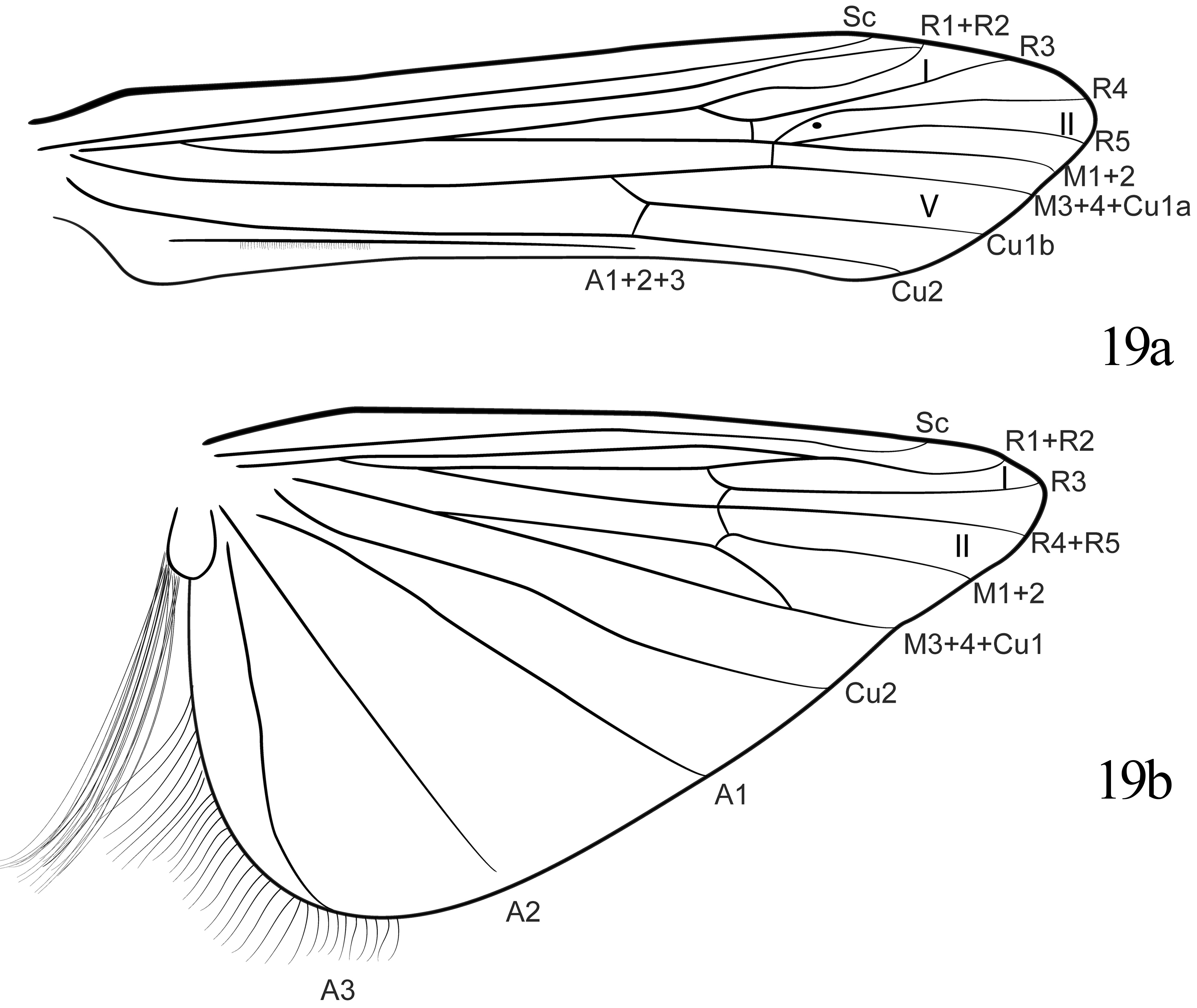
Thorax: Prothorax less than half as long as head; pronotum with pair of transversely elongate setal warts. Mesothorax broad, mesonotum with median longitudinal dark line reaching mesoscutellum; mesoscutellum almost circular, with pair of setal warts and small posterior depressions. Metathorax half as long as mesothorax, without setae. Tibial spur formula 2-4-4; external spurs of median and posterior tibiae shorter than internal spurs. Forewings each with fork I arising on apical quarter of discoidal cell, R1 and R2 fusing at wing margin ( Fig 19a View FIGURE 19 ). Hind wings each with fork I arising on apical tenth of discoidal cell, R1 and R2 merging about one-third length of R2 from wing margin; anal lobe with brush-like tuft of long setae ( Fig 19b View FIGURE 19 ).
Abdomen: Simple, without differentiated structures.
Male genitalia: Segment IX on each side, in lateral view with anterior margin sinuous and posterior margin slightly projected at midheight; midlateral sutures separating each side of segment into 3 parts, dorsal part and ventral part overlapping and occupying more than half of height of segment, middle part isolated in upper half of posterior margin ( Fig 20a View FIGURE 20 ); dorsal part not projecting above segment X ( Fig 20b View FIGURE 20 ). Preanal appendages, in lateral view, short, slender, apically blunt ( Fig 20a View FIGURE 20 ); elongate-oval in dorsal view, with numerous setae ( Fig 20b View FIGURE 20 ). Segment X with apex foot-like in lateral view, slightly upturned ( Fig 20a View FIGURE 20 ); triangular, broad at base, apically pointed and with narrow and deep median notch in dorsal view ( Fig 20b View FIGURE 20 ). Inferior appendages each with two articles: Basal article conical, with base twice as broad as apex, slightly curved mesad; apical article short, with small conical spines apically; in ventral view with internal margin slightly sinuous ( Fig 20c View FIGURE 20 ). Phallus tubular, in lateral view curved about 70 caudad near base ( Fig 20d View FIGURE 20 ); in ventral view straight ( Fig 20e View FIGURE 20 ); endotheca membranous, with small conical spines; phallotremal sclerite C-like in lateral view ( Fig 20d View FIGURE 20 ), V-like in ventral view ( Fig 20e View FIGURE 20 ).
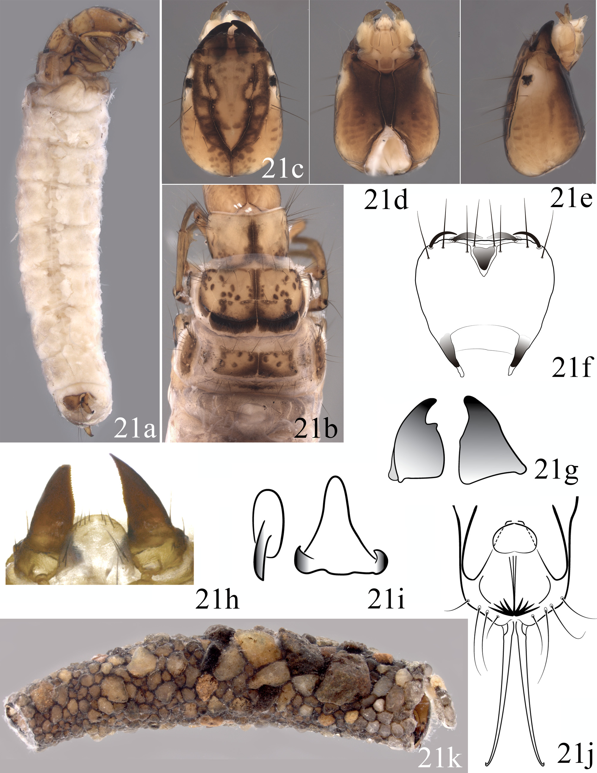
Final Instar Larva: Body length 11.5–16.7 mm (n = 4). Background color of sclerites yellow and abdomen light yellow in alcohol ( Fig 21a View FIGURE 21 ).
Head: In dorsal view subrectangular, broadest one-fourth distance from base; yellow with distinctively broad black Y-shaped lines along frontoclypeal and short coronal sutures, lighter brown with pale areas between slightly visible carinae and frontoclypeal sutures, and anterior portion of frontoclypeus with pair of diagonal black spots with pale areas posteriorly ( Fig 21c View FIGURE 21 ); in ventral view ventral apotome and adjacent regions black; ventral apotome 3.1 times as long as broad, tapered to posterior apex, separating genae entirely ( Fig 21d View FIGURE 21 ). Eyes rimmed by light areas ( Fig 21e View FIGURE 21 ). Labrum with concave apical margin slightly broader than concave basal margin, convex apicolateral margin broader than convex basolateral margin; setae 2 and 3 each robust, with row of smaller setae (papiliforms) between them, with setae 3, 4, 5, 6 straight and less rigid thickness ( Fig 21f View FIGURE 21 ). Mandibles robust, asymmetrical, mesal tooth of right mandible vestigial ( Fig 21g View FIGURE 21 ).
Thorax: Pronotum yellow, with setae on anterolateral corners, anterior margin concave, black median longitudinal line broad and reaching posterior margin, with pair of regions of other dark spots near posterior margin. Mesonotum divided into 3 pairs of sclerites: On each side anteromesal sclerite (sa 1) yellow with 3 rows of dark spots converging anterolaterally, one broad stripe on mesal suture, and small spots in middle of sclerite; posteromesal sclerite (sa 2) yellow with transverse dark area on posterior margin and occupying more than one-third of sclerite, dark spot on mesal suture, and other spots in middle of sclerite; lateral sclerite (sa 3) yellow with 3 rows of dark spots converging on anterior angle and with many setae on anterior margin. Metanotum divided into 5 sclerites: Anteromesal pair (sa 1) yellow with darker margins especially posteromesally, dark spot near mesal suture anteriorly and 4 other spots near middle and laterally, with setae on anterior margin,; lateral pair (sa 3) brown with setae on anterior and lateral margins; posteromesal sclerite (fused sa 2 sclerites) yellow, transversely elongate, straight, with few setae laterally ( Fig 21b View FIGURE 21 ). Legs yellow.
Abdomen: Abdominal gill formula as in Fig 5 View FIGURE 5 . Tergite IX subtriangular. Anal prolegs without teeth on external margins of claws.
Pupa: Body length 14.0– 15.5 mm (n = 3). Body brown in alcohol.
Head: Mandibles broad, more than two times as long as wide, distal portion narrowed, serrate on entire internal margin. Labrum subquadrate, with anterolateral angles rounded ( Fig 21h View FIGURE 21 ).
Thorax: Mesotarsi each with fringe of long setae.
Abdomen: Segments III–VII each with pair of small oval anterior hook plates, each hook plate with one hook oriented posterad; segment V with pair of triangular posterior hook plates, each with 2 posterior hooks oriented anterad ( Fig 21i View FIGURE 21 ). Terminal processes long, slender, and slightly divergent from base ( Fig 21j View FIGURE 21 ).
Case: Length 10.7–20.1 mm (n = 7), composed of various sizes of grains of coarse sand, slightly curved, broadening from posterior to anterior ( Fig 21k View FIGURE 21 ).
Etymology: The specific epithet pays homage to the author of the genus Marilia , the German naturalist and naturalized Brazilian Fritz Müller, an important scholar in Brazilian science, and an enthusiast for Darwinian ideas.
Bionomics: This species was found in regions of high altitude (1,233 m elev.) in fields and Araucaria forest. The river was impacted by plantations and pasture, with widths of 15 to 20 m, depths of 0.15 to 1.0 m, and rocky substrate with many macrophytes.
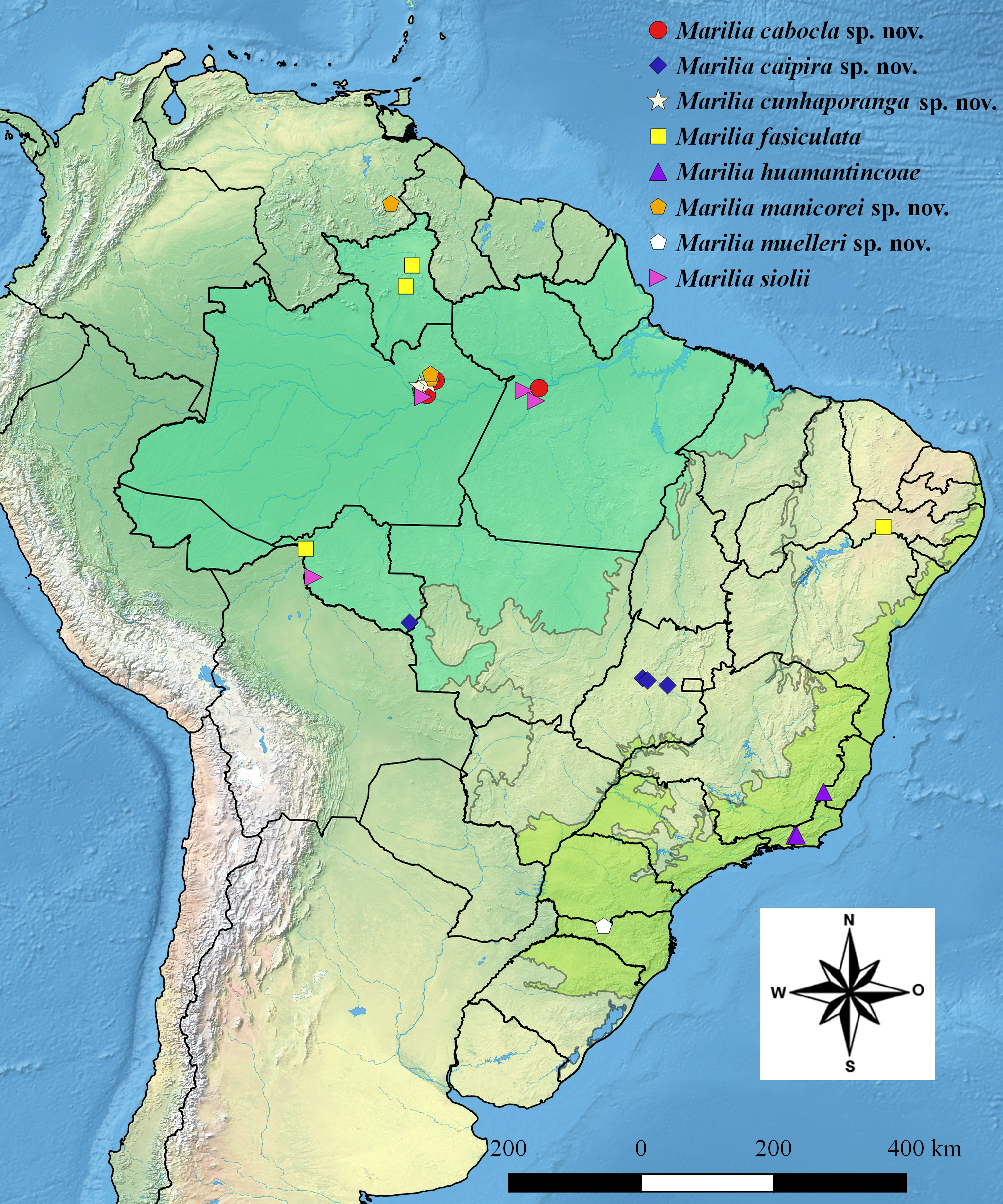
Distribution: BRAZIL: Santa Catarina.
Holotype male: BRAZIL, Santa Catarina, Água Doce: #16, Rio Roseiras , 26°42’35.3”S 51°36’24.0”W, 1233 m alt, 17.ix.2011, A.M.O Pes, R. Boldrini, P. Cruz, N. Hamada leg. 1 male and its exuviae [alcohol] ( INPA-TRI 000083 ). GoogleMaps
Paratypes: BRAZIL, Santa Catarina, Água Doce: #16, Rio Roseiras , 26°42’35.3”S 51°36’24.0”W, 1233 m alt, 17.ix.2011, A.M.O Pes, R. Boldrini, P. Cruz, N. Hamada leg. 1 male intersex and its exuviae [inferior appendages and phallus not developed] ( INPA-TRI 000084 ); 6 larvae and 3 pupae [alcohol] ( INPA-TRI 000085 ); 1 larva [alcohol] ( MZSP); 1 larva [alcohol] ( DZRJ). GoogleMaps


| R |
Departamento de Geologia, Universidad de Chile |
| MZSP |
Sao Paulo, Museu de Zoologia da Universidade de Sao Paulo |
No known copyright restrictions apply. See Agosti, D., Egloff, W., 2009. Taxonomic information exchange and copyright: the Plazi approach. BMC Research Notes 2009, 2:53 for further explanation.
|
Kingdom |
|
|
Phylum |
|
|
Class |
|
|
Order |
|
|
Family |
|
|
Genus |