
Maemonstrilla hyottoko, Grygier & Ohtsuka, 2008
|
publication ID |
https://doi.org/ 10.1111/j.1096-3642.2007.00381.x |
|
publication LSID |
lsid:zoobank.org:pub:942A51C7-EE79-4C60-8B80-9EA8835889CC |
|
persistent identifier |
https://treatment.plazi.org/id/03EBB12D-F756-0C09-FF39-EF28EE8D7B18 |
|
treatment provided by |
Felipe |
|
scientific name |
Maemonstrilla hyottoko |
| status |
|
MAEMONSTRILLA HYOTTOKO SPECIES GROUP
Diagnosis: Cephalothorax with reticulate pattern of cuticular ridges. Antennules, lateral sides of metasomal somites and urosomal segments, dorsum of telson, and caudal rami also so ornamented, except in M. simplex sp. nov. Dorsal surface of metasomal pedigers and first three urosomal segments, as well as outer face of thoracopods (coxa and exopod), densely denticulate or spinulose, except in M. simplex . No spine-like scales posteriorly near dorsal midline on first and second free pedigers. Outer basis seta of leg 3 at least as long as exopod. No inner seta on first exopodal segment of legs 1–4; inner seta of first endopodal segment of these legs absent or represented by socket-like or button-like structure. Leg 5 a long, thin rod with two distal setae, one apical and one slightly subapical; endopodal lobe absent (or reportedly tiny and unarmed in one instance). Posterior part of genital compound somite with ventral protrusion.
MAEMONSTRILLA HYOTTOKO SP. NOV.
FIGURES 2–6 View Figure 2 View Figure 3 View Figure 4 View Figure 5 View Figure 6 , 29 View Figure 29
Diagnosis: Reticular ridges of cephalothorax sporadically spinulose and with many short side branches; complex cuticular figures present within wide anterior and posterior reticular meshes, but not within narrow meshes in between. On antennule, 2v-setae prominent and straight, 2v 2 and 2v 3 being particularly long; 4v 1 seta about half as long as 3-seta, and latter over three times longer than other 4-setae. Oral papilla prominent. Pair of tubular pores anterior to oral papilla, with four spines on lip of each tube. Dorsal posterior margins of genital compound somite and penultimate segment each bearing six clusters of 3–4 large denticles; denticles immediately preceding these bigger than those over rest of dorsal surface. Posteroventral part of genital compound somite produced into large spur.
Etymology: Noun in apposition, from the Japanese festival mask called ‘Hyottoko’, which has a conical, protruding mouth similar to the oral papilla in this species.
Material examined: Eleven females collected by M. J. Grygier at Sesoko Island (type locality): holotype (used for microscopic drawings; vial and slide KMNH IvR 700 202) and seven paratypes, 13.viii.1989 (co-occurring with M. spinicoxa and M. turgida , see below) [of paratypes, four intact (ZMA Co. 205908), three used for SEM (SO lab), but one of latter also used for microscopic drawings (slide of legs: KMNH IvR 700 203)]; one intact paratype, 14.ix.1988 (NHM Reg. No. 2006. 1911); one intact paratype, 2.x.1992 (USNM 1093747) (co-occurring with M. turgida , see below); one intact paratype, 22.v.1996 (LBM Reg. No. 1430000926). In addition, one intact paratype female collected by S. Ohtsuka (LBM Reg. No. 1430000927), south coast of Ishigaki Island, 30.iv.1994. Four nontype females tentatively assigned to this species, all intact: three collected by A. Murase off Kabira, Ishigaki Island (co-occurring with M. spinicoxa and M. turgida , see below), 14.iv.1996 (LBM Reg. No. 1430000928); one collected by S. Kubota (KMNH IvR 700 204), Hirara Port, Miyako Island, 5.v.1993.
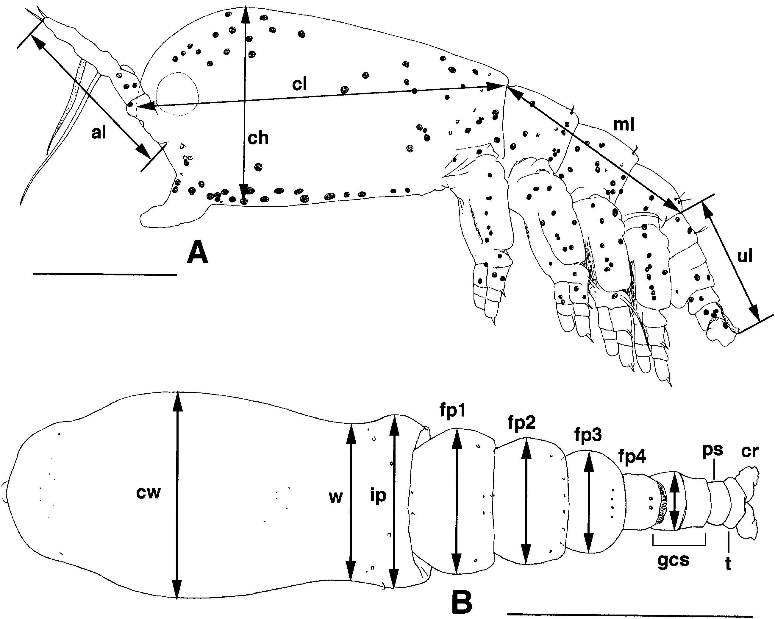
Description: Cephalothorax distinctly bulbous anteriorly ( Fig. 2A View Figure 2 ); incorporated first pediger widening again in dorsal view behind distinct ‘waist’ (as in Fig. 1B View Figure 1 ). Measurements taken from seven or (for some) eight specimens from Sesoko Island. Body length in lateral view (sum of lengths of cephalothorax, metasome and urosome as defined in Fig. 1A View Figure 1 ) 1.15–1.60 mm, with these body regions contributing 49.6–52.1, 28.1–31.2 and 19.2–20.7%, respectively. Height and greatest width of cephalothorax 49.2–53.9 and 50.8–54.3% of cephalothorax length, respectively. Antennule length 40.1–46.1% of cephalothorax length. Width of incorporated first pediger 69.8– 81.9% of greatest width; widths of succeeding three free pedigers and genital compound somite relative to that of incorporated pediger 81.1–89.5, 60.3–73.4, 53.4–56.3 and 32.2–37.5%, respectively. Ovigerous spines (seven pairs measured) 26.5–35.3% as long as body (as seen in dorsal view). Specimen from south coast of Ishigaki with wider first and second free pedigers (97.3 and 85.9% as wide as incorporated pediger, respectively) and slightly longer ovigerous spines (38.9% as long as body as seen in dorsal view). Non-type, tentatively included specimens from off Kabira and from Hirara port comprising two smaller specimens (body length as short as 1.07 mm) and ones exhibiting higher cephalothorax height/length ratios (57.9–64.5%).
Although mostly reticulate as mentioned in diagnosis, cephalothorax smooth in area posterodorsal to antennules and on posterior half of ventral side (except for band behind oral papilla) ( Fig. 2A View Figure 2 ). Reticular meshes broad in posterior third and anterior sixth of cephalothorax, very narrow in intervening region; their borders and interiors as described in diagnosis.
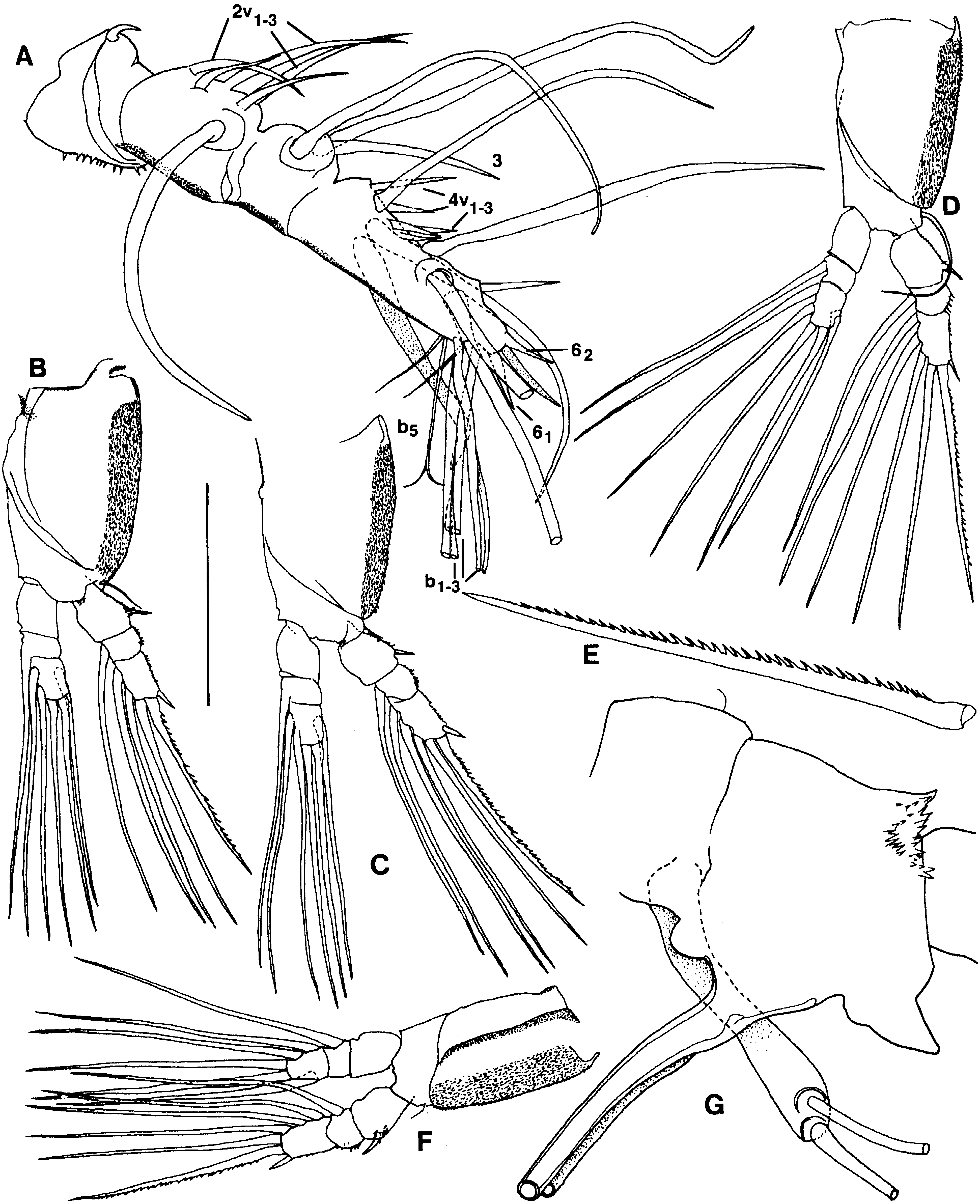
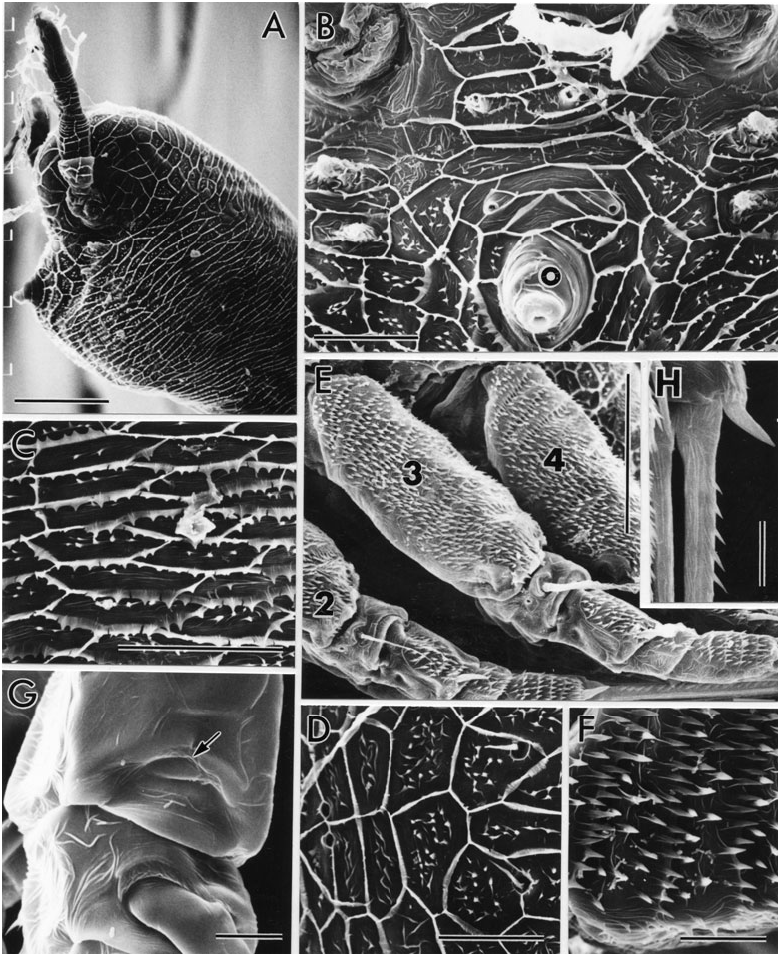
Antennule clearly four-segmented, but proximal region of first segment consisting of complexly folded arthrodial membrane and sclerites for muscle attachment ( Fig. 3A View Figure 3 ); reticular ridges becoming lower and simpler distally. All setal elements identified by Grygier & Ohtsuka (1995) present ( Fig. 4A View Figure 4 ). Spiniform 2v-setae and 3-seta much longer than 4-setae; row of spinules present on at least 2v-seta, possibly on other spiniform setae. Outer distal b 1-3 setae each branched at least twice in asymmetrically dichotomous manner; b 5 seta bifid. Apical 6-setae similar to each other in size.
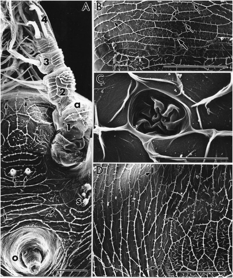
Oral papilla ( Fig. 3A View Figure 3 ) very large, funnel-shaped, directed ventrally and preceded by pair of simple pores. Pair of tubular pores, with four spines on lip of each pore, situated further anteriorly between posterior ends of bases of antennules. Cluster of three knob-like scars behind base of each antennule, middle knob smaller than others.
Forehead region ( Fig. 3A View Figure 3 ) with pair of circles formed of cuticular ridges just anterodorsal to antennule bases, smaller circle on midline just posterior to these, and pair of pores anterodorsal to large circles (pair of hair-like sensilla seen arising from these pores in some paratypes). Subcircular pit with wrinkled thin cuticle on dorsal midline of forehead, 10 Mm in largest diameter ( Fig. 3B, C View Figure 3 ). In an SEM specimen, two pores observed behind this structure, and three pores on each side further laterally (some shown in Fig. 3B View Figure 3 ); numbers and positions of these pores not necessarily constant in other specimens. Lateral cups of naupliar eye about same size as ventral cup, about 80 Mm in diameter .
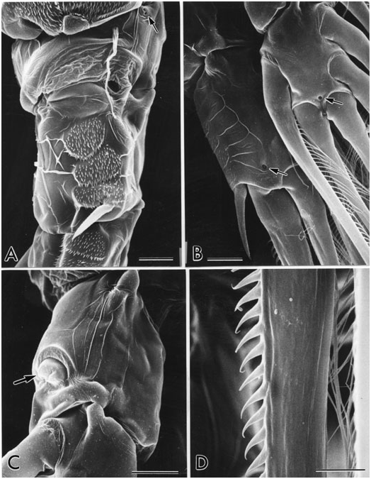
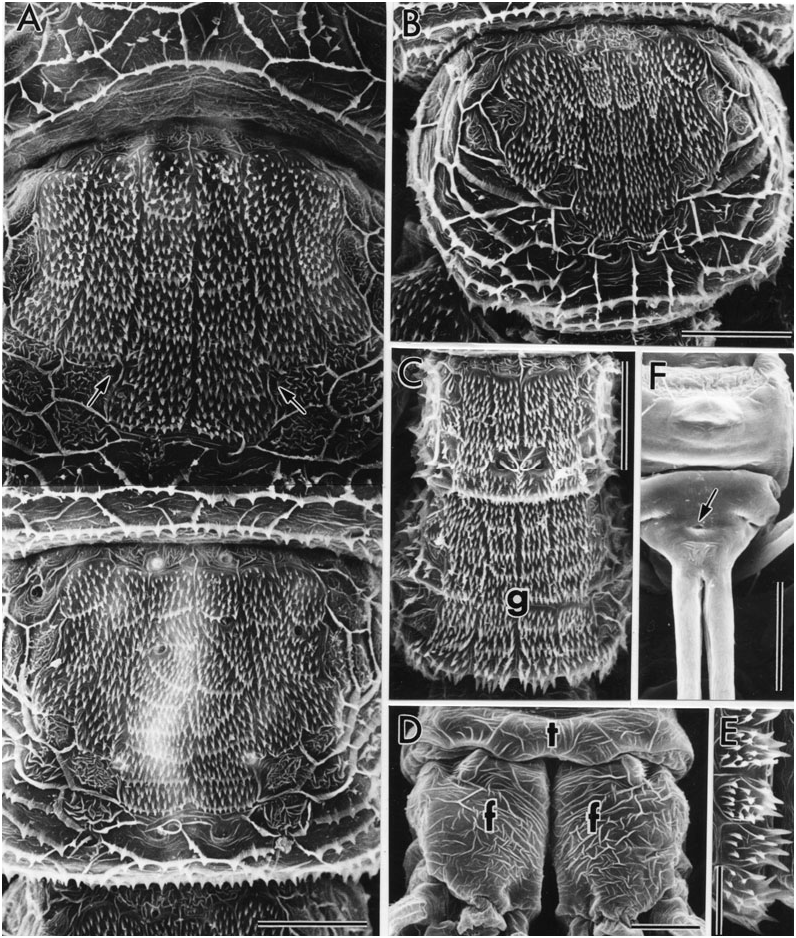
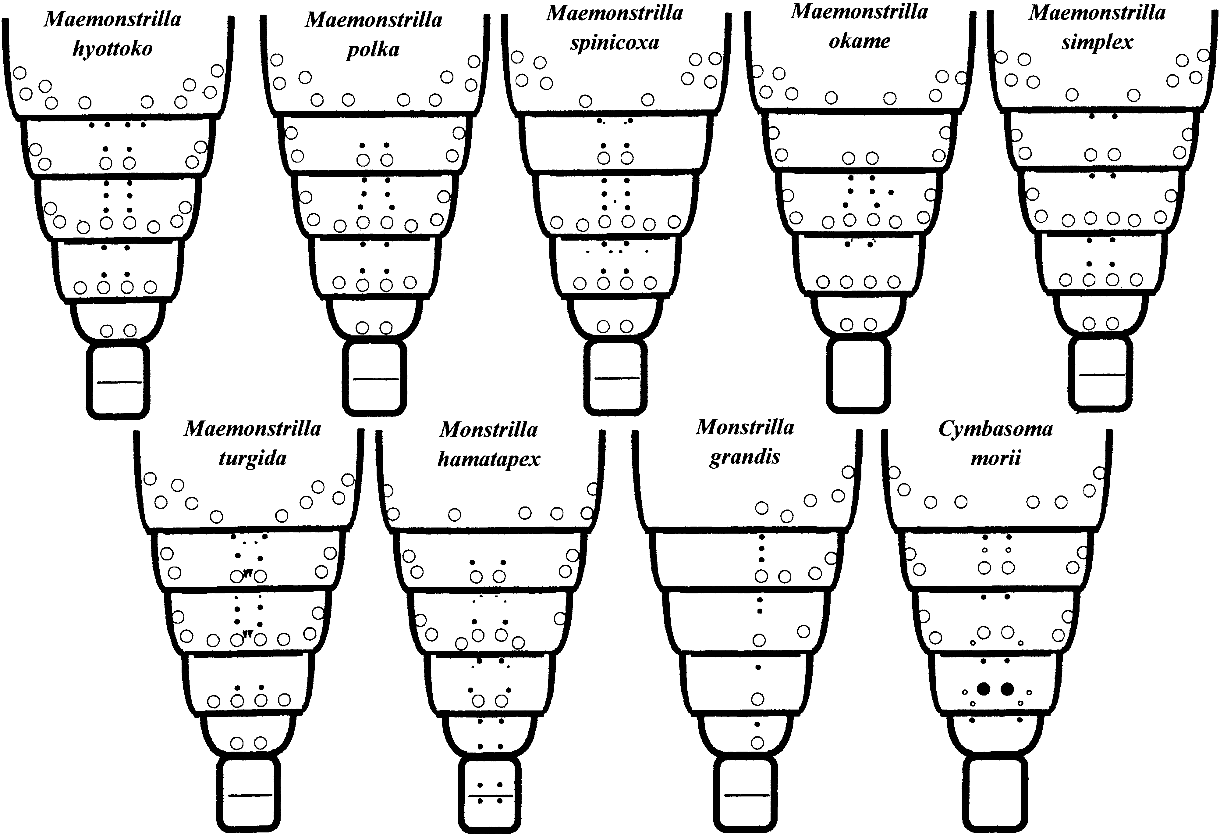
Three pairs of dorsal pores on cephalothorax at posterior boundary between narrow and broad reticular meshes ( Fig. 3D View Figure 3 ). Arrangement of dorsal pores and dorsal and lateral pit setae from rear of cephalothorax to urosome shown in Figure 29 View Figure 29 (see also Fig. 5A–D View Figure 5 ). Dorsal pores on free pedigers 1–3 only. Dorsal pit setae in one widely separated pair on incorporated pediger; in one, two, two and one pair, respectively, on succeeding four free pedigers. In addition, four pairs of dorsolateral and lateral pit setae on incorporated pediger and two pairs each on first and second free pedigers, anterior pair on each segment located more ventrally than posterior one. Dorsum of first free pediger ( Fig. 5A View Figure 5 ) covered with sharp, posteriorly directed denticles arrayed in six longitudinal rows of rectangular patches, these patches numbering 3-3-7-7-3-3, respectively; those of second free pediger arranged the same ( Fig. 5A View Figure 5 ); denticle patches of third free pediger fewer, 3-2-4-4-2-3 ( Fig. 5B View Figure 5 ); and those of fourth free pediger 2-3-2-2-3-2 ( Fig. 5C View Figure 5 ). Dorsum of genital compound somite ( Fig. 5C View Figure 5 ): anterior third with six rows of three denticle patches each; middle third with grid of cuticular ridges; posterior third with six rows of denticle patches arrayed 1-2-2-2-2-1, followed by band of six arrays of considerably larger denticles, with largest three or four denticles in each array extending beyond somite’s posterior margin. Penultimate segment ( Fig. 5C View Figure 5 ) with transverse band of four denticle patches followed by six arrays of large denticles like those on genital compound somite.
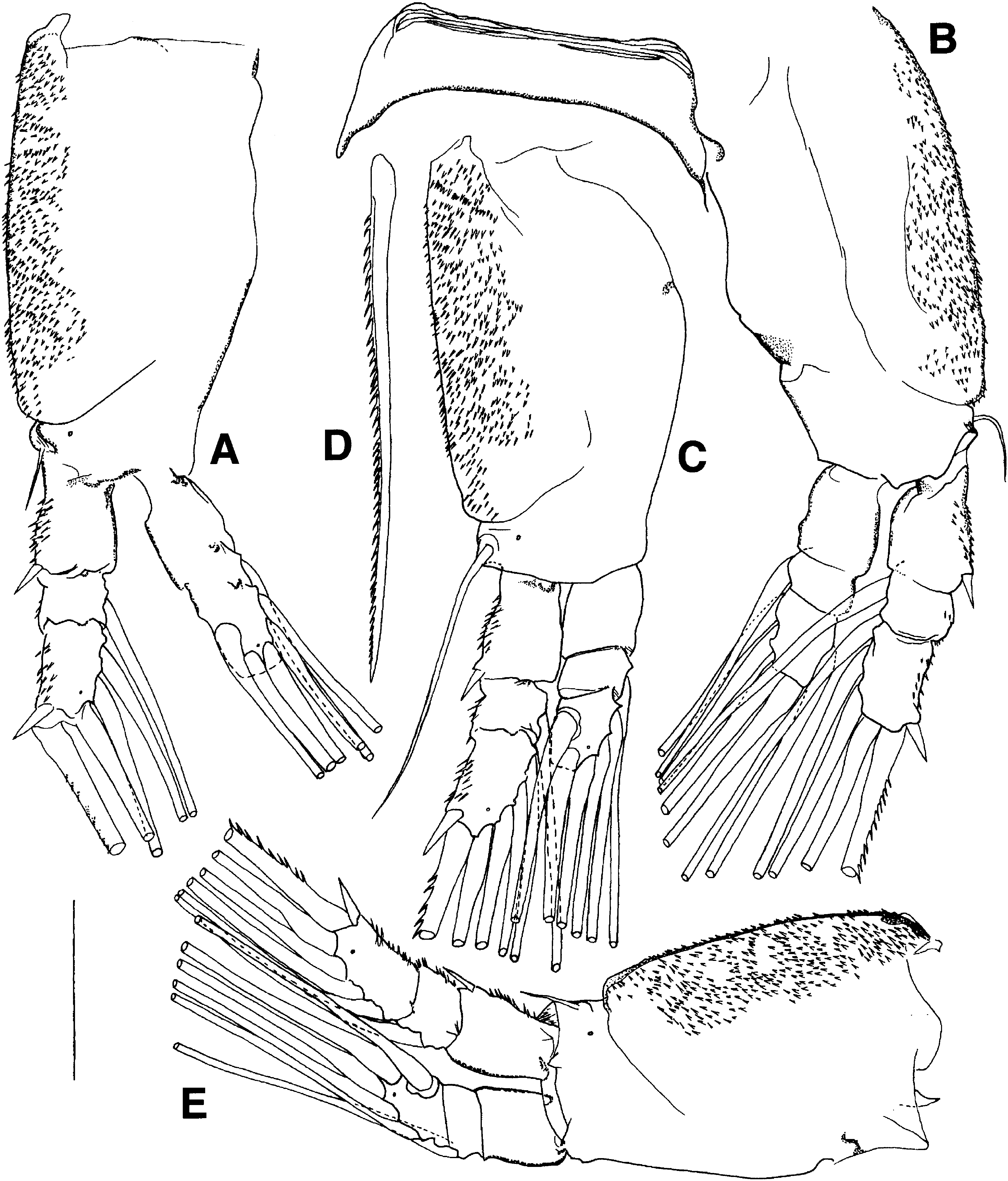
Legs 1–4 each with two arcuate sclerites laterally at base of leg ( Fig. 2B View Figure 2 ). Intercoxal sclerites low and wide with ventrally produced outer corners ( Fig. 2C View Figure 2 ). Protopod with longitudinal groove on anterior face; suture separating coxa and basis extending diagonally across posterior face and continuing around onto outer part of anterior face ( Fig. 4B–D, F View Figure 4 ). Sharp, distally pointing denticles on outer faces of coxa and exopod ( Figs 5C View Figure 5 , 6A View Figure 6 ), as large as denticles on posterior margin of genital compound segment. Pore just in front of lateral seta on basis ( Fig. 6A View Figure 6 ); this seta short and hair-like in legs 1, 2 and 4, but much longer and plumose in leg 3 ( Fig. 4D View Figure 4 , plumosity omitted). Posterior faces of rami lightly reticulated ( Fig. 5C View Figure 5 ); distal pore on anterior face of third segment of each ramus (e.g. Fig. 6C View Figure 6 ). No inner seta on first exopodal or first endopodal segment ( Fig. 6B View Figure 6 ). Outer spiniform seta on first and third exopodal segment in legs 1–4 simple, that of third segment bigger. Sparse spinules as well as two rows of long setules found on natatory setae ( Fig. 6C View Figure 6 ): one inner seta on second segment of each ramus; two inner setae on third segment of each ramus in leg 1, but three inner setae on exopod in legs 2–4; one outer seta on third segment of endopod, and two apical setae on each ramus ( Fig. 4B–D, F View Figure 4 ). Outer apical seta of exopod with outer row of widely spaced, talon-like denticles instead of setules ( Figs 4E View Figure 4 , 6C View Figure 6 ). Leg 5 rod-like, distally slightly clavate, with two apical setae ( Figs 2B View Figure 2 , 4G View Figure 4 ).
Ovigerous spines arising from conical projection of ventral surface of anterior half of genital compound segment ( Fig. 2B, C View Figure 2 ). Crescent-shaped slit leading to copulatory opening at posterior base of cone ( Fig. 2D View Figure 2 ); slit flanked by pair of small knobs projecting from cone. Tips of ovigerous spines found at any point between legs 2 and midlength of cephalothorax; spines cylindrical and smooth, but slightly thickened and wrinkled at two-thirds length, tips naked and tapered. Posteroventral protrusion of genital compound somite large, subconical, spur-like ( Figs 2D View Figure 2 , 4G View Figure 4 ).
Caudal rami ( Figs 5C View Figure 5 , 6D View Figure 6 ) lightly reticulate dorsally and with ventromedial condyle articulating to telson; anal slit visible in telson. Each ramus with ventral distal pore and six setae (at least five of them plumose), one arising ventrolaterally and the other five apically; most dorsal apical seta (seta VII of Huys & Boxshall, 1991) shorter than others, appearing smooth in some individuals but setulose in two specimens (perhaps setules orientated dorso-ventrally).
MAEMONSTRILLA POLKA SP. NOV.
FIGURES 1A View Figure 1 , 7 View Figure 7 , 8A View Figure 8 , 9–12 View Figure 9 View Figure 10 View Figure 11 View Figure 12 , 13A View Figure 13 , 29 View Figure 29
Diagnosis: Spherical spots of red pigment throughout body. Cuticular ridges of cephalothorax sporadically spinulose but lacking side branches, discontinuous in anterior ventrolateral region; reticulations uniform in size, enclosing clusters of small, cuneiform cuticular figures. Spiniform 2v-setae and 3-seta on antennule about twice as long as 4-setae; apical 6-setae minutely spinulose. Outer proximal part of coxa of legs 1–4 produced into two low, oval lobes armed with spinules slightly larger than those on rest of coxa. Posterior dorsal margin of genital compound somite and penultimate segment each slightly produced into six flanges bearing minute spinules of same size and density as those over rest of dorsum. Posteroventral part of genital compound somite produced into apron-like flap.
Etymology: Derived from Polish via English, as in ‘polka dots’, referring to the characteristic red spots throughout the body and legs (cf. Figs 1A View Figure 1 , 11 View Figure 11 , 13A View Figure 13 ); to be treated as a Latin adjective.
Material examined: Three ovigerous females collected by S. Ohtsuka on south coast of Ishigaki Island (type locality; co-occurring with M. simplex and M. turgida , see below), 30.iv.1994: holotype intact (KMNH IvR 700 205), used for photography; one paratype intact, but egg mass broken (KMNH IvR 700 215); other paratype used for light microscopic drawings and SEM, egg mass with ovigerous spines in vial and four legs on slide (vial and slide: KMNH IvR 700 216), rest of body on SEM stub (SO lab).
Description: Cephalothorax bulbous anteriorly, in lateral view tapering evenly from rounded front to rear ( Figs 1A View Figure 1 , 7A View Figure 7 , 13A View Figure 13 ). In dorsal view, incorporated pediger widening only slightly behind ‘waist’. Spherical spots of red pigment throughout body and legs 1–4 ( Figs 1A View Figure 1 , 11 View Figure 11 , 13A View Figure 13 ).
Measurements taken from holotype and non-SEM paratype; widths approximate due to difficulty in orientating these ovigerous specimens for microscopic examination in dorsal view. Body length in lateral view (sum of lengths of cephalothorax, metasome and urosome as defined in Fig 1A View Figure 1 ) 2.56–2.66 mm, with these body regions contributing 51.6–52.7, 28.6–29.0 and 18.3–19.8%, respectively. Height and greatest width of cephalothorax 45.9–51.3 and 49.4–50.7% of cephalothorax length, respectively. Antennule length 42.5–46.15% that of cephalothorax. Width of incorporated pediger 72.4–79.2% of greatest width. Widths of succeeding three free pedigers relative to that of incorporated pediger 84.2–88.3, 74.7–75.1 and 63.0– 63.2%, respectively.
Meshwork of cephalothorax and lateral parts of metasomal pedigers and urosome formed of finer cuticular ridges than in M. hyottoko , uniformly polygonal over most of surface ( Figs 7A View Figure 7 , 9A–D View Figure 9 ). Abundant small cuticular figures occurring inside meshes, and often fine lineations as well. Polygonal pattern largely replaced by maze-like pattern of discontinuous cuticular figures anterolaterally on cephalothorax ( Fig. 7B View Figure 7 ). Oval, ridge-bounded region between antennules with denser, finer cuticular wrinkling than in regions to its front and rear ( Fig. 9A View Figure 9 ). Oral papilla ( Fig. 9A View Figure 9 ) and ventral side of cephalothorax posterior to it smooth, with very fine cuticular lineations. Dorsal side of telson ( Fig. 10C View Figure 10 ) and caudal rami weakly reticulated.
Antennule clearly four-segmented. Basal part of first segment as in M. hyottoko . Distal three segments with fine cuticular ridges forming two rows of ladderlike reticulations ( Fig. 7D View Figure 7 ). Setation generally as in M. hyottoko ( Fig. 8A View Figure 8 ). 1-seta arising from medial protrusion of first segment. Spiniform 2v-setae and 3-seta longer than 4-setae, but relatively shorter than in M. hyottoko . Outer distal b 5 -seta shown by SEM to bifurcate three times ( Fig. 7D View Figure 7 ); b 4 - and b 6 -setae simple; a few setules apparently present on b 2 -seta. Apical 6-setae subequal in size in dissected paratype, but lateral one 50% longer than medial one in undissected paratype; both 6-setae covered with abundant minuscule spinules.
Oral papilla large, bent anteriorly at midlength in SEM paratype ( Fig. 7B View Figure 7 ) and in holotype ( Fig. 13A View Figure 13 ). Two pairs of simple pores just behind and just in front of oral papilla, respectively, and two more pairs further anteriorly between scar clusters ( Fig. 9A View Figure 9 ). Medial anterior pair probably homologous to tubular pores of M. hyottoko . Three pairs of scars just posterior to bases of antennules, in form of fluted mounds ( Fig. 7C View Figure 7 ). Pair of seta-like sensilla arising from pores between and dorsal to antennules on ‘forehead’ ( Fig. 9A View Figure 9 ); at least seven pores dorsal to these; no pit similar to that in M. hyottoko observed. Naupliar eye as in M. hyottoko .
Presence of any dorsal pores along cephalothorax unconfirmed. Incorporated pediger with five pairs of dorsal and lateral pit-setae ( Figs 9B View Figure 9 , 29 View Figure 29 ). Dorsum of free pedigers covered with very small, posteriorly directed spinules; faint cuticular ridges divide spinulose region into six rows of irregularly quadrangular fields, arranged 3-3-5-5-3-3 on first and second free pedigers ( Fig. 10A View Figure 10 ), 3-2-4-5-2-3 on third ( Fig. 10B View Figure 10 ). Anterior pores on dorsum of first free pediger unconfirmed by SEM, but pair of pores present just beyond midlength, as well as posterior pair of pit-setae ( Fig. 10A View Figure 10 ). Second free pediger with three pairs of pores (anterior, one-third of way from front, and twothirds of way from front), and two pairs of posterior pit-setae ( Fig. 10A View Figure 10 ). First two free pedigers (but not third and fourth) with two pairs each of lateral pitsetae placed similarly to most lateral pairs of cephalothorax (and as in M. hyottoko ) ( Fig. 9C View Figure 9 ). Third free pediger with pair of anterior pores, another pair at two-thirds length, and two pairs of posterior pit-setae ( Fig. 10B View Figure 10 ). Fouth free pediger (first urosome segment, bearing legs 5) with dorsal surface divided by ridges into spinulose patches as follows ( Fig. 10B View Figure 10 ): 4-4-2-2- 4-4, with non-spinulose area anteromedially and additional pair of spinulose fields behind posterior pair of pit-setae; no dorsal pores.
Compound genital somite with spinulose, transverse dorsal ridge at midlength ( Fig. 9D View Figure 9 ; dorsal view not illustrated). Six longitudinal rows of three square, spinulose reticulations each found anterior to this ridge. Six longitudinal rows of four rectangular meshes each found behind this ridge: first mesh in each row short and smooth; second mesh square with smooth anterior half and spinulose posterior half; third and fourth meshes square and spinulose, with fourth slightly produced into plate overhanging posterior edge of somite ( Fig. 10C View Figure 10 ). Penultimate somite with six longitudinal rows of three square, spinulose regions each; posterior margin as in genital compound somite ( Fig. 10C View Figure 10 ).
Latero-basal arcuate sclerites ( Fig. 9C View Figure 9 ), intercoxal sclerites, and protopodal segmentation of legs 1–4 as in M. hyottoko . Outer side of each coxa with about 15 spinulose fields separated by narrow bare strips ( Fig. 10D View Figure 10 ); spinules much smaller and more numerous than in M. hyottoko , all pointing distally; posterior basal part of coxa produced into two low, oval lobes bearing slightly larger spinules (see also Fig. 11B, D, E View Figure 11 ). Basis with lateral seta (small and hair-like in legs 1, 2 and 4, long and plumose in leg 3; Figs 11 View Figure 11 , 12A View Figure 12 ) and anterolateral pore (double pore in at least one examined leg; Fig. 12A View Figure 12 ). Each segment of each ramus articulated to more proximal limb segment by prominent, anteromedial condyle at base ( Fig. 12B, C View Figure 12 ); third segment of each ramus with distal pore on anterior side ( Figs 11A, D View Figure 11 , 12B View Figure 12 ). Patches of distally directed spinules on outer face of exopodal segments; three such patches on first segment, two on second, five on third ( Fig. 12A View Figure 12 ). Distal edge of each patch marked by row of slightly larger spinules and continuing as simple ridge onto posterior side of exopod. Setation of legs 1–4 as in M. hyottoko ( Fig. 11 View Figure 11 ). First endopodal segment with button-like vestigial seta or socket-like structure on inner side ( Figs 11 View Figure 11 , 12C View Figure 12 ). Outer spiniform setae of first and third exopodal segments similar in size ( Fig. 11 View Figure 11 ), at least proximal one bearing minute spinules. Outer apical seta of exopod with long setules along inner side and row of conical, curved denticles along outer side ( Figs 11C View Figure 11 , 12D View Figure 12 ); these denticles relatively shorter and more closely spaced than in M. hyottoko . Leg 5 as in M. hyottoko ( Fig. 9E View Figure 9 ).
Posterior part of genital compound somite protruding ventrally as simple, apron-like flap ( Fig. 9D View Figure 9 ). Anteriorly directed ovigerous spines broken off in examined paratype, and not visible due to adhering egg masses in other specimens, but medial copulatory pore visible posteriorly at base of prominence bearing them (not illustrated). Caudal rami (not illustrated) as in M. hyottoko , but socket of dorsal apical seta somewhat set off from rest of ramus. In undissected paratype, dorsal apical seta seen to be plumose, with medium-short setules spaced as in other caudal setae.
Eggs: Large egg mass somewhat oval in lateral view with more pointed anterior end, laterally compressed with flat sides, and reaching ventrally further than the tips of the leg setae ( Fig. 13A View Figure 13 ). In lateral view, egg mass appearing bigger than copepod itself (e.g. 2.07 mm long, 1.28 m high), but width slightly less than half that of cephalothorax (within which all eggs were formerly stored). Mean egg diameter 34.4 Mm (n = 15).
MAEMONSTRILLA SPINICOXA SP. NOV.
FIGURES 8B, C View Figure 8 , 14–16, 21A View Figure 21 , 29 View Figure 29
Diagnosis: Cephalothoracic reticulation of unbranched fine ridges with occasional minute spinules; additional minute spinules sparsely distributed within meshes. Spiniform antennular 2- and 3-setae less than twice as long as 4-setae; apical 61 -seta distinctly shorter than 62 -seta. Oral papilla prominent and straight; base narrower than in preceding two species. Proximal outer part of coxa of legs 1–4 produced into two low, rounded lobes bearing numerous large, bluntly rounded denticles; remainder of coxa with nubbly texture as seen by light microscopy, and outer side of exopod armed with denticles smaller than those on lobes. Genital compound somite with large, rounded anteroventral protrusion and small, rounded posteroventral spur.
Etymology: Noun in apposition, referring to the two spinose lobes on the coxa of legs 1–4.
Material examined: Four females: holotype ( KMNH IvR 700 217) in vial with left legs 1–4 mounted on slide, collected by M. J. Grygier at Sesoko Island (type locality), 13.viii.1989 ; paratype (SO lab) used for microscopical drawings, then SEM, collected by A. Murase off Kabira , Ishigaki Island, in light-trap at 18 m depth, 14.iv.1996 ; paratype ( USNM 1093748 View Materials ), intact, collected by S. Kubota at Shirahama , Iriomote Island, 11–13.xi.1992 ; paratype ( LBM Reg. No. 1430000929), intact, collected by M. J. Grygier at Sesoko Island , 14.x.2003 .
Description: In lateral view ( Fig. 14A), cephalothorax distinctly bulbous anteriorly, widening again in dorsal view at rear behind distinct ‘waist’ (incorporated pediger; Fig. 14E). Measurements taken from all three non-SEM specimens, but dorsal view not available for dissected holotype. Body length in lateral view (sum of lengths of cephalothorax, metasome and urosome as defined in Fig. 1A View Figure 1 ) 1.48–1.70 mm, with these body regions contributing 53.0–55.3, 26.3–29.5 and 17.5–19.5%, respectively. Height and greatest width of cephalothorax nearly equal, 43.4–46.3 and ·
44.3–44.7% of cephalothorax length, respectively. Antennule length 43.7–49.6% that of cephalothorax. Width of incorporated pediger 76.3–82.2% of greatest width. Widths of succeeding three free pedigers and genital compound somite relative to that of incorporated pediger 76.1–81.8, 67.0–70.2, 53.4–54.9 and 32.5–33.4%, respectively.
Cephalothorax ornamented with delicate, polygonal reticulation without side branches ( Fig. 14A, B, D), polygons higher than long on sides but more or less equilateral on dorsum. Occasional minute spinules on mesh-bounding ridges in dorsolateral region around midlength, but not elsewhere; only a few isolated spinules, or groups of two or three linked spinules, found within meshes ( Fig. 14D), but rearwards, dorsal meshes containing more complex figures and lateral meshes containing swirls of fine ridges. Posterolateral reticulations on tergites of free pedigers 1–3 like those on dorsum of cephalothorax, but swirls of fine ridges found within anterodorsolateral meshes on these tergites ( Fig. 15A, B View Figure 15 ).
Antennules 4-segmented ( Figs 8B, C View Figure 8 , 14C), with clear boundaries between first three segments (that between second and third segment on outer side only, as seen by SEM) and incomplete or partial articulation (constriction only, as seen by SEM) between third and fourth segments; fourth segment very indistinctly divided into proximal and distal parts. Ornamentation of antennule including weak, fine reticulation ( Fig. 14C). Full setal armature as set forth by Grygier & Ohtsuka (1995) present, except for subapical, fine b 6 -seta (not seen in three examined antennules). Neighbouring b 5 -seta bifid. All spiniform setae (series 1–6) appearing simple by light microscopy; 2- and 3-setae not more than twice as long as 4-setae, and apical 61 -seta distinctly shorter than 62 -seta.
Oral papilla prominent, narrowly conical ( Fig. 14A, B); two pairs of simple pores preceding it, and one pair behind, all three pairs similarly far from midline and none produced as tubes (not illustrated). Three pairs of small, knob-like scars in cluster behind bases of antennules ( Fig. 14B). Cups of naupliar eye well separated, 60–73 Mm in diameter. Pair of hair-like sensilla on ‘forehead’ between antennules; more dorsally, anterior pair of closely set large pores, pair of minute pores widely flanking them, and three pores on midline behind (middle one might be sensillum or sensory pit). Another pair of large pores far laterally behind antennular bases. Two pairs of dorsal pores, one at two-thirds length and flanked by anterior end of swirly mesh area, other halfway between there and posterior margin (none of these pores or sensilla illustrated). Rear of cephalothorax (incorporated pediger) with widely spaced pair of dorsal pit setae, three pairs more laterally arranged in equilateral triangles, and at least one pair further ventrolaterally ( Fig. 29 View Figure 29 ).
Distribution of more posterior cuticular organs ( Fig. 29 View Figure 29 ) based on both light microscopy and SEM of specimen from Ishigaki Island. First free pediger with pair of large, close-set pores under overhang of cephalothorax, pair of minute pores between them, pair of dorsal pores at midlength, and one pair (at least) of posterior pit setae ( Fig. 15A View Figure 15 ). Second free pediger with two anterior pairs of large pores (mostly hidden in SEM photo, Fig. 15A View Figure 15 ), two pairs of small middorsal pores with very small pore on midline at their centre, and (at least) two pairs of posterior pit setae. Second free pediger also with two pairs of lateral pit setae, as in M. hyottoko , but only posterior counterpart of them seen on first free pediger (SEM field of view, Fig. 14F, not reaching far enough forward). Third free pediger with anterior pair of large dorsal pores (hidden in SEM photo, Fig. 15B View Figure 15 ), pair of dorsal pores at about two-thirds length, and two pairs of posterior pit setae in transverse row. Fourth free pediger with one pair of posterior pit setae ( Fig. 15C View Figure 15 ).
Trunk somites with dorsal ornamentation of fields of small spinules, much as in M. hyottoko , with six rows of spinule fields arranged as follows on free pedigers 1–4 ( Fig. 15A–C View Figure 15 ): 3-3-5-5-3-3, 3-4-6-6-4-3, 3-2-4-4-2-3 and 3-3-3-3-3-3. On first free pediger, each row of fields widening posteriorly, like fish-tail; on second, most fields coming to point posteriorly, but most posterior field of outer and inner rows truncate posteriorly; above-mentioned middorsal pores found in notches of second and fifth fields of innermost rows, and on midline at anterior end of fourth fields ( Fig. 15A View Figure 15 ). On third free pediger, outermost rows of spinule fields partly separated from others by areas of swirly ornamentation ( Fig. 15B View Figure 15 ); above-mentioned dorsal pores in notches at outer base of fourth pair of fields of innermost rows. Genital compound somite ( Fig. 15C View Figure 15 ) with spinule fields arranged 3-3-3-3-3- 3 in anterior half, rearmost field of each row continuing onto transverse ridge; 2-2-2-2-2- 2 in posterior half, plus six sets of several large denticles each on rear margin, preceded by some small denticles. Penultimate somite with 3-3-3-3-3-3 arrangement of spinule fields ( Fig. 15C View Figure 15 ); posteriormost denticles in each field like others in front rank, larger than others but regular in second rank, and larger than others but irregular in third rank (on rear margin of somite). Telson unornamented; caudal rami with weak anteroposterior striations on dorsal side.
Intercoxal sclerites of legs 1–4 low and wide. Two low, rounded knobs at outer posterior base of each coxa, each knob bearing 17–18 large, pointed denticles ( Figs 14E, F, 16 View Figure 16 ). Anterior to knobs, row of four spinule fields found, each field outlined distally and posteriorly by close-set row of spinules (imparting a scale-like appearance to the array); similar spinule fields found in two rows more distally, but observed clearly only in leg four ( Fig. 14F): five fields in anterior row, four in posterior row, the distalmost posterior field lacking the bounding palisade. By light microscopy, these spinule arrays appear nubbly, not coarsely spinulose as in M. hyottoko . Outer margin of exopod armed with denticles smaller than those on knobs but much larger than spinules on rest of coxa ( Fig. 15E View Figure 15 ). Pore present in front of outer basis seta; this seta long and plumose in leg 3, short and hairlike in legs 1, 2 and 4 ( Fig. 16 View Figure 16 ). Leg setation as in M. hyottoko , and minute pore found on anterior side of third segment of each ramus ( Fig. 16 View Figure 16 ). Outer spiniform setae on exopodal segments 1 and 3 similar in size, that of segment 1 (at least) with double row of minute serrations. Outer side of outer apical seta of third segment with row of large, distally orientated, somewhat widely spaced denticles ( Figs 15F View Figure 15 , 16 View Figure 16 ). Proximal segment of endopod with invaginated, socket-like structure or projecting, button-like setal vestige ( Figs 14G, 16 View Figure 16 ); proximal segment of exopod lacking any trace of inner seta. Leg 5 as in M. hyottoko ( Fig. 14A).
Genital compound somite with large, rounded protrusion anterior to base of ovigerous spines, as well as small, rounded posteroventral spur ( Fig. 15D View Figure 15 ). Ovigerous spines directed anteriorly, with blunt tips reaching beyond first pair of legs to rear of cephalic part of cephalothorax ( Fig. 14A), those of Sesoko paratype 49.2% as long as body length.
Caudal rami with six setae arranged as in M. hyottoko (not illustrated), dorsal apical seta about twothirds as long as others, apparently simple.
Eggs: Those attached to ovigerous spines of SEM specimen nearly spherical, 24 Mm in diameter ( Figs 14A, 21A View Figure 21 ).
MAEMONSTRILLA OKAME SP. NOV.
FIGURES 17 View Figure 17 , 18A, B View Figure 18 , 19 View Figure 19 , 20 View Figure 20 , 21B–F View Figure 21 , 29 View Figure 29
Diagnosis: Cuticular meshwork of cephalothorax consisting of high, thin ridges; at least anterolaterally and laterally, edges of ridges thorny. Cuticle within meshes ornamented with many specks and short, fine arcs (lateral meshes with several minute spinules only). Antennules with same defining setal characters as M. hyottoko (see above), but 4v 1 seta longer, at least 70% as long as 3-seta. Oral papilla small, directed more anteriorly than ventrally, region lateral and posterior to it expanded like puffed cheeks. Genital compound somite lacking obvious dorsal suture. Spinules of outer face of coxa of legs 1–4 arranged, in part, in about eight transverse rows. Small posteroventral bump on compound genital somite.
Etymology: Noun in apposition, named for the Japanese festival mask called ‘Okame’ because of a putative resemblance between the copepod’s ‘face’ and the mask’s small mouth and puffed cheeks.
Material examined: Nine females collected by M. J. Grygier at Sesoko Island (type locality): holotype ( KMNH IvR 700 218), 22.v.1996, intact, used for microscopic drawings; two non-ovigerous paratypes (SO lab), 22.v.1996, used for SEM; one ovigerous paratype ( ZMA Co. 205909), 22.v.1996, intact but its egg mass decayed without hatching; one nonovigerous paratype ( USNM 1093749 View Materials ), 22.v.1996, intact; two ovigerous paratypes ( KMNH IvR 700 219, IvR 700 220), collected 2.v.1996, 20:00 h, fixed on 5 May after death and partial decay, latter used for microscopic drawings with right legs 1–4 mounted on slide, hatched nauplii from both used in failed SEM effort; one ovigerous paratype ( LBM Reg. No. 1430000930), 13.v.1996, 19:45–20:00 h, used for microscopic drawings with right legs 1–4 mounted on slide, rest of body in vial, hatched nauplii used for SEM; one paratype ( KMNH IvR 700 221), 22.xii.1996, intact, used for microscopic drawings .
Description: Anteriorly bulbous cephalothorax distinctly swollen at and behind small, anteriorly directed oral papilla, entire anteroventral side resembling a face with pursed lips and bulging cheeks ( Fig. 17A View Figure 17 ). In dorsal view, slight expansion (incorporated pediger) behind ‘waist’ towards rear of cephalothorax. Measurements taken from six specimens in lateral and dorsal view (one from 2 May, three from 22 May, one from 22 December, one from 13 May). Body length in lateral view (sum of lengths of cephalothorax, metasome and urosome as defined in Fig. 1A View Figure 1 ) 1.20–1.67 mm, with these body regions contributing 49.3–54.2, 27.9–30.8 and 17.4– 20.7%, respectively. Height and width of cephalothorax 50.0–61.2 and 48.6–53.7% of cephalothorax length, respectively. Antennule length 29.6–47.3% that of cephalothorax. Width of waist of cephalothorax 63.3–75.6% that of greatest width; width of incorporated pediger 72.9–87.8% of greatest width. Widths of succeeding three pedigers and genital compound somite relative to that of incorporated pediger 82.6–84.6, 65.2–74.5, 53.7–56.1 and 31.0– 33.2%, respectively.
Oral papilla preceded immediately by pair of pores and, further anteriorly at same distance from midline, another pair of pores at ends of small tubes ( Fig. 17B View Figure 17 ). Probably only two pairs of scars present. Naupliar eye extremely large, lateral and ventral cups approximately equal, 102 ¥ 126 Mm diameter in one specimen, 106 ¥ 134 Mm in another. Lateral meshes of cephalothoracic reticulation higher than wide ( Fig. 17C View Figure 17 ), but antero- and posterodorsal ones more equally dimensioned ( Fig. 17D View Figure 17 ). Mesh borders and interiors as described in diagnosis.
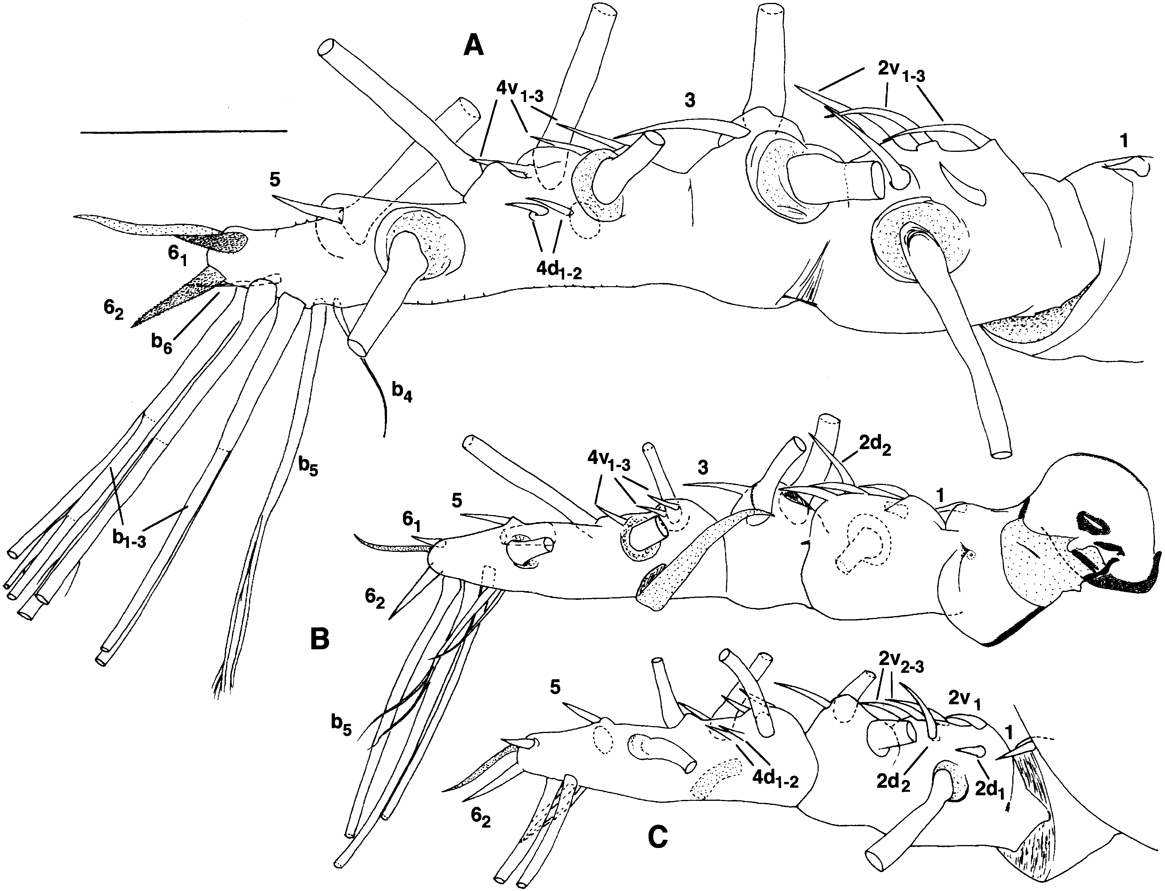
Antennule with distinct first joint, second joint (between second and third segments) distinct ventrally, third joint represented by constriction only ( Figs 17A View Figure 17 , 18A View Figure 18 ). Cuticular reticulations present, height of ridges decreasing distally. Spiniform 2v 1-3 - setae, 3-seta and 4v 1 -seta very long, with two rows of short, spiky setules, 4v 1 -seta at least 70% as long as 3-seta ( Fig. 18A View Figure 18 ). Position and size of other setae and aesthetascs as in M. hyottoko ; in one case (not Fig. 18B View Figure 18 ) b 5 -seta clearly bifid.
Anterior part of cephalothorax with pair of anterodorsal setae followed by closely spaced pair of pores ( Fig. 17D View Figure 17 ). Most of rest of cephalothorax not examined for such structures, but transverse row of at least four pores present mid-dorsally just in front of ‘waist’. Incorporated pediger with at least four pairs of dorsolateral and lateral pit setae, medial-most ones being very widely separated from each other ( Figs 19A View Figure 19 , 29 View Figure 29 ); rear dorsal meshes not much ornamented. On first free pediger ( Fig. 19A View Figure 19 ), spinule field formula apparently 3-3-5-5-3-3, but boundaries between fields hard to see, each field being bounded posteriorly by comb of spinules of same size as others in field; spinulose region surrounded by reticulate region, its meshes full of squiggly figures. Pores on this segment not clearly seen, possibly a tiny anterior one just right of midline and a large one at midlength just left of midline, definitely a pair at two-thirds length. Pit setae including dorsal posterior pair and two lateral pairs, anterior one of which located further ventrally. On second free pediger ( Fig. 19A View Figure 19 ), spinule fields and surrounding meshes as above, former with formula 3-3-8-7-3-3 (some of the 7 being triangular in shape). Anterior pair of large pores, another pair at one-third length, single pore far to right of these, and final pair at two-thirds length; two pairs of posterodorsal pit setae and two lateral pairs, anterior of latter being further ventral. On third free pediger ( Fig. 19B View Figure 19 ), spinule fields better defined than above, formula 3-2-4-4-2-3; one pair of large anterior pores, possibly another pair (or 1 + 2) in front of them; two pairs of posterodorsal pit setae. On all three of these pedigers, lateral meshes of tergites containing several small cuticular figures each. On fourth free pediger ( Fig. 19C View Figure 19 ), posterior pair of pit setae present, spinule fields as 1-4-3-3-4-1 (lateralmost single field at same level as second field of other rows; third field in innermost rows considered to include spinule areas behind pit setae).
Genital compound somite with no or very little hint of dorsal division ( Fig. 19C View Figure 19 ), ventral side with medial pore at base of ovigerous spines, two pairs of lateral pores (or combinations of pore and groove), and posterior small, medial bump ( Fig. 19F View Figure 19 ). Spinule fields (as seen in dorsal view; possibly more lateral fields exist) with formula 7-7-7-7, with four fields in each row anterior to the purported dorsal division. Rearmost spinules on this segment about twice as long as other spinules ( Fig. 19C View Figure 19 ). In dorsal view, anterior half of penultimate segment and all of telson ornamented with weak, wavy, transverse ridges ( Fig. 19D View Figure 19 ). Posterior part of penultimate segment with transverse array of six spinule fields, each ending posteriorly in 2–4 largish spines ( Fig. 19E View Figure 19 ). Caudal rami with more-orless rectilinear pattern of short, weak ridges ( Fig. 19D View Figure 19 ), armed with six setae, five of them long and biserially plumose, but dorsalmost seta shorter with single, dorsal row of setules.
Segmentation and setation of legs 1–4 as in M. hyottoko ( Fig. 20 View Figure 20 ). Outer anterior face of coxa of legs 1–4 spinulose ( Fig. 17E View Figure 17 ), these spinules about twice as large as dorsal ones on trunk somites, arranged in about eight transverse combs with many others between combs ( Fig. 17F View Figure 17 ). Two ‘pads’ of unexplained nature at distal end of spinulose area, another found subapically anterior to these (barely visible in Fig. 17E View Figure 17 , leg 2). Basis seta of leg three much longer than that of legs 1, 2 and 4; single pore anterior to base of this seta in each leg ( Fig. 20 View Figure 20 ). Outer margin of all three exopodal segments spinulose; about four rows on third segment ( Fig. 17E View Figure 17 ). No inner seta on first segment of either ramus, at most a ‘scar’ on endopod ( Fig. 17G View Figure 17 ). Tiny pore distally on anterior face of third segment of each ramus ( Fig. 20A, C, E View Figure 20 ). Outer apical seta of third exopodal segment with widely spaced, rather weak, distally directed denticles ( Figs 17H View Figure 17 , 20D View Figure 20 ). Intercoxal plates about 40% wider than leg coxae, and 5–6 times wider than their height at midline ( Fig. 20B View Figure 20 ). Leg 5 as in M. hyottoko (not illustrated).
Ovigerous spines arising from conical ventral protruberance of anterior half of genital double somite ( Fig. 19F View Figure 19 ), 24.1–36.7% of body length in lateral view. Spines tapering to fine distal points with slight irregular thickening at midlength.
Eggs: Spherical, 29 Mm in mean diameter in one ovigerous specimen (n = 10), 37 Mm in another (n = 9).
Nauplius: Oval, 38 Mm long, 25 Mm wide, with smooth dorsal surface and large, trapezoidal, labral bulge in middle of ventral side between bases of limbs ( Fig. 21B View Figure 21 ). Antennule possibly three-segmented, but small, proximal ‘first segment’ not seen clearly ( Fig. 21C, D View Figure 21 ). Second segment moderately elongate and curved, with two long medial setae (one near base and other one distal and setulose). Third segment smaller than second, with setulose seta on inner apical corner, two outer subapical setae (at least outermost of these setulose), and subapical outer spinule. Protopod of antenna not well seen; coxal endite rounded with a few blunt teeth on margin ( Fig. 21C View Figure 21 ). Endopod two-segmented; first segment short, cylindrical; second segment long, straight, and thin, with short sensillum or duct on outer apical corner, next to long, hook-like seta bent inwards at up to right angle to axis of segment ( Fig. 21C, D View Figure 21 ; presence of any subapical sensillum not confirmed). Exopod foursegmented ( Fig. 21D View Figure 21 ); first segment short; second segment elongate, rectangular, with four spinules arranged 1-2-1 on anterior margin and long inner distal seta with setules; third segment short, its armament, if any, unconfirmed; tiny fourth segment with two long setae, at least inner one with setules. Mandible with short, unarmed coxa and oblong basis, neither seen clearly (e.g. Fig. 21C View Figure 21 ). Exopod twosegmented, both segments very short, bearing one and two long setae, respectively, at least that of first segment setulose ( Fig. 21E View Figure 21 ). Endopod (see Discussion) consisting of small hook with spur on its outer side and simple seta inserting at its base posteriorly ( Fig. 21E, F View Figure 21 ). Furcal setae (‘balancers’) arising from raised cylindrical sockets, examined one apparently bifid near its base (not illustrated).
MAEMONSTRILLA SIMPLEX SP. NOV.
FIGURES 18C, D View Figure 18 , 22 View Figure 22 , 23 View Figure 23 , 29 View Figure 29
Diagnosis: Cephalothorax less bulbous and more cylindrical than in other species of this genus. Its polygonal reticulation simple and completely nonspinulose. No reticulation or spinulation noted on other body regions by light microscopy (SEM not attempted due to scarcity of material). Articulations between antennular segments 2–4 not distinct. Spiniform 2 v- and 3-setae more than three times longer than 4-setae; 1-seta and all 2-setae appearing minutely spinulose even by light microscopy, and 6-setae differing greatly in size. Oral papilla relatively smaller than in other species except M. okame , preceded by parallel grooves and pair of tubular pores. Basis of legs 1–4 slightly protruding on inner side. Two large, rounded, ventral lobes on anterior and posterior parts of genital compound somite.
Etymology: Named for the simple appearance of the trunk cuticle.
Material examined: Three females: Holotype ( KMNH IvR 700 222) and ovigerous paratype ( KMNH IvR 700 223) collected by S. Ohtsuka on south coast of Ishigaki Island , 30.iv.1994, holotype used for microscopic drawings with all legs 1–4 mounted on slide, body in vial, paratype also used for microscopic drawings; one paratype ( LBM Reg. No. 1430000931), collected by M. J. Grygier from Kabira Bay, Ishigaki Island, 9.iii.1993, intact.
Description: Cephalothorax only moderately bulbous, cylindrical with rounded anterior end and slightly tapered posterior half ( Fig. 22A, B View Figure 22 ). In dorsal view, modest ‘waist’ evident before incorporated pediger. Dorsal and lateral measurements made from all three specimens. Body length in lateral view (sum of lengths of cephalothorax, metasome and urosome as defined in Fig. 1A View Figure 1 ) 1.80–1.98 mm, with these body regions contributing 58.6–59.9, 25.7–26.6 and 14.2– 15.5%, respectively; thus, overall size larger, cephalothorax relatively longer, and urosome relatively shorter than in other members of this species group. Height and greatest width of cephalothorax close to equal, 40.4–42.8 and 41.1–42.1% of cephalothorax length, respectively, lower than in other members of this species group. Antennule length 45.1–48.3% that of cephalothorax. Width of cephalothorax at waist 70.8–83.0% of greatest width, that of incorporated pediger 86.0–91.9%. Widths of succeeding three free pedigers and genital compound somite relative to that of incorporated pediger 72.9–78.0, 63.5–70.2, 49.5– 55.6 and 30.6–35.5%, respectively. Ovigerous spines 67.4–71.9% as long as cephalothorax.
Cephalothorax reticulate posterior to level of naupliar eye ( Fig. 22A, B View Figure 22 ). Reticulations polygonal, similar in shape and size throughout, smooth inside and bounded by fine, simple, cuticular riges. No reticulations noted elsewhere on body. No beds of denticles or spinules noted on dorsum of trunk by light microscopy, nor any projecting armament along rear margins of genital compound somite and penultimate somite. Outer face of coxa and exopod in legs 1–4 also seemingly smooth, except for three transverse rows of spinules observed on outer side of third exopodal segment of left leg 1 ( Fig. 23A View Figure 23 ).
Antennules ( Fig. 18C, D View Figure 18 ) with three small sclerites for muscle attachment amidst wrinkled arthrodial membrane at base of first segment. Second to fourth segments not clearly separated from each other. All setal elements as shown in Grygier & Ohtsuka (1995) present on right antennule of examined specimen, some distal elements lost from left antennule. Two apical 6-setae greatly differing in size, 61 being smaller and 62 apparently bearing some long setules. 2 v- setae and 3-setae more than three times longer than 2 d- setae and 4-setae; 1-seta and 2-setae with two rows of minute spinules discernible by light microscopy, 3-seta and 4-setae with even smaller spinules, and 5-seta seemingly smooth.
Oral papilla conical, nearly straight and protruding ventrally ( Fig. 22A, B View Figure 22 ), relatively the smallest among the species of Maemonstrilla introduced in this paper except that of M. okame . Anteroventral surface of cephalothorax between oral papilla and bases of antennules longitudinally wrinkled, with pair of conical tubular pores, presumably homologous to tubular pores of M. hyottoko ( Fig. 22C View Figure 22 ). Postantennular scars expressed as two pairs of small, radially rayed knobs level with conical pores, each pair accompanied by pore (or minute third knob?) ( Fig. 22C View Figure 22 ). ‘Forehead’ transversely wrinkled, with pair of hair-like sensilla among wrinkles ( Fig. 22D View Figure 22 ); buttonlike structure present anterodorsally (presumably homologous to wrinkled pit in M. hyottoko ), followed by two medial pores and two pairs of more lateral pores. One pair of pores close behind oral papilla ( Fig. 22C View Figure 22 ). Three cups of naupliar eye well developed, diameters 110–139 Mm, lateral ones separated from each other and slightly larger than ventral cup.
More posterior pores and pit setae shown in Figure 22A View Figure 22 (in part) and Figure 29 View Figure 29 . Cephalothorax with five pairs of dorsolateral and lateral pit setae on incorporated pediger (mostly only their pits observed). First free pediger with one dorsal and two lateral pairs of pit setae, and second free pediger with two dorsal and two lateral pairs; more anterior of lateral pit setae also most ventral on these two somites. Third free pediger with two pairs of dorsal pit setae, and fourth free pediger with one pair. Each free pediger with pair of anterior dorsal pores hidden under rear margin of preceding segment or cephalothorax. Second free pediger with additional pair of double pores at one-third length and pair of simple pores at two-thirds length. Third free pediger with pair of dorsal pores at about two-thirds length.
Legs 1–4 ( Fig. 23 View Figure 23 ) with low and wide intercoxal sclerite, as wide as or wider than legs, with very slight projection at midline and distally directed processes at outer corners. Inner margin of basis rounded and slightly protruding. Outer seta and pore of basis, as well as segmentation and setation of rami, and pore in distal segment of each ramus, as in M. hyottoko . Outer spiniform setae on exopod simple, that on third segment slightly larger than that on first. Inner distal corner of first endopodal segment with protruding, socket-like structure ( Fig. 23A, D View Figure 23 ). Outer margin of outer apical exopodal seta with row of small, sharp, distally pointing, closely spaced denticles ( Fig. 23B View Figure 23 ). Leg 5 as in M. hyottoko ( Fig. 22E View Figure 22 ).
Dorsum of genital compound somite with transverse partial articulation ( Fig. 22F View Figure 22 ). Ventrally, posterior part and portion anterior to bases of ovigerous spines each produced into round swelling ( Fig. 22E View Figure 22 ). Anteriorly directed ovigerous spines cylindrical but tapering to naked tips from slightly swollen, wrinkled region at two-thirds length, reaching perhaps just beyond first legs ( Fig. 22A, E View Figure 22 ). Caudal rami armed as in M. hyottoko , dorsal seta originally with setules, but these now all broken off ( Fig. 22F View Figure 22 ); ventral pore present.
Eggs: Mean diameter of eggs from ovigerous specimen from south coast of Ishigaki Island 32 Mm (n = 15).


No known copyright restrictions apply. See Agosti, D., Egloff, W., 2009. Taxonomic information exchange and copyright: the Plazi approach. BMC Research Notes 2009, 2:53 for further explanation.
|
Kingdom |
|
|
Phylum |
|
|
Class |
|
|
Order |
|
|
Family |
|
|
Genus |