
Macrocyprina youngi, Brandão, 2005
|
publication ID |
https://doi.org/ 10.5281/zenodo.5402815 |
|
persistent identifier |
https://treatment.plazi.org/id/E5718798-FFFA-FFA5-776E-D4A4FDC7FCF7 |
|
treatment provided by |
Marcus |
|
scientific name |
Macrocyprina youngi |
| status |
sp. nov. |
Macrocyprina youngi View in CoL n. sp.
( Figs 1-5 View FIG View FIG View FIG View FIG View FIG )
TYPE MATERIAL. — Holotype: Praia da Tartaruga, Armação de Búzios , Rio de Janeiro, 22°52 ’S, 41°54’W, on sponges over rocky shore, between 1 to 5 m, 27. III.2000, S. N. Brandão, E. N. Calderon & C. A. Echeverría coll., adult (in alcohol 70%, MNRJ 15700 View Materials ) GoogleMaps . Paratypes: same locality, 9, 22, 29 juvs ( MNRJ 15701 View Materials ) GoogleMaps ; same locality, 1, 4, 5 juvs (MNHN-Os 592) GoogleMaps ; Praia de João Fernandes, Armação de Búzios , Rio de Janeiro, on macroalgae, 27. III.2000, S. N. Brandão, E. N. Calderon & C. A. Echeverría coll., 1, 2, 6 juvs, 3 LV, 1 RV, 3 RLV ( MNRJ 16833 View Materials ) .
DIMENSIONS. — Holotype ( MNRJ 15700 View Materials ): LV, h 0.50 mm, l 1.23 mm ; RV, h 0.53 mm, l 1.25 mm. Paratypes ( MNRJ 15701 View Materials ) spm 2,: LV, h 0.53 mm, l 1.28 mm ; RV, h 0.53 mm, l 1.30 mm; spm 3,: RV broken; LV, h 0.50 mm, l 1.38 mm; spm 4,: LV, h 0.50 mm, l 1.35 mm; RV, h 0.53 mm, l 1.38 mm; spm 6,: LV, h 0.53 mm, l 1.35 mm; RV, h 0.55 mm, l 1.38 mm; spm 8,: LV, h 0.53 mm, l 1.33 mm; RV, h 0.53 mm, l 1.35 mm; spm 9,: LV, h 0.50 mm, l 1.28 mm; RV, h 0.53 mm, l 1.30 mm;: LV, h 0.50 mm, l 1.35 mm; RV broken; spm 11,: LV, h 0.50 mm, l 1.28 mm; RV, h 0.53 mm, l 1.30 mm.
ETYMOLOGY. — Named in honour to Dr Paulo S. Young (†), who was a brilliant Brazilian carcinologist, and who was a mindful advisor
DISTRIBUTION. — Recent. Only known from the type locality. Brazil, Rio de Janeiro, Armação de Búzios, between 1 and 5 m.
DIAGNOSIS. — Carapace fairly large, elongate in lateral outline, its lateral surface with three or four small subcircular patches and other irregular patches in variable positions. Male appendages V strongly asymmetrical, right appendage larger than left; left appendage bell shaped, with base and two podomeres, last podomere not recurved, profusely setose. Zenker’s organ with tiny terminal bulb; hemipenis with subtriangular posterior lamella, with one angle posteroventrally projected.
DESCRIPTION
Carapace fairly large, elongate in lateral outline, inequilateral ( Fig. 1A, B View FIG ), with fairly ramified radial pore canals ( Fig. 1C, D View FIG ). Greatest height at midlength, slightly posterior to muscle scars. Length approximately 2.5 times maximum height. Lateral surface with three or four small subcircular, white, opaque patches; other irregular patches in variable positions, but mainly near margins. Dark brown cuticle around central opaque patch, and at anterodorsal and posterior regions of valve surface. Dorsal margin evenly low-arched, with indistinct dorsal angle. Posterodorsal margin fairly straight. Anterior margin ( Fig. 1C View FIG ) unevenly rounded. Ventral margin rounded anteriorly; with shallow indentation, and only slightly upswung posteriorly. Posterior angle rounded ( Fig. 1D View FIG ), dorsal and ventral margins forming an angle of about 50°. Zone of concrescence ( Fig. 1C, D View FIG ) of moderate width, irregular. Adductor muscle scar pattern
A
B
( Fig. 1E View FIG ) with three dorsal scars and eight scars of variable sizes situated below. Carapace ovateoblong in ventral and dorsal views ( Fig. 1F, G View FIG ).
Antenna I ( Fig. 2A View FIG ) with seven slender, elongate podomeres, with long, thin, flexible setae. Podomeres II and III fused with incomplete suture. Podomere VII with four setae, dorsodistal seta thinner than others.
Antenna II ( Fig. 2B View FIG ) robust, with six podomeres. Podomere III, medially ( Fig. 2C View FIG ), with six long, subequal setae located ventrally at proximal margin; laterally, with dorsoproximal scale with three setae; ventrally at distal margin with four setae ( Fig. 2D View FIG ). Podomere IV with two thin setae proximally at dorsal margin; with one thin seta near ventrodistal edge; medially, dorsodistal angle
E D
( Fig. 2E, F View FIG ) with three ventral setae, two of them sexually dimorphic setae (simple in female and candle-shaped in male); sexually dimorphic setae subequal in length in male, but in female more ventral seta about two thirds length of more dorsal seta. Male also with one thin seta dorsal to sexually dimorphic setae. Podomere V subtriangular ( Fig. 2G View FIG ); distal angle with four claws and one seta; dorsoproximal angle with one claw and one seta.
A
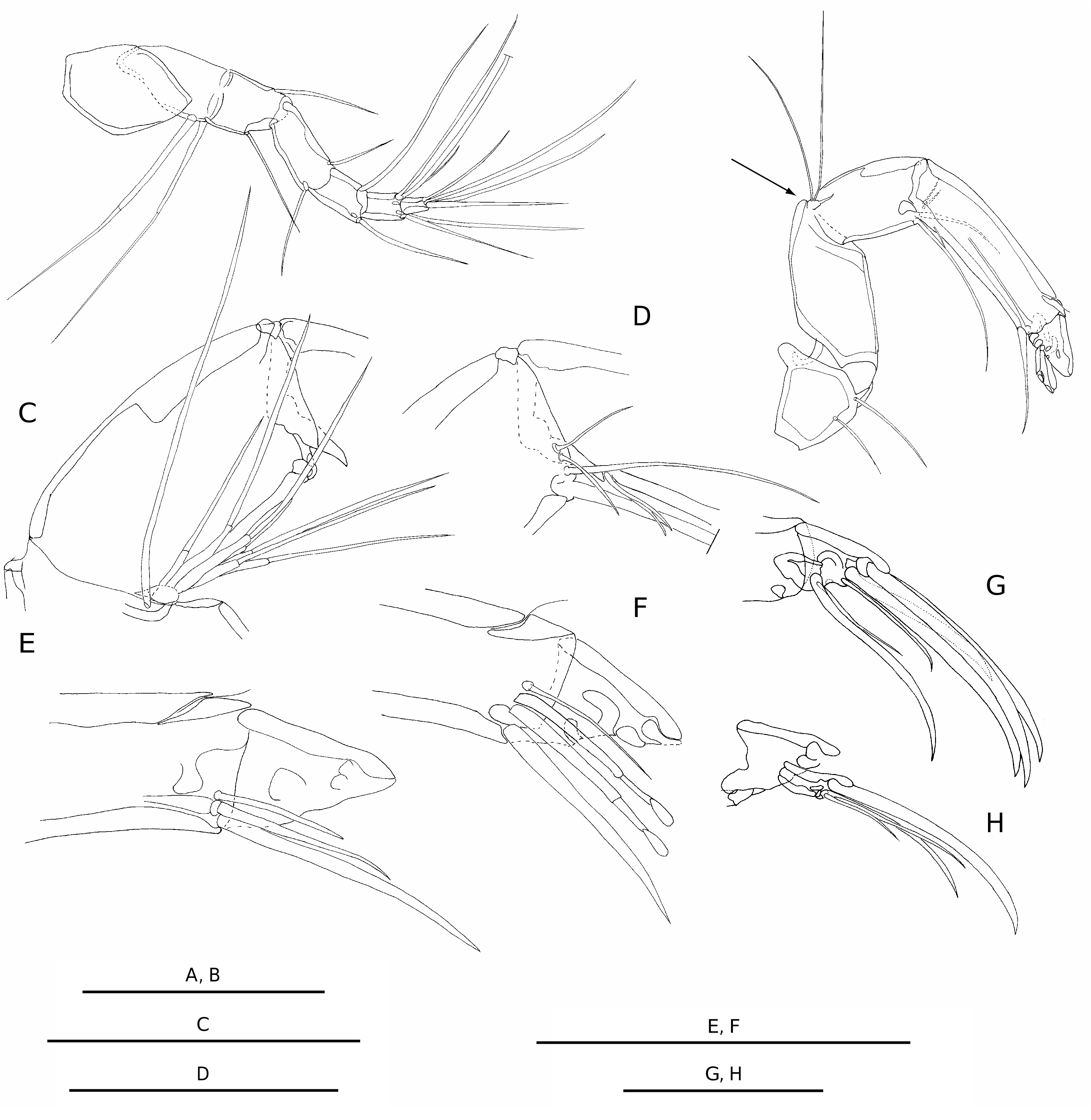
Mandible ( Fig. 3A View FIG ) with a broad masticatory jaw ( Fig. 3B View FIG ) armed with one dorsal, conical tooth followed by six tricuspidate teeth and several setae and pegs. Podomere I of palp with exopodite ( Fig. 3C View FIG ) with a dorsoproximal peg on medial surface, and six or seven distal setae. Podomere I with three setae at ventrodistal angle. Podomere II ( Fig. 3D View FIG ) with eight ventrodistal setae. Podomere III ( Fig. 3E View FIG ) with three middorsal setae; six ventrodistal setae. Podomere IV ( Fig. 3F View FIG ) with about three thick claws and two setae at distal margin.
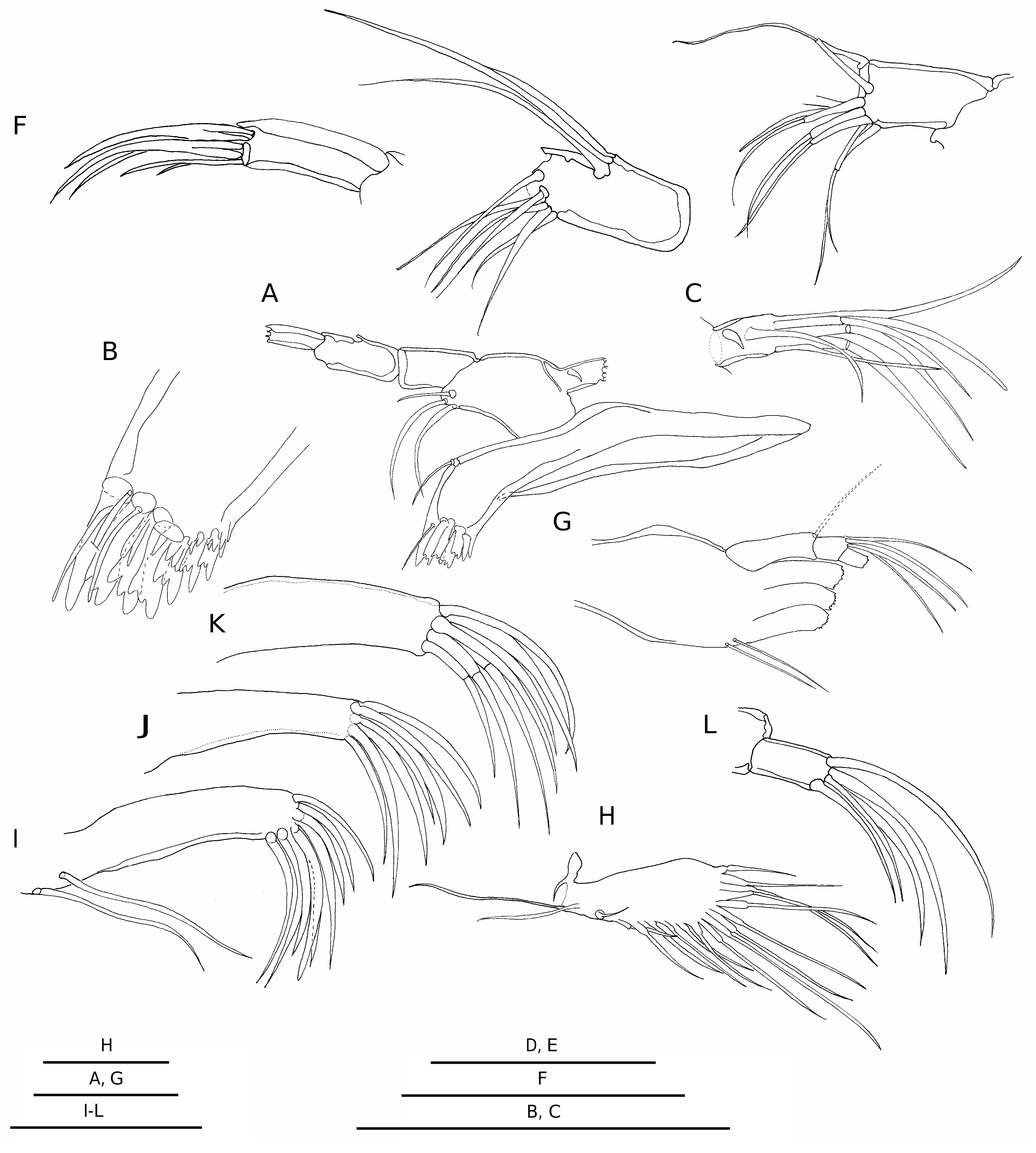
Maxilla I ( Fig. 3G View FIG ) with three slender endites, each armed distally with a dense fringe of subequal claws. Endite I ( Fig. 3H View FIG ) with 10 distal claws, and two ventral setae; endites II and III ( Fig. 3I, J View FIG ) with seven distal claws each. Dorsal vibratory plate ( Fig. 3K View FIG ) with two proximal setae; and with a series of long setae along posterior and ventral margins. Palp slender, flexible with three podomeres. Podomere II with four dorsodistal setae. Podomere III ( Fig. 3L View FIG ) with five distal setae. Anterior lobe of appendage V similar in males and females, with nine to 11 setae ( Fig. 4B View FIG ). Female left and right appendages V symmetrical, slender, with thick distal claws ( Fig. 4E View FIG ). Podomere IV with three distal claws (middistal one longest). Male appendages V very asymmetrical ( Fig. 4A, C View FIG ). Right appendage ( Fig. 4A View FIG ) robust, highly sclerotized, considerably larger, about 1.5 time length of left appendage. Palp hook-shaped, ventrodistal angle of podomere I with two thick pegs; one short, thin seta; and one thick, short, cylindrical structure, similar to distal aesthetasc of hook. Dorsodistal angle with one seta. Podomere II recurved at approximately 100°; with one robust, distal aesthetasc; without thin aesthetascs (also without visible gaps in dorsal and ventral cuticle). Left appendage ( Fig. 4C View FIG ) slightly sclerotized, highly modified. Podomere I with convex ventral margin; distal setae at lateral surface; ventrodistal angle with two pegs and one short, thin seta. Podomere II ( Fig. 4D View FIG ) short, but conspicuous, not recurved, lateral surface hirsute, covered with numerous short setulae; with well developed aesthetasc on dorsodistal angle; without thin, ventral aesthetascs (also without visible gaps in dorsal and ventral cuticle). Suture between podomeres I and II conspicuous, but not complete.
Podomeres I and II of appendage VI ( Fig. 4F View FIG ) fused, with an incomplete suture, recurved, forming a knee; with one seta at posterodistal angle (inside the knee); anterior margin (outside the knee) with two setae proximal to suture, and three setae distal to suture. Distal margin of podomere III with a fanlike tuft of setulae. Podomeres III and IV with tufts of very short, fine setulae at dorsal margin. Podomere VI with two thick claws and one seta; dorsodistal claw slightly smaller than middistal claw; length of short seta about one third length of middistal claw.
Appendage VII ( Fig. 4G View FIG ) with basal segment recurved, forming a knee; and apparently composed of two podomeres (I + II), separated by a weak, incomplete suture; posterodistal angle (inside the knee) with one seta. Dorsal margin with one seta proximal to suture; and three setae distal to suture, two of them modified. Podomere III with one ventrodistal seta, and one dorsodistal setula. Podomere IV with one ventrodistal seta. Podomere V with two ventrodistal setae. Podomere VI with two ventrodistal setae, the most ventral one longest; and one reflexed seta of the same length of podomeres III to VI together. Furcal rods ( Fig. 4H View FIG ) symmetrical, long, thick, curved at tips, with conspicuous suture between rami and distal setae; with short proximal setae; distal two thirds of posterior margin of each ramus with thin, short barbs.
Male genitalia: hemipenis ( Fig. 5A, B View FIG ) oblongovate, posterior lamella subtriangular, one angle posteroventrally projected. Hemipenis approximately half of length of body. Zenker’s organ ( Fig. 5C, D View FIG ) long, approximately three quarters of body length, with tiny terminal bulb; vas deferens arranged in three loose loops, about equal in length to Zenker’s organ and situated around it.
REMARKS
Macrocyprina youngi n. sp. presents a ventral, huge aesthetasc medial to the hook of male right appendage V ( Fig. 4A View FIG ). This aesthetasc is similar to the distal aesthetasc of the podomere II of the same appendage. In the other species of Macrocyprididae the aesthetasc ventral to the hook is, as Maddocks (1990: 16) describes it “delicate, sometimes hard to see”.
The male appendages are known for 15 species of Macrocyprina : M. africana ; M. barbara ; M. bermudae ; M. captiosa ; M. dispar ; M. hartmanni ; M. hawkae ; M. hortuli ; M. madagascarensis ; M. moza ; M. propinqua ; M. schmitti ; M. skinneri ; M. succinea ; M. swaini . The male left appendages V of these species, with the exception of M. hawkae , are hook shaped, being similar to the respective right appendages. The male left appendage V of M. youngi n. sp. is remarkably different from these species, the ventral margin of the podomere I is concave, the podomere II is not recurved, and the lateral surface of the podomere II is densely setose. Macrocyprina hawkae presents a similar
A C D
modified male left appendage V, but the podomere II is vestigial ( Maddocks 1990: fig. 28.15, 16). Macrocyprina youngi n. sp. is very similar to M. hawkae , which inhabits the Caribbean Sea, not only on the modified left appendage V (which was until now unique to M. hawkae ), but also on carapace features, hemipenis and Zenker’s organ. These two species differ in the following characters: Macrocyprina hawkae is less elongate, higher in proportion to length, with a more arcuate dorsal margin than M. youngi n. sp.; M. hawkae has larger lateral patches, tending to fuse with each other, and occupying great part of the lateral surface ( Maddocks 1990: figs 16.11, 12, 17.11, 12), while M. youngi n. sp. has smaller and subcircular patches; the hirsute podomere II of male left appendage V is vestigial in M. hawkae , while in M. youngi n. sp. this podomere is conspicuous; the posterior lamella of the hemipenis of M. hawkae is irregularly shaped, while M. youngi n. sp. presents a subtriangular posterior lamella.
The other 10 species were described from carapaces, and sometimes also from females appendages. M. caiman , M. jamaicae , M. okinawae , M. parcens and M. vargata are higher in proportion to length, with a more triangular outline than M. youngi n. sp.; M. bonaducei and M. rattrayi have more angulate anteroventral margins and deeper ventral indentations; M. quadrimaculata has a more straight ventral margin, with four lateral patches situated more dorsally, while M. youngi n. sp. has three large patches situated more ventrally and other smaller patches in variable positions. Macrocyprina belizensis , from Caribbean, and M. noharai , from West Pacific, are more similar to M. youngi n. sp. But M. belizensis has a smaller size, the carapace is higher in proportion to length, with a more arched dorsal margin, with a deeper ventral indentation and larger opaque patches. Macrocyprina noharai has more equilateral valve, with a less arched dorsal margin, more narrowly rounded anterior margin, and with the middle and posterior lateral opaque patches tending to fuse, while the patches of M. youngi n. sp. are distinctly separated.
The carapace of Macrocyprina sp. 1 recorded by Dias-Brito et al. (1988) is higher in proportion to length, has a more arcuate dorsal margin, and a more straight ventral margin than M. youngi n. sp. The carapace of Macrocyprina sp. 2 recorded by Coimbra (1995) and Coimbra et al. (1992, 1999) has a more broadly concave ventral margin; a more convex dorsal margin; and a greater carapace length (1.6 mm vs 1.3 mm) than M. youngi n. sp. Furthermore, the posterodorsal margin of left valve of Macrocyprina sp. 2 is slightly concave, and in M. youngi n. sp. it is continuously convex.


| RV |
Collection of Leptospira Strains |
No known copyright restrictions apply. See Agosti, D., Egloff, W., 2009. Taxonomic information exchange and copyright: the Plazi approach. BMC Research Notes 2009, 2:53 for further explanation.