
Macrocyprina rocas, Brandão, 2005
|
publication ID |
https://doi.org/ 10.5281/zenodo.5402815 |
|
persistent identifier |
https://treatment.plazi.org/id/E5718798-FFF3-FFBF-7484-D7C4FB3DFDF7 |
|
treatment provided by |
Marcus |
|
scientific name |
Macrocyprina rocas |
| status |
sp. nov. |
Macrocyprina rocas View in CoL n. sp.
( Figs 6-10 View FIG View FIG View FIG )
TYPE MATERIAL. — Holotype: Rocas Atoll , 3°51.680’S, 33°49.604’W, on sponges and macroalgae, 19 m, P. S. Young, P. C. Paiva & A. A. Aguiar coll., 16.X.2000, adult in alcohol 70% ( MNRJ 15702 View Materials ) GoogleMaps . Paratypes: same locality, 10, 5, 8 juvs ( MNRJ 15703 View Materials ) GoogleMaps ; same locality, 3 adults, 2 juvs (MNHN-Os 593) GoogleMaps ; Rocas Atoll , 3°51.356’S, 33°49.559’W, on sediment and macroalgae, 16 m, 16.X.2000, P. S. Young, P. C. Paiva & A. A. Aguiar coll., 1, 4 juvs ( MNRJ 15704 View Materials ) GoogleMaps ; Rocas Atoll , Ilha do Cemitério, intertidal region, 9.XI.2001, C. S. Serejo & M. C. Rayol coll., 1 LV, 1 RV ( MNRJ 16834 View Materials ) ; Rocas Atoll , outside of the Atoll, northwestern region, on macroalgae, 9 m, 3.I.2001, F. B. Pitombo & R. Barroso coll., 1 juv. ( MNRJ 15705 View Materials ) .
DIMENSIONS. — Holotype (MNRJ 15702): LV, h 0.39 mm, l 1.06 mm; RV, h 0.40 mm, l 1.08 mm. Paratypes (MNRJ 15702) spm 3,: LV, h 0.40 mm, l 1.08 mm; RV, broken valve, l 1.09 mm; spm 2,: LV, h 0.40 mm, l 1.10 mm; RV, h 0.40 mm, l 1.13 mm; spm X,: LV, h 0.40 mm, l 1.08 mm; RV, h 0.40 mm, l 1.08 mm; spm 4,: LV, h 0.40 mm, l 1.05 mm; RV, h 0.40 mm, l 1.08 mm; spm 5,: LV, h 0.40 mm, l 1.05 mm; RV, h 0.41 mm, l 1.08 mm; spm 6,: LV, h 0.41 mm, l 1.05 mm; RV, h 0.43 mm, l 1.08 mm; smp 7,: LV, h 0.41 mm, l 1.10 mm; RV, h 0.44 mm, l 1.10 mm; sex unknown: LV, h 0.41 mm, l 1.08 mm; RV, h 0.43 mm, l 1.08 mm.
ETYMOLOGY. — The name refers to the type locality, Rocas Atoll, and is used in apposition.
DISTRIBUTION. — Recent. Only known from the type locality. Brazil, Rocas Atoll, between 9 and 19 m.
DIAGNOSIS. — Carapace elongate, fairly small, its lateral surface with one to four central opaque patches. Male appendages V very asymmetrical, right appendage larger than left; left appendage V bellshaped, with base and two podomeres, podomere II not recurved, profusely setose. Zenker’s organ with small terminal bulb; posterior lamella of hemipenis elongated dorsoventrally.
DESCRIPTION
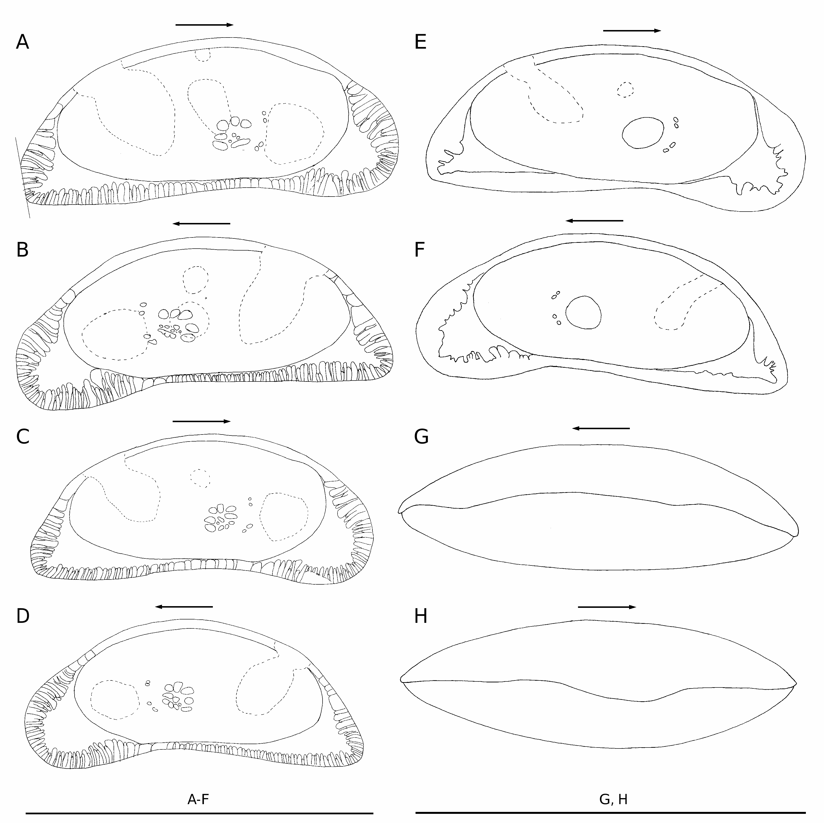
Carapace yellow-brown in fresh specimens, fairly small ( Fig. 6 View FIG A-F), elongate in lateral outline, inequilateral, with ramified radial pore canals. Greatest height at midlength, slightly posterior to muscle scars. Length approximately 2.7 times maximum height. Lateral surface with one to four central opaque patches. Only posterodorsal patch (the largest one) present in all specimens, this patch is diagonally elongated, reaching the dorsal margin. Most specimens with one anterior medium-sized patch. Approximately half of specimens with a small centrodorsal patch in both valves, patch on right valve always stronger than patch on left valve; other specimens with this patch only on right valve. Few specimens with one centroventral medium-sized patch, with part of this patch on the adductor muscle scars.Cuticle present on lateral surface of carapace lightly coloured, not dark brown. Dorsal margin evenly low-arched, with indistinct dorsal angle. Posterodorsal margin fairly straight. Anterior margin of carapace unevenly rounded.Ventral margin sinuous, with conspicuous, broad indentation. Posterior angle rounded, dorsal and ventral margins forming an angle of about 55°. Zone of concrescence very broad. Adductor muscle scar pattern ( Fig. 7B View FIG ) with three large dorsal scars and about nine or 10 scars of variable sizes situated below. Carapace ovate-oblong in dorsal and ventral views ( Fig. 6G, H View FIG ).
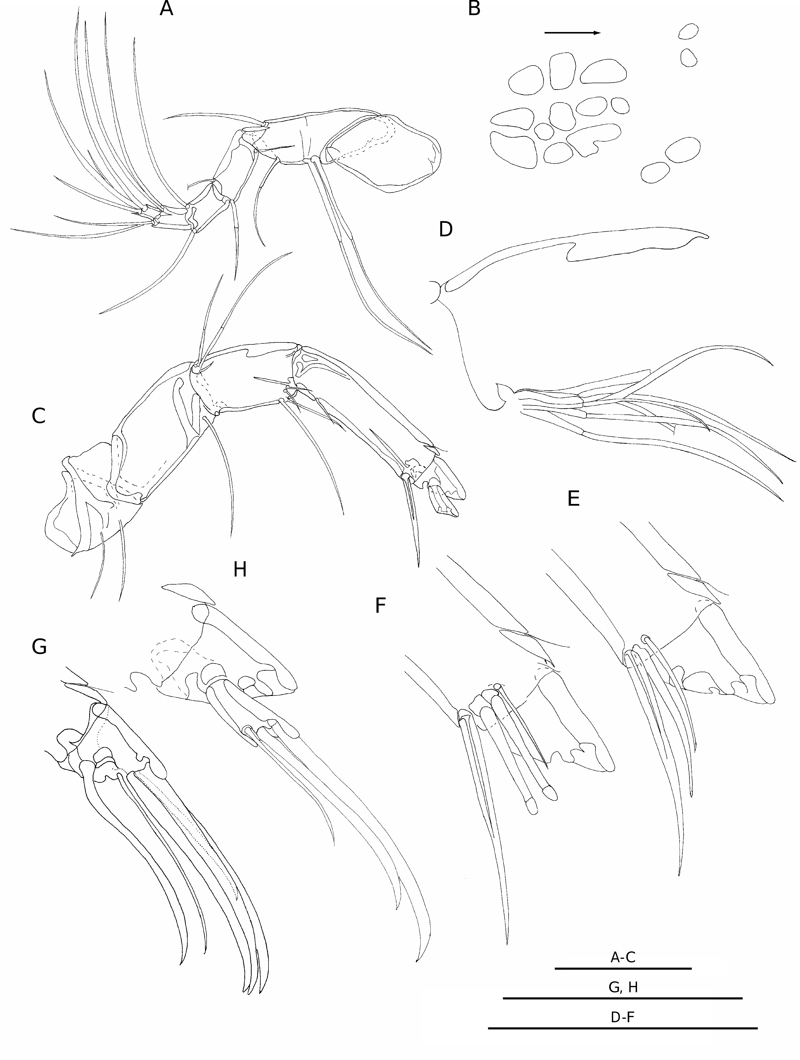
Antenna I ( Fig. 7A View FIG ) with seven slender, elongate podomeres, with long, thin, flexible setae. Podomeres II and III fused with incomplete suture. Podomere VII with four setae, dorsodistal seta thinner than others.
Antenna II ( Fig. 7 View FIG C-H) robust, with six podomeres. Podomere III, medially ( Fig. 7D View FIG ), with six long, subequal setae located ventrally at proximal margin; laterally, with dorsoproximal scale with three setae; ventrally at distal margin with four setae. Podomere IV with two thin setae at dorsal margin; with one thin seta near ventrodistal edge; medially, dorsodistal angle with three ventral setae, two of them sexually dimorphic (simple in female and candle-shaped in male) ( Fig. 7E, F View FIG ); sexually dimorphic setae subequal in length in male, but in female more ventral seta about two thirds length of more dorsal seta. Male also with one thin seta dorsal to sexually dimorphic setae. Podomere V subtriangular ( Fig. 7G View FIG ); distal angle with four claws and one seta; dorsoproximal angle with two setae.
Mandible ( Fig. 8A View FIG ) with a broad masticatory jaw ( Fig. 8B View FIG ) armed with one dorsal, conical tooth followed by six tricuspidate teeth and several setae and pegs. Podomere I of palp with exopodite with a dorsoproximal peg on medial surface, and six or seven distal setae. Podomere I with three ventrodistal setae. Podomere II with six ventrodistal setae. Podomere III with three middorsal setae; and six ventrodistal setae. Podomere IV with about three thick distal claws.
Maxilla I ( Fig. 8C View FIG ) with three slender endites, each armed distally with a dense fringe of subequal claws. Endite I with two ventral setae. Dorsal vibratory plate with two proximal setae; and with a series of long setae along posterior and ventral margins. Palp slender, flexible with three podomeres. Podomere II with three dorsodistal setae. Podomere III with three distal setae.
Anterior lobe of appendage V similar in males and females, with ten setae ( Fig. 8 View FIG D-F). Female left and right appendages V symmetrical, slender, with thick distal claws ( Fig. 8D View FIG ). Podomere IV
A C
with three distal claws (middistal one longest). Male appendages V very asymmetrical ( Fig. 8E, F View FIG ). Right appendage ( Fig. 8E View FIG ) robust, highly sclerotized, considerably larger, about 1.5 times length of left appendage. Palp hook-shaped, ventrodistal angle of podomere I with two thick pegs; none or one short, thin seta; and none or one thick, short, cylindrical structure, similar to distal aesthetasc of hook. Dorsodistal angle with one seta. Podomere II recurved at approximately 125°; with one robust, distal aesthetasc; without dorsal, thin aesthetasc (also without visible gap in dorsal cuticle). Left appendage ( Fig. 8F View FIG ) slightly sclerotized, highly modified. Podomere I with convex ventral margin; distal seta at lateral surface; ventrodistal angle with two pegs and one short, thin seta. Podomere II short, but conspicuous, not recurved, lateral surface covered with numerous short setulae; with well developed aesthetasc on dorsodistal angle; without thin, ventral aesthetascs (also without visible gaps in dorsal and ventral cuticle). Suture between podomeres I and II conspicuous, but not complete.
Podomeres I and II of appendage VI ( Fig. 9A) fused, with an incomplete suture, and recurved, forming a knee; with one seta at posterodistal angle (inside the knee); anterior margin (outside the knee) with two setae proximal to suture, and three setae distal to suture. Distal margin of podomere III with a fanlike tuft of setulae. Podomeres III and IV with tufts of very short, fine setulae at dorsal margin. Podomere VI with two thick claws and one seta; dorsodistal claw slightly smaller than middistal claw; length of short seta about half the length of middistal claw. Appendage VII ( Fig. 9B) with basal segment recurved, forming a knee; and apparently composed of two podomeres (I + II), separated by a weak, incomplete suture; posterodistal angle (inside the knee) with one seta. Dorsal margin with one seta proximal to suture; and three setae distal to suture, two of them modified. Podomere III
A
C D
with one ventrodistal seta, and a tuft of very short setulae at distal margin. Podomere IV with one ventrodistal seta. Podomere V with two ventrodistal setae. Podomere VI with two ventrodistal setae, the most ventral longest; and one reflexed seta of the same length of podomeres III to VI together.
Furcal rods ( Fig. 9C) symmetrical, long, thick, curved at tips, with conspicuous suture between rami and distal setae; with short proximal setae; distal two thirds of posterior margin of each ramus with thin, short barbs.
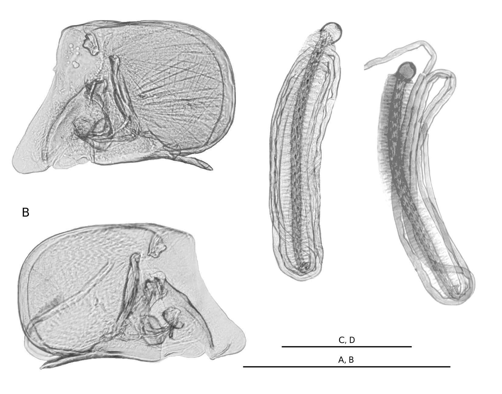
Male genitalia: hemipenis ( Fig. 10A, B View FIG ) oblongovate, posterior lamella elongated. Hemipenis approximately half of length of body. Zenker’s organ ( Fig. 10C, D View FIG ) long, approximately three quarters of body length, with small terminal bulb; vas deferens arranged in three loose loops, about equal in length to Zenker’s organ and situated around it.
REMARKS
An age-related variation in number and size of lateral patches is observed in specimens of Macrocyprina ; the juveniles usually present fewer and smaller patches than adults ( Maddocks 1990: 25). Only adults of M. rocas n. sp. (the appendages were used to determine the adulthood) were analysed in this study in order to describe the variation of the patches. Therefore, the described variation is not related to size and ontogenetic stages. The largest specimen analysed (spm 2, male: LV length 1.10 mm, RV length 1.13 mm) presents only the posterodorsal patch on the two valves, and a small central patch on right valve ( Fig. 6E, F View FIG ); while another specimen of approximately the same size (spm 7, male: LV length 1.10 mm, RV length 1.10 mm) presents four well developed patches on both valves ( Fig. 6A, B View FIG ); and one smaller specimen (spm 6, female: LV length 1.05 mm, RV length 1.08 mm) presents three patches on each valve ( Fig. 6C, D View FIG ).
The male appendages are known for 15 species of Macrocyprina (enumerated in Remarks of M. youngi n. sp.). The male left appendages V of these species, with the exception of M. hawkae , are hook shaped, being similar to the respective right appendages. The male left appendage V of M. rocas n. sp. is remarkably different ( Fig. 4C, D View FIG ) from these species (details of these differences described to M. youngi n. sp. also apply to M. rocas n. sp.).
Beside the differences on the male left appendage V, M. hawkae has a more triangular carapace, presents larger lateral patches and less ramified radial pore canals than M. rocas n. sp.
The valves of M. belizensis , M. caiman , M. jamaicae , M. noharai and M. parcens are higher in relation to length, with more arched dorsal margins, and thinner zones of concrescence. The valves of M. bonaducei have a deeper ventral indentation, and are higher in proportion to length. Macrocyprina okinawae and M. vargata have more subtriangular and equilateral valves than M. rocas n. sp. The carapace of M. quadrimaculata has a more straight ventral margin, and smaller lateral patches, which are located in a more dorsal position. Finally, M. rattrayi has more subtriangular valves.
The outline of Macrocyprina sp. 1 recorded by Dias-Brito et al. (1988) is more subtriangular, with a more straight ventral margin than M. rocas n. sp. Macrocyprina sp. 2 recorded by Coimbra (1995: 38) and Coimbra et al. (1999: 373) is larger, with valves more elongated, less triangular, and presenting a straight posterodorsal margin, while the posterodorsal margin of M. rocas n. sp. is concave to slightly concave.
Macrocyprina youngi n. sp. is larger than M. rocas n. sp. The cuticle present on the lateral surface of the carapace of M. youngi n. sp. is dark brown, on M. rocas n. sp. it is lightly coloured. Macrocyprina youngi n. sp. presents the valve with a more acute posterior angle than M. rocas n. sp. The ventral margin of M. youngi n. sp. is upswung and straight posteriorly, while that of M. rocas n. sp. is sinuous. The outlines of the male right appendages V are different in both species. The basal region of podomere I of male left appendage V of M. youngi n. sp. is thicker than that of M. rocas n. sp.; podomere II is subrectangular in M. youngi n. sp., and subtriangular in M. rocas n. sp. The terminal bulb of Zenker’s organ of M. youngi n. sp. is proportionally smaller than terminal bulb of M. rocas n. sp.


| RV |
Collection of Leptospira Strains |
| R |
Departamento de Geologia, Universidad de Chile |
No known copyright restrictions apply. See Agosti, D., Egloff, W., 2009. Taxonomic information exchange and copyright: the Plazi approach. BMC Research Notes 2009, 2:53 for further explanation.