
Longior surieli, Morffe & García & Adams & Hasegawa, 2020
|
publication ID |
https://doi.org/ 10.11646/zootaxa.4877.1.5 |
|
publication LSID |
lsid:zoobank.org:pub:2930F98B-0EBD-45BF-B596-8DE8149A1C7C |
|
DOI |
https://doi.org/10.5281/zenodo.4562881 |
|
persistent identifier |
https://treatment.plazi.org/id/514587AB-2545-752E-FF62-FEB6F0341F14 |
|
treatment provided by |
Plazi |
|
scientific name |
Longior surieli |
| status |
sp. nov. |
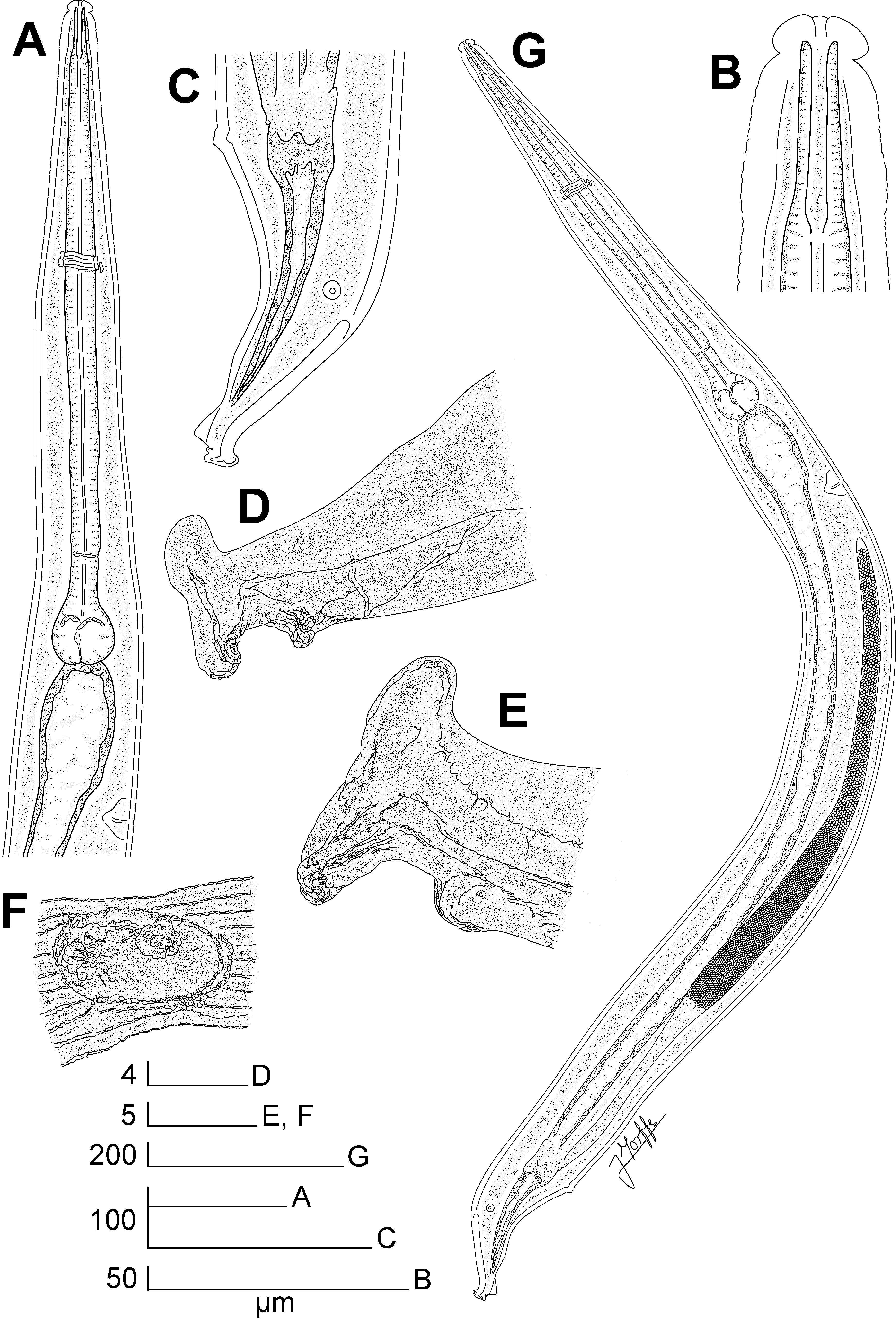
Longior surieli n. sp.
Fig. 1 View FIGURE 1 A–F, Fig. 2 View FIGURE 2 A–G, Fig. 3 View FIGURE 3 A–E
Type material. ♀ holotype, Dominican Republic, La Vega province, Reserva Científica “Ébano Verde”, Sendero El Arroyazo-Casabito ; in Antillanax dominicanus ; 19°04.42’N, 70°34.18’W; 16/II/2014; G. de los Santos, C. Suriel coll.; CZACC 11.7150 View Materials . Paratypes GoogleMaps 4♀♀, same data as holotype, CZACC 11.7151 –11.7154 GoogleMaps ; 15♂♂, same data as holotype, CZACC 11.7155 –11.7169 GoogleMaps ; ♀, same data as holotype, MNHNSD 05.001 View Materials GoogleMaps ; ♂, same data as holotype, MNHNSD 05.002 View Materials GoogleMaps .
Description. Female. Body long and slender, widening gradually posterior to head, reaching its maximum width at level of the vulva, then narrowing gradually towards tail. Cuticle thin. Sub-cuticular striae present. Cervical cuticle unarmed, with wide annuli (ca. 8 µm) from the base of the first cephalic annule to ca. the level of the first third of procorpus. Lateral alae well-developed, extending from ca. a body-width posterior to the basal bulb to the level of the anus. Head bearing eight paired, digitiform cephalic papillae, which originate from the external edge of the head, at ca. half of its height. A cuticular, veliform annular lip surrounding the trirradiate oral opening. Amphids lateral, at level of the base of the cephalic papillae. First cephalic annule comparatively long, truncate, slightly dilated. Stoma long and slender, surrounded by an oesophageal collar. Oesophagus consists of a muscular, sub-cylindrical procorpus, its base similar in diameter to the isthmus. Basal bulb rounded, valve-plate well-developed. Intestine simple, sub-rectilinear, its fore region slightly dilated. Rectum comparatively long. Anus as a crescent-like ventromedian slit, anteriorly directed, not prominent. Nerve ring encircling procorpus at ca. 30% of its length. Excretory pore located at ca. half of a body-width posterior to the basal bulb. Vulva a median transverse slit, its lips slightly prominent, located at level of midbody. Vagina muscular, forwardly directed. Genital tract monodelphic-prodelphic, occupying ca. 30% of the body length. Ovary distally reflexed at ca. a body-width posterior to the excretory pore, distal flexure ca. one body-width in length. Oocytes in a single row. Eggs ellipsoidal, bearing eight rough longitudinal ridges in the shell that do not reach the poles. Uterus contains 2– 3 eggs at a time. Tail conical, subulate, ending in a fine tip.
Male. Body shorter and comparatively slender than females. Posterior region ventrally curved. Cuticle unarmed, markedly annulated up to the level of the nerve ring, annuli ca. 2 µm. Rest of body with less conspicuous annuli (ca. 1 µm), up to level of the dorsal cuticular thickening. From the base of the head to a distance of ca. 30 µm, the cervical annuli increase their diameter to a maximum of ca. 30 µm, forming a dilatation at the cephalic region. Lateral alae narrow, extending from the last third of the oesophageal region to a distance of ca. 100 µm before the ventromedian pair of copulatory papillae. Head set-off from body by a deep groove, bearing eight ellipsoidal, slightly flattened cephalic papillae arranged as two sub-dorsal and two sub-ventral pairs. Six digitiform lips originate from the internal edge of the cephalic papillae and project to the center of the oral aperture. Lips are arranged as one lip dorsal, one ventral, two sub-dorsal and two sub-ventral; the latter flanking the dorsal and ventral lips, respectively. Amphids lateral, as crescent-like pores. Oesophagus consists of a muscular, sub-cylindrical procorpus, diminishing its diameter toward its posterior half, almost equal to the diameter of the isthmus at the level of their junction. Basal bulb rounded, valve-plate well-developed. Intestine simple, its fore region slightly inflated. Nerve ring encircling procorpus at ca. 45% of its length. Excretory pore located at ca. 1.5 body-widths posterior to the basal bulb. Monorchic, testis outstretched, its distal end located at a distance of a little less than a body-width behind the excretory pore. Vas deferens with three distinguishable regions: an anterior region with granular content; a median, slightly swollen region, also granular (granuli slightly shorter in diameter) and a posterior region that diminishes its diameter through the cloaca. Spicule absent. A ventral bursa-like structure present. The tail tip presents a hammer-like appearance due to a ventral bending and a dorsal knob-like protuberance. Dorsal cuticle of the tail end is thickened and smooth. Such thickening forms a cuticular crest at the tail tip that extends terminally and laterally at the ventral bending. Phasmids pore-like, lateral, located at level of the beginning of the bursa. Four pairs of copulatory papillae, two pre-cloacal and two post-cloacal. A ventromedian large pair consist of duplex papillae very close each other on an ellipsoidal protuberance (appear to be a single papilla in lateral view) at ca. 120–170 µm from the tail tip. A sub-lateral pair of papillae is at ca. the level of the beginning of the dorsal cuticular thickening. The remaining two post-cloacal pairs consist of small papillae: one pair ventral, located at the tip of a papilliform protuberance just posterior to the bursa and one pair ventral, sub-terminal, close to the tail tip, below and in contact with the terminal cuticular crest.
Differential diagnosis. Longior surieli n. sp. presents lateral alae extending from ca. a body width posterior to the basal bulb to the level of the anus, similar to the rest of the species of the genus. This feature differentiates it from L. semialata , which possess lateral alae that extend from the level of the vulva to just before the anus ( Hunt 1981). In addition, the oesophagus is comparatively shorter in L. surieli n. sp. (b = 4.11–4.52 vs. 3.30–3.70) and the tail is longer than L. semialata (c = 5.31–5.83 vs. 5.80–8.60).
Longior surieli n. sp. has a body shorter than L. longicollis , L. longior and L. zumpimito n. sp. (2.550 –2.850 mm vs. 2.980 –3.640 mm vs. 3.500 –4.525 mm vs. 3.110 –3.920 mm). The tail of L. surieli n. sp. is comparatively longer than in the aforementioned species (c = 5.31–5.83 vs. 9.03–9.33 vs. 6.24–8.54 vs. 7.93–9.58). Also, the body of L. surieli n. sp. is comparatively more robust than L. longior and L. zumpimito n. sp. (a = 14.47–15.83 vs. 23.61–28.28 vs. 18.10–22.64).
Longior surieli n. sp. is longer than L. elieri and L. panamensis (2.550 –2.850 mm vs. 1.650 –1.950 mm vs. 1.670 –2.060 mm). The body of L. surieli n. sp. is comparatively more robust than L. elieri (a = 14.47–15.83 vs. 17.11–18.44) and its oesophagus is comparatively longer (b = 4.11–4.52 vs. 3.65–3.98). Longior similis differs from L. surieli n. sp. by its tail being comparatively shorter (c = 6.22–6.99 vs. 5.31–5.83) and a genital tract that comprises ca. 40% of the body length, instead of ca. 30% in L. similis . The excretory pore of L. surieli n. sp. is located at ca. a half of a body-width behind the basal bulb, more posterior than in L. lamothei n. sp., with the excretory pore just posterior to the basal bulb.
Males of L. surieli n. sp. can be easily differentiated from the aforementioned species by having a swelling next to the head, formed by a group of cuticular annuli, their diameter increased. It can be differentiated from L. longi-collis by its more robust body (a = 11.19–15.50 vs. 21.65–23.79) and a comparatively shorter tail (c = 38.33–58.00 vs. 30.66–32.83). The oesophagus (b = 2.94–3.75 vs. 3.79–3.97) and the tail (c = 38.33–58.00 vs. 62.50–72.00) of L. surieli n. sp. are comparatively longer than L. zumpimito n. sp. Longior surieli n. sp. is slightly longer than L. lamothei n. sp. and L. similis (1.000 –1.550 vs. 0.850 –1.030 vs. 0.860 –1.370 mm). Also, L. lamothei n. sp., L. similis and L. longior lack the hammer-like structure at the tail tip, which is present in L. surieli n. sp.
Type locality. Sendero El Arroyazo-Casabito, Reserva Ecológica “Ébano Verde”, La Vega province, Dominican Republic .
Type host. Antillanax dominicanus (Doesburg, 1953) ( Coleoptera : Passalidae ).
Site. Hind gut.
Etymology. Species named after colleague and friend Carlos Suriel, researcher from the Museo de Historia Natural “Eugenio de Jesús Marcano”, Dominican Republic, and collector of the hosts.


| CZACC |
Coleccion Zoologia, Academia de Ciencias de Cuba |
No known copyright restrictions apply. See Agosti, D., Egloff, W., 2009. Taxonomic information exchange and copyright: the Plazi approach. BMC Research Notes 2009, 2:53 for further explanation.
|
Kingdom |
|
|
Phylum |
|
|
Class |
|
|
Order |
|
|
Family |
|
|
Genus |