
Lissodendoryx (Ectyodoryx) coloanensis, Fernandez, Julio C. C., Cárdenas, César A., Bravo, Alejandro, Lôbo-Hajdu, Gisele, Willenz, Philippe & Hajdu, Eduardo, 2016
|
publication ID |
https://doi.org/ 10.11646/zootaxa.4092.1.4 |
|
publication LSID |
lsid:zoobank.org:pub:51F44763-E817-4E58-A4AC-525E63B6D27B |
|
DOI |
https://doi.org/10.5281/zenodo.5615609 |
|
persistent identifier |
https://treatment.plazi.org/id/03EF87BE-FFC7-FFB0-419B-F8A0FB087138 |
|
treatment provided by |
Plazi |
|
scientific name |
Lissodendoryx (Ectyodoryx) coloanensis |
| status |
sp. nov. |
Lissodendoryx (Ectyodoryx) coloanensis View in CoL sp. nov.
( Tab. 2 View TABLE 2 ; Figs 8–9 View FIGURE 8 View FIGURE 9 )
Holotype. IZUA–POR 168, Bahia Nash, Isla Santa Inés, Francisco Coloane Marine Protected Area, Magellan Strait, Chile (53°41’S / 72°20’W), 20 m depth, coll. C.A. Cárdenas, May 2007. Fragment from holotype deposited under MNRJ 17608.
Diagnosis. Globular Lissodendoryx (Ectyodoryx) composed of a dense mass of juxtaposed slender hollow tubes (ca. 0.5 mm in diameter each), which may anastomose; terminally microspined tylotes (150–198/4–5), acanthostyles (I. 190–300/7.2–9, II. 84 –115/6–8), and arcuate isochelae (I. 26 –31.2, II. 19–22).
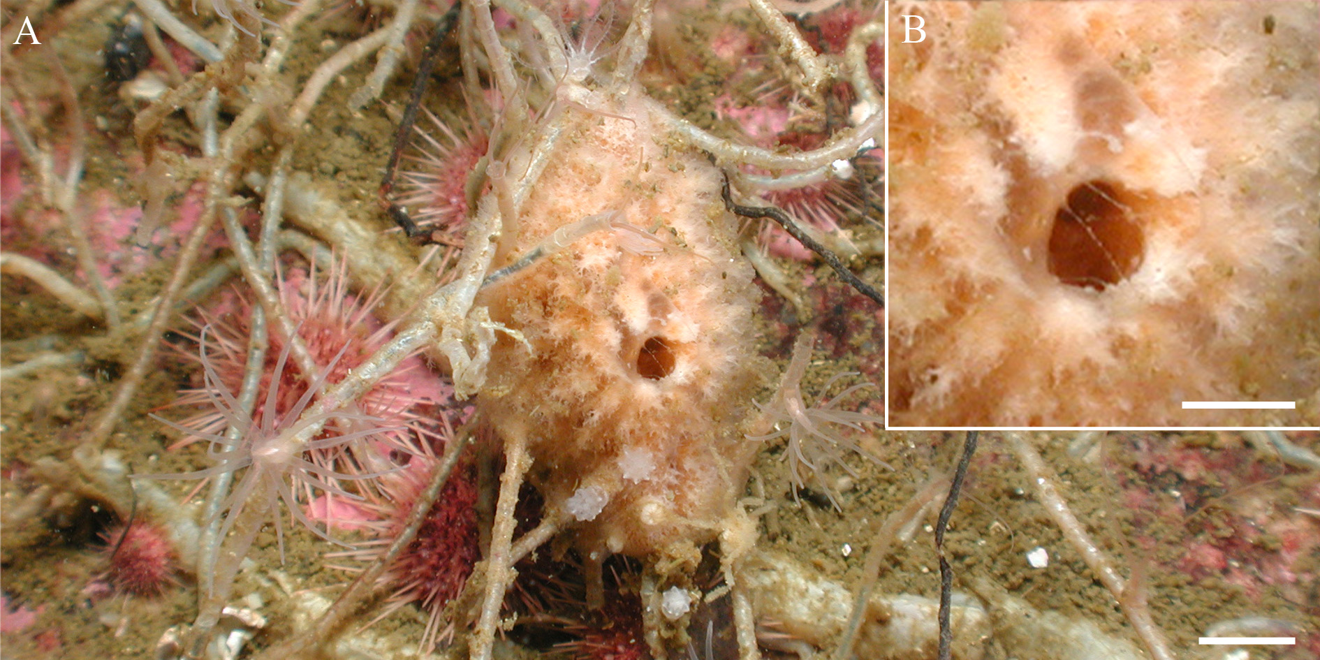
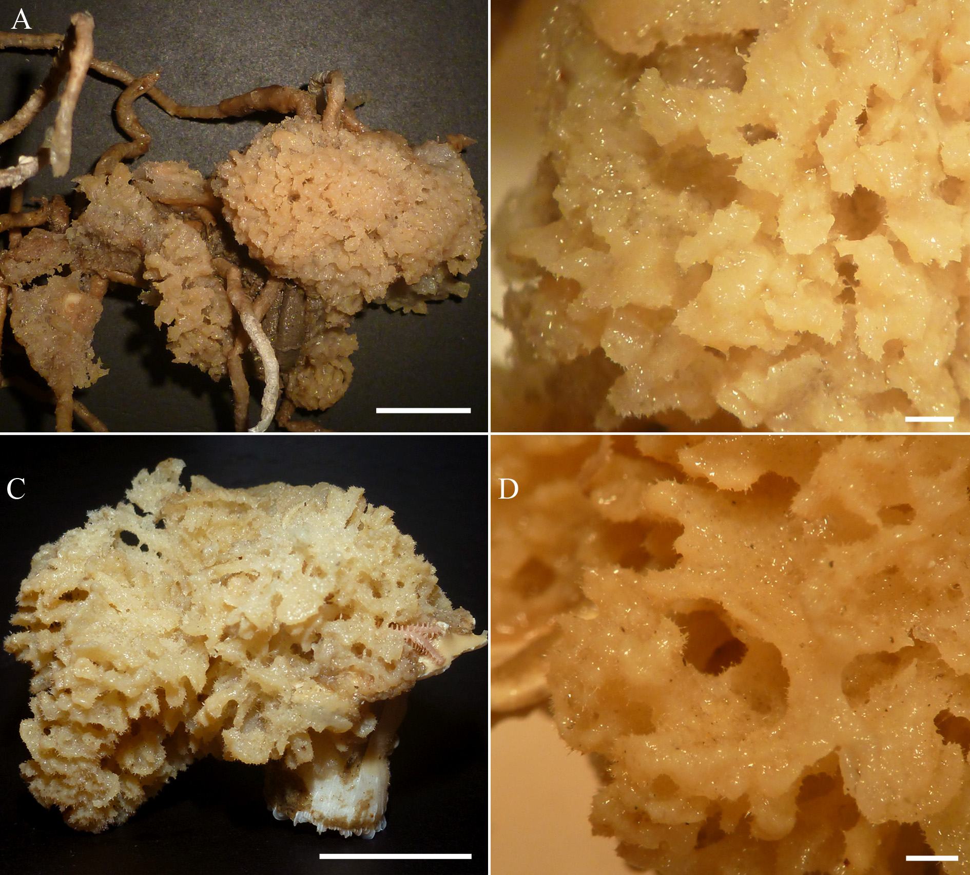
Description. Globular ( Figs 8 View FIGURE 8 A–B), 4 cm in diameter x 3 cm high; hispid surface ( Fig. 8 View FIGURE 8 C); body made up of a dense mass of juxtaposed small slender hollow tubes (ca. 0.5 mm in diameter each ‘fiber’), which may anastomose ( Fig. 8 View FIGURE 8 B); larger, simple openings (possibly oscula, up to 0.3 cm in diameter) are spread at the surface; colour in life beige, and in ethanol, lighter or darker beige; consistency rather compressible and delicate; rough texture.
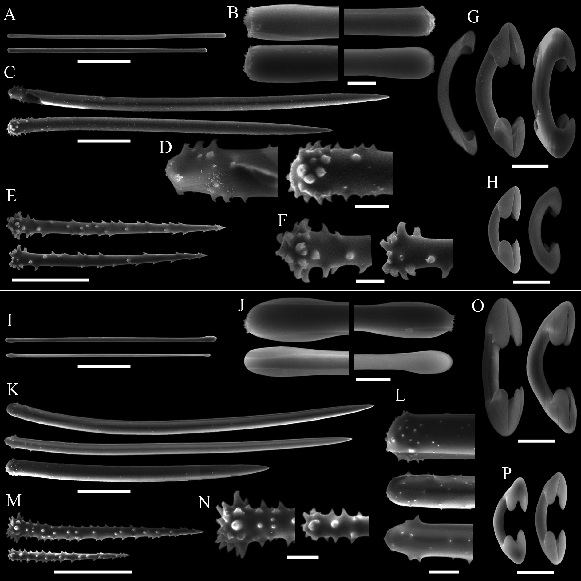
Skeleton. Sub-anisotropic reticulation ( Fig. 9 View FIGURE 9 A) of larger acanthostyles forming ascending pauci- to multispicular tracts (up to seven spicules across) in the choanosome, reaching the surface and piercing it by up to 200 µm. These acanthostyles also constitute secondary orthogonal tracts, one spicule long, and up to three in thickness ( Fig. 9 View FIGURE 9 B). Spongin in fibres is not apparent. These ascending spicule tracts can inter-cross or anastomose in the inner parts of the sponge, but are free from each other near the surface. Smaller acanthostyles echinate the main choanosomal tracts and the nodes of the reticulation. Tylotes are strewn at the surface, also tangentially ( Fig. 9 View FIGURE 9 C). Two kinds of arcuate isochelae occur throughout the choanosome and ectosome, the smaller category being the most abundant. Large choanosomal cavities are present, round or more elongated in section, up to 2000 µm long.
Spicules. Megascleres ( Tab. 2 View TABLE 2 ): Tylotes ( Figs 9 View FIGURE 9 D–E), straight, microspined at both ends , sometimes slightly aniso-tylote; tyles only slightly swollen and elongated, 150– 187. 2 (14) –198/4– 4.7 (0.5)–5. Acanthostyles I ( Figs 9 View FIGURE 9 F–G), straight to slightly curved, stout and somewhat fusiform; base slightly constricted, frequently bearing a subtle neck and discreet tyle, sometimes only irregularly round; apex sharpening gradually; spines (not abundant), up to 1.2 µm high, straight, concentrated on the basal portion of the spicule, a few spicules (variably thick) bear much less if any spines at all, 190– 267.5 (34.3) –300/7.2– 8 (0.6) –9. Acanthostyles II ( Figs 9 View FIGURE 9 H–I), frequently straight, base swollen (up to 3 µm thicker than the shaft); gradually sharpening apex; abundant spines up to 3 µm high, straight, spread all over the spicule, 84– 100 (10) –115/6– 7. 5 (0.6) –8. Microscleres ( Tab. 2 View TABLE 2 ): Arcuate isochelae I ( Fig. 9 View FIGURE 9 J), shaft curved, smooth, relatively thick; alae small but slightly elongated, young forms slender with markedly reduced alae, 25– 29. 3 (2) –31. Arcuate isochelae II ( Fig. 9 View FIGURE 9 K), same as the preceding one, but smaller, 19– 19.7 (0.9) –22. The Sturges algorithm confirmed the occurrence of two size classes of isochelae.
Ecology. The sponge was growing on rocky substrate, over tubes of the polychaete Chaetopterus variopedatus , and next to Tedania sp. ( Tedaniidae ) and another unidentified haplosclerid sponge.
Distribution. Provisionally endemic from its type locality at Isla Santa Inés (Magellan Strait, Chile).
Etymology. The specific epithet is derived from the new species’ occurrence in Chile’s Francisco Coloane Marine Protected Area.
Remarks. Lissodendoryx (Ectyodoryx) coloanensis sp. nov. is distinguished from Lissodendoryx (E.) spp. occurring in the SE Pacific, additional allied biogeographic provinces, as well as L. (E.) ballena sp. nov., due to its two categories of arcuate isochelae combined with terminally microspined tylotes ( Tab. 2 View TABLE 2 ). The spicule set of L. (E.) coloanensis sp. nov. is rather similar to that of L. (E.) corrugata sp. nov. ( Fig. 9 View FIGURE 9 and Fig. 7 View FIGURE 7 , respectively). Nevertheless, the former can be distinguished by its shorter and more slender acanthostyles ( Tab. 2 View TABLE 2 ), besides a relatively distinct outer morphology and consistency ( Fig. 8 View FIGURE 8 e Figs 4–5 View FIGURE 4 View FIGURE 5 , respectively).


No known copyright restrictions apply. See Agosti, D., Egloff, W., 2009. Taxonomic information exchange and copyright: the Plazi approach. BMC Research Notes 2009, 2:53 for further explanation.