
Lebiasina minuta, Netto-Ferreira, 2012
|
publication ID |
https://doi.org/ 10.1590/S1679-62252012000300002 |
|
persistent identifier |
https://treatment.plazi.org/id/03CA87F0-FFBE-FF8B-FC32-FAA2FCF7FDEB |
|
treatment provided by |
Felipe |
|
scientific name |
Lebiasina minuta |
| status |
sp. nov. |
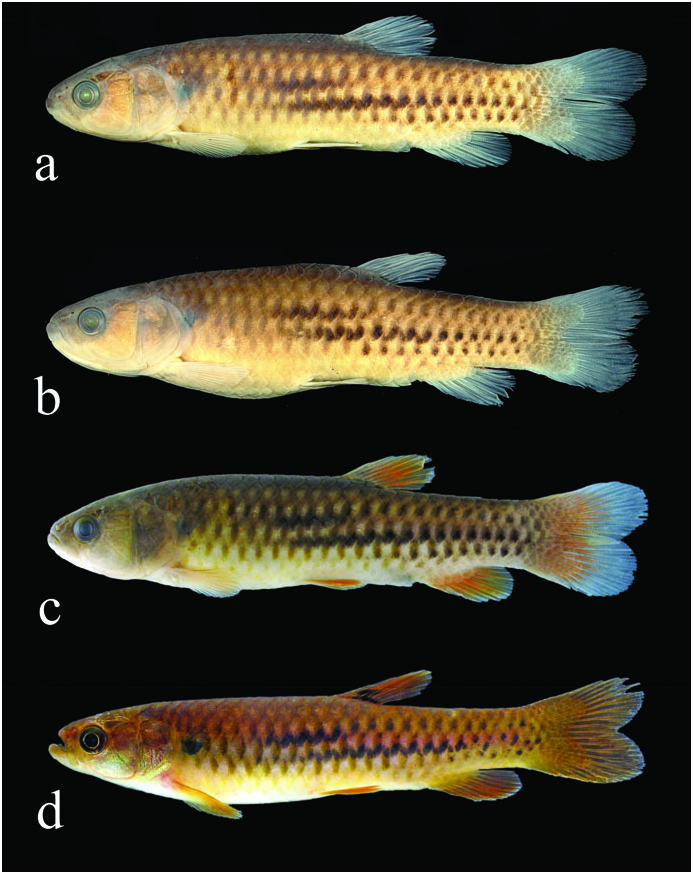
Lebiasina minuta View in CoL , new species.
Fig. 6 View Fig
Holotype. MZUSP 110693 View Materials , 62.1 mm SL, Brazil, Pará, Altamira, rio Xingu drainage, rio Treze de Maio , rio Iriri basin, near BR-163, 08°47’03”S 54°58’29”W, 22 Jan 2009, A. L. Netto-Ferreira, J. L. O. Birindelli, L. M. Sousa, P. Hollanda-Carvalho. GoogleMaps
Paratypes. Brazil, Pará, Altamira: ANSP 192062 View Materials , 5 View Materials , 58.2-62.5 mm SL, MZUSP 101423 View Materials , 94 View Materials , 14.8-68.4 mm SL; 2 cs 55.7-58.0 mm SL, same data as holotype; MZUSP 101412 View Materials , 1 View Materials , 54.0 mm SL, rio Xingu drainage, rio Treze de Maio , rio Iriri basin, near BR-163 road, upstream of dam, 08°47’03”S 54°58’29”W, 22 Jan 2009, A. L. Netto-Ferreira, J. L. O. Birindelli, L. M. Sousa & P. Hollanda-Carvalho GoogleMaps .
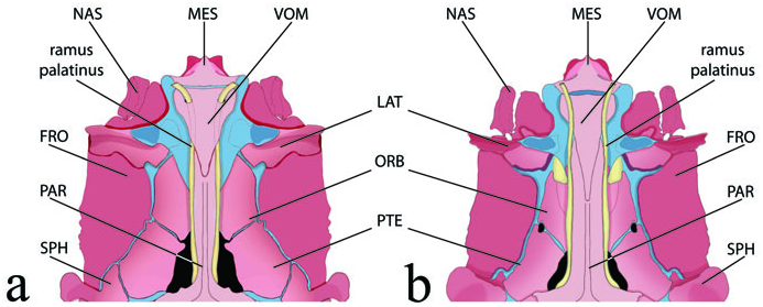
Diagnosis. Lebiasina minuta is readily distinguished from all lebiasinins (except for L. melanoguttata ) by the absence of the primary and secondary stripes, as well as the caudal blotch; the presence of three longitudinal series of dark blotches at the base of the scales; the pair of foramina on the vomer through which the ramus palatinus of the facial nerve passes ( Fig. 2a View Fig ); the large laminar extrascapular bone in contact with the fifth infraorbital and overlaying the anterodorsal portion of the opercle; and the nearly equal caudal-fin lobes. Lebiasina minuta differs from L. melanoguttata in the presence of a dark blotch at the base of the middle dorsal-fin rays, the second infrapharyngobranchial being edentulous, the yellowish overall coloration of the body, the dark olive green eyes, and the dark blotches of longitudinal series 3 and 4 coalescing where scales of adjacent longitudinal series overlap (vs. dark blotch absent; second infrapharyngobranchial with conical teeth; the reddish overall color of body and eyes; and the dark blotches of longitudinal series not coalescing).
Description. Morphometric data of the holotype and paratypes presented in Table 3. Lateral view of holotype, preserved paratype, recently preserved holotype and live paratype in Fig. 6 View Fig (a, b, c, and d, respectively). Body cylindrical, elongate. Dorsal profile of head convex from tip of upper lip to vertical through anterior nostrils; nearly straight from latter point to base of scales covering posterior half of parietals. Body profile nearly straight from that point to base of dorsal fin, then distinctly concave to origin of anteriormost dorsal procurrent ray of caudal fin. Ventral profile of head and trunk distinctly convex from lower lip to pelvic-fin origin; nearly straight from that point to anal-fin origin; convex along anal-fin base, and nearly straight from anal-fin terminus to origin of anteriormost ventral procurrent ray of caudal fin.
Mouth sub-superior. Premaxillary with single row of 13(2) tricuspid teeth. Posterior terminus of maxilla reaching to, or extending slightly beyond, anterior margin of orbit. Maxillary with 5(2) tricuspid or conical teeth.Anteriormost tooth usually largest. Dentary with two series of teeth; outer series with 16(2) pedunculate tricuspid teeth with central cusp distinctly longer than marginal cusps, gradually decreasing in size posteriorly. Inner teeth series with several minute conical teeth extending from symphysis to coronoid process. Branchiostegal rays 4(2); three rays attaching to anterior ceratohyal and one to posterior ceratohyal.
Scales cycloid, circuli restricted to border of scales, several radii converging to center of scale and strongly anastomosed in center, and forming several cells. Lateral line with 25(5), 26*(14) or 27(1) scales, of which only 5*(20) are perforated. Longitudinal rows of scales between dorsal and pelvic fins 7*(20). Predorsal scales 12(1), 13(6), or 14*(11). Circumpeduncular scales 12*(20).
Pectoral-fin rays i,11(6), 12*(8), 13(5), or 14(1). Tip of pectoral fin falling far short of pelvic-fin insertion. Pelvic-fin rays i,7*(21). Supraneurals 11(2), anterior to neural spine of centra 5 to 15(2). Dorsal-fin rays ii,8*(21). First dorsal-fin pterygiophore inserting behind neural spine of centrum 15(1) or 16(1). Distal margin of extended dorsal fin rounded. Dorsal-fin origin distinctly closer to caudal-fin origin than to tip of snout. Base of last dorsal-fin ray located distinctly anterior to vertical through anal-fin origin. Anal-fin rays iii,8*(20) or iii,9(1), with last ray adnate. Distal border of extended anal fin rounded. First anal-fin pterygiophore inserted posterior to haemal arch of centrum 23(1) or 24(1). Adipose fin absent. Caudal fin furcate with upper lobe slightly longer than lower lobe; both lobes rounded. Caudal-fin principal rays ii,8/ii,7(3), ii,8/i,8*(12) or i,9/i,8(4). Dorsal caudal-fin procurrent rays 8(1) or 9(1); ventral caudal-fin procurrent rays 8(2). Precaudal vertebrae 22(1) or 23(1); caudal vertebrae 15(2).
Color in alcohol. Background color predominantly tan. Head densely pigmented from upper lip to origin of scales overlying portion of parietals. Dark brown pigmentation extending from that point to insertion of caudal fin, over mid-dorsal series of scales and immediately bordering scale rows. Maxilla, infraorbitals and opercular series becoming lighter ventrally. Lower lip darkly pigmented. Ventral portion of head with scarce pigmentation. Opercular membrane with small scattered chromatophores.
Trunk dark dorsally, becoming lighter ventrally from third longitudinal series of scales.Abdominal region light yellowish, with minute dark chromatophores from isthmus to anal-fin origin. Humeral blotch rounded, conspicuous in both juvenile and smaller adult specimens. Primary and secondary longitudinal stripes absent. Three longitudinal series of dark blotches at base of scales of longitudinal series 3 to 5; dark blotches of series 3 and 4 coalescing. Caudal blotch absent. Dark pigmentation present along rays and intervening membranes of all fins.
Color in life. Same as in alcohol, except for dark olive green eyes and yellowish fins; yellowish ventral portion of head; pearly coloration posterior to the dark blotches on longitudinal scale series 3 and 4 ( Fig. 6d View Fig ).
Sexual dimorphism. As in most lebiasinids, male specimens of Lebiasina minuta demonstrate several modifications in the anal fin as described for L. melanoguttata . These modifications are less pronounced in L. minuta perhaps as a function of the small body size of this species.
Distribution. Lebiasina minuta is known from two tributaries of the rio Curuá (rio Xingu basin) in Serra do Cachimbo ( Fig. 4 View Fig ).
Etymology. The specific epithet minuta (=small) is in reference to the small size of adult specimens of Lebiasina minuta . Despite the large sample size of this species, no specimen larger than 68.4 mm SL was collected. In addition, most specimens larger than 50 mm SL were mature. An adjective.

No known copyright restrictions apply. See Agosti, D., Egloff, W., 2009. Taxonomic information exchange and copyright: the Plazi approach. BMC Research Notes 2009, 2:53 for further explanation.