
Ituglanis macunaima, Datovo & Landim, 2005
|
publication ID |
https://doi.org/ 10.1590/S1679-62252005000400002 |
|
publication LSID |
lsid:zoobank.org:pub:A57EF6E1-22BA-496E-8081-552021DAB61D |
|
persistent identifier |
https://treatment.plazi.org/id/9181B1B3-284E-450D-B23E-BD3FA3453F77 |
|
taxon LSID |
lsid:zoobank.org:act:9181B1B3-284E-450D-B23E-BD3FA3453F77 |
|
treatment provided by |
Carolina |
|
scientific name |
Ituglanis macunaima |
| status |
sp. nov. |
Ituglanis macunaima View in CoL , new species
Figs. 1-9 View Fig View Fig View Fig View Fig View Fig View Fig View Fig View Fig View Fig
Holotype. MZUSP 88452 View Materials , 30.5 View Materials mm SL; Brazil, Mato Grosso, Cocalinho; rio Araguaia basin, corixo da Saudade (corixinho); 14 o 17’20.6"S 51 o 9’12.1"W; A. Datovo, A. Oliveira, C. R. Moreira, J. C. Nolasco, J. L. Birindelli, M. I. Landim & O. T. Oyakawa; 10 Oct 2004. GoogleMaps
Paratypes. LIRP 5642 View Materials , 6 View Materials , 24.7 View Materials - 31.5 View Materials mm SL, 1 c&s (29.1mm SL) and MZUSP 86237 View Materials , 7 View Materials , 23.4 View Materials - 31.1 View Materials mm SL, 1 c&s (25.5 mm SL); same data as holotype GoogleMaps . MZUSP 86251 View Materials , 2 View Materials , 22.4 View Materials - 25.5 View Materials mm SL, 1 c&s (25.5 mm SL); Brazil, Mato Grosso, Cocalinho; rioAraguaia basin, rio Cristalino ; 14 o 12’45"S 51 o 18’21"W; A. Datovo, A. Oliveira, C GoogleMaps . R. Moreira, J. C. Nolasco, J.L.Birindelli, M.I. Landim, & O. Oyakawa; 14 Oct 2004 . MZUSP 86272 View Materials , 1 View Materials , 26.4 View Materials mm SL; Brazil, Mato Grosso, Cocalinho; rioAraguaia basin, rio Cristalino drainage; Corixão do Meio 14 o 11’14.3"S 51 o 14’58"W; A. Datovo, A. Oliveira, C GoogleMaps . R. Moreira, J. C. Nolasco, J. L. Birindelli, M. I. Landim, & O . T. Oyakawa; 14 Oct 2004 .
Diagnosis. Ituglanis macunaima is distinguished from all congeners by the following characters in combination: posterior fontanel absent (vs. present in all other Ituglanis except some I. epikarstikus ); pectoral-fin rays usually i,4 (vs. i,5 or more in all other Ituglanis except I. parahybae ); pelvic-fin rays usually i,4 (vs. i,3 or pelvic fin absent in I. parahybae ); branched caudal-fin rays usually 5,5 (vs. 5,6 or more in all other Ituglanis except I. nebulosus ); mottled color pattern formed by irregular to roughly roundish dark brown spots distributed on whitish background (vs. distinct color pattern in all other Ituglanis except some I. eichorniarum , I. gracilior , and I. proops ). Although data were not available from all other Ituglanis species , the following reductive characters in combination further diagnose I. macunaima : reduced supraorbital canal with pores s1 and s2 lacking (vs. s1 present in I. amazonicus , I. eichorniarum , I. gracilior , I. herberti , I. nebulosus , I. parkoi , and I. proops ; and s2 present in I. proops ; not seen in I. guayaberensis , I. laticeps , and I. metae ); reduced infraorbital canal with pores i1 and i3 lacking (vs. both present in I. proops ; not seen in I. guayaberensis , I. laticeps , and I. metae ); 2-3 pleural ribs (vs. 5 or more in I. bambui , I. epikarsticus , I. parahybae , I. passensis , I. proops , and I. ramiroi ; not seen in I. guayaberensis and I. laticeps ); 35-38 vertebrae (vs. 39 or more in I. amazonicus , I. eichorniarum , I. gracilior , I. herberti , I. metae , I. parahybae , I. parkoi , I. proops ; not seen in I. guayaberensis and I. laticeps ).
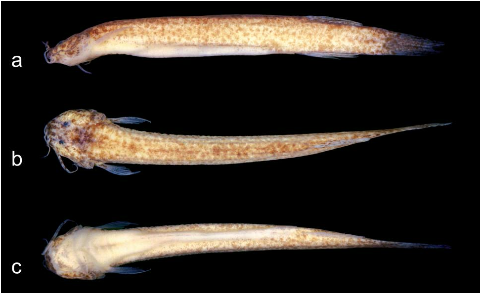
Description. Morphometrics given in Table 1. Refer to Fig. 1 View Fig for general aspects.
External morphology. Body elongate. Dorsal and ventral profiles of body straight or slightly convex in trunk and straight on caudal peduncle. Cross section of trunk nearly oval and becoming gradually more compressed posterior to pectoral girdle.
Head wide and strongly depressed, often slightly concave with swollen lateral cephalic musculature (adductor mandibulae and dilatator operculi muscles); cordiform in dorsal view. Dorsal profile of head straight; ventral profile ranging from straight to somewhat convex. Eyes slightly anteroposteriorly elongated and dorsally placed on anterior half of head; orbital rim not free. Thin and translucent skin covering eye, not adhered to surface of eyeball.Anterior nostril surrounded by tubular flap continuous with nasal barbel base; posterior nostril opening slightly larger than anterior one and with crescent thin flap on its anterior border.
Mouth subterminal and curved. Inferior lip with lateral tegumentar folds continuous with rictal barbel base. Nasal barbel emerging from lateral region of anterior nostril and usually reaching center of cranial crown. Maxillary barbel reaching pectoral-fin base. Rictal barbel usually somewhat shorter than nasal and crossing posterior edge of interopercular patch of odontodes. Branchial membranes thick, united to isthmus only anteriorly and forming small free fold across isthmus. Median most branchiostegal rays barely visualized through skin.
Opecular patch of odontodes rounded, posteriorly detached from head surface and dorsolaterally placed. Interopercular patch of odontodes narrow, elongate, posteriorly curved, and placed fully anterior to opercular patch. Odontodes markedly erected from integument in most specimens.
Pectoral-fin rays i,4 (two specimens i,3 on one side of body; two i,5 on one side and two on both sides). Origin of pectoral fin just posterior to branchial membrane. First pectoral-fin ray distinctly longer than remaining rays and continued distally as filament. Remaining rays gradually shorter than lateral one, making distal margin of pectoral fin obliquely straight. Inconspicuous axillary pore present. Pelvic-fin rays, i,4 (three specimens i,3 on one side of body), origin anterior to origin of dorsal fin; posterior margin convex. Bases of pelvic fins close to each other. Tip of adpressed pelvic fin not reaching anal-fin origin. Urogenital and anal openings at posterior half of pelvic-fin length. Dorsal-fin rays ii,6 (one specimen ii,5, two ii,7, one iii,5, three iii,6), located on posterior one-third of trunk; distal margin convex. Anal- fin rays ii,5 (one specimen ii,4, one iii,4), origin of fin approximately at vertical through origin of dorsal fin; distal margin convex. Caudal fin with posterior margin ranging from convex to nearly straight. Principal caudal-fin rays 12: i,5 on both dorsal and ventral lobes (two specimens i,4 on dorsal lobe). Unsegmented rays of unpaired fins hardly visible through skin (see Osteology).
Color in alcohol. General color pattern mottled, formed by irregular to roughly circular dark brown spots distributed on whitish background. Skin separated into two principal layers. Outer layer thicker, translucent and without pigmentation, allowing external examination of inner tissue pattern. Tiny brown chromatophores distributed on inner integumentary layer. Most chromatophores grouped into brown spots, but some scattered between spots. Spots ranging from one to two times eye diameter and, in most cases, have irregular shapes and margins; few specimens with roughly rounded spots; one specimen more densely pigmented with larger and coalesced spots. Pigmentation more densely scattered on dorsum, becoming gradually smaller, clearer and sparser towards ventral region. Spots rarely contact each other and never appear to form stripes. On ventral surface of body, spots occur only posterior to origin of anal fin; most of trunk without brown pigmentation. On head, spots smaller and even more irregular than on body. Maxillary and nasal barbels nearly banded; diffuse spots occur on ventral surface of head, rictal barbel base, and between interopercular odontodes. Small irregular spots appear on proximal portions of fins, more intensely on caudal fin. Background color – which ranges from white to pale yellow – provided by color of musculature and connective tissues visible through translucent layer of skin. Head with large irregular dark blotch on posterior part of cranial roof. Its origin on dark pigments present on membrane that covers brain, and externally visible by transparent laminar bones of cranial roof.
Osteology. Mesethmoid with anterior margin straight; shaft not as wide as cornua ( Fig. 2 View Fig ). Lateral ethmoid without lateral projections. Frontal and parieto-supraoccipital fully joined by sutures; anterior and posterior fontanels absent. Co-ossified sphenotic-prootic-pterosphenoid with anterior projection with infraorbital sensory canal opening. Vomer arrow-shaped and with long posterior process. Synchondrosis between orbitosphenoid and sphenotic-prootic-pterosphenoid above and below anterior most trigeminofacialis foramen. Parasphenoid with two anterior and one posterior processes. Co-ossified basioccipital-exoccipital posteriorly fused to Weberian capsule and without anterior processes. Weberian capsule with small lateral opening and tiny pores on entire surface.
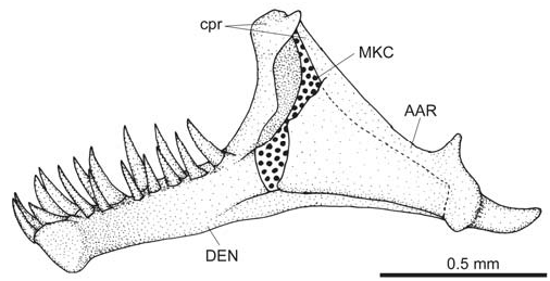
Autopalatine with deep medial notch, posteriorly expanded with wide lateral process and separate socket-type articulations with lateral ethmoid and with vomer ( Fig. 2 View Fig ). Premaxilla rectangular with two regular rows of conical teeth. Maxilla nearly as long as premaxilla and with well developed anteroventral process. Angulo-articular with elevated coronoid process ( Fig. 3 View Fig ). Dentary with two regular rows of conical teeth and long coronoid process with posteromedially curved tip.
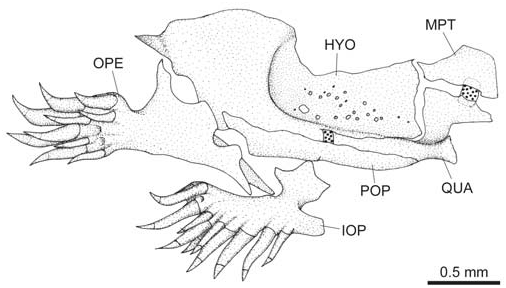
Hyomandibula with conspicuous lateral depression for levator arcus palatini insertion and perforated by pores on dorsomedian region ( Fig. 4 View Fig ). Dorsal portion of quadrate laminar and nearly triangular. Metapterygoid articulating with hyomandibula and with quadrate through cartilaginous block.
Opercle with 10-15 odontodes ( Fig. 4 View Fig ). Interopercle with 10-15 odontodes, none on anteroventral projection. Odontodes progressively larger and more curved towards posterior region of patches.
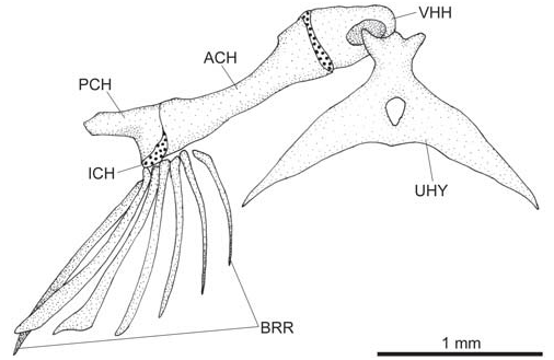
Urohyal with thin, pointed and elongated lateral process and two small dorsal condyles ( Fig. 5 View Fig ). Deep depressions on ventral hypohyal for articulation with urohyal condyles. Branchiostegal rays 7.
Basibranchials: first absent; second and third ossified and stem-like; fourth flattened and fully cartilaginous, larger in width than in length ( Fig. 6 View Fig ). Hypobranchials: first ossified and stem-like; second and third flattened, cartilaginous and with ossified anterolateral process; fourth absent. Ceratobranchials: first, second and third with posterior laminar expansions; fourth flattened; fifth posteriorly divergently curved with small teeth on anterior half. Epibranchials: first with long anterior process; second with two discrete processes; third with one posterior process; fourth flattened with no process; fifth absent or not evident. Pharyngobranchials: first and second absents; third stem-like; fourth firmly attached to tooth plate. Upper pharyngeal tooth plate with long ventromedially oriented conical teeth.
First complete hemal canal on fourth or fifth vertebra past Weberian complex. Long and medially curved parapophysis on vertebrae with complete hemal canal but incomplete hemal spine. First complete hemal spine on 14th or15th vertebra Total vertebrae 35-38. Pleural ribs 2-3.
Cleithrum roughly recteangular or triangular in ventral view ( Fig. 7 View Fig ). Scapulocoracoid restricted to lateral region of cleithrum.
Pelvic girdle delicate and poorly ossified ( Fig. 8 View Fig ). Basipterygium with no cartilaginous tips on anterior processes; posteromedial margins fully cartilaginous. Thin pelvic splint parallel to first pelvic ray.
Dorsal fin with 7-8 basal radials distributed between neural spines of 22-24th to 26-28th vertebrae; unsegmented dorsal-fin rays 2. Anal fin with 6 basal radials distributed between hemal spines of 23-24th to 27-28th vertebrae; unsegmented anal-fin rays 1-2.
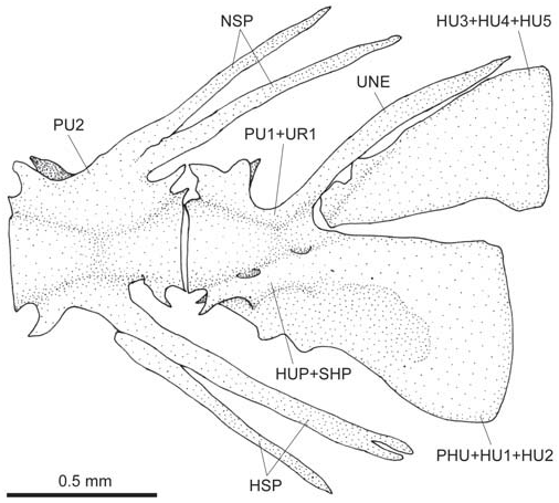
Preural centrum with each half of hemal and/or neural spines often not aligned, sometimes forming double spine ( Fig. 9 View Fig ). Upper caudal plate composed of uroneural and one single triangular element (co-ossified hypurals 3-5). Lower caudal plate as one element (co-ossified hypurals 1-2 and parhypural), which is fused to compound caudal centrum (preural 1 + ural 1). Hypurapophysis complex (hypurapophysissecondary hypurapophysis) of “ type B” (Lundberg & Baskin, 1969). Procurrent caudal rays 11-12 dorsal, 8-10 ventral.
Laterosensory system. Cephalic laterosensory canals with simple (non-dendritic) tubes ending in simple pores ( Fig. 2a View Fig ). All canals continuous and connected to each other. Supraorbital canal present mostly in frontal bone and bearing only pores s3 and s6 placed on interorbital region; pores s1 and s2 absents. Infraorbital canal present mostly in soft tissue and restricted to branches and pores i10 and i11 located ventroposteriorly to eyeball; pores i1 and i3 absents. Otic canal without pores and passing through sphenotic-prooticpterosphenoid. Postotic (temporal) canal with branches and pores po1 (mostly in pterotic bone) and po2 (mostly in posttemporosupracleithrum); pores placed anterodorsally to opercular patch of odontodes. Body with short lateral line canal with only pores ll1 and ll2 located dorsoposteriorly to pectoral-fin base.
Etymology. From the modernist Brazilian masterpiece by Mário de Andrade – “Macunaíma: o herói sem nenhum caráter” – meaning the hero without any character, in reference of the absence of any exclusive (taxonomic) character for the new species. Mário de Andrade’s Macunaíma was based in folk Amazonian indian myth, and also presents infantile features, in allusion to the paedomorphic characters of the new species. Treated here as a noun in apposition.
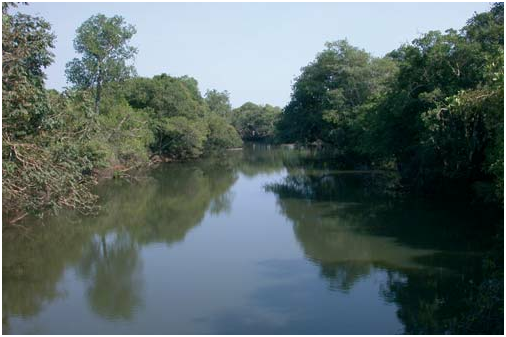
Distribution and habitat notes. The new species is known only from three localities from two tributaries (rio Cristalino and corixo da Saudade) of the left margin of the rio Araguaia basin ( Fig. 10 View Fig ). The new species was found always associated with leaf litter, or other decomposing vegetal matter, accumulated in shallow (not deeper than 1 m), slow flowing portions of the river. At the type-locality ( Fig. 11 View Fig ), specimens where found also associated with a partially decomposed log, resting on the litter bed. These specimens were found more externally on crevices in the bark, together with an undescribed species of Microglanis . Several specimens of an undescribed species of Centromochlus were found more deeply associated in the log.


| R |
Departamento de Geologia, Universidad de Chile |
| T |
Tavera, Department of Geology and Geophysics |
No known copyright restrictions apply. See Agosti, D., Egloff, W., 2009. Taxonomic information exchange and copyright: the Plazi approach. BMC Research Notes 2009, 2:53 for further explanation.