
Ischnopelta vellozia Rosso & Campos
|
publication ID |
https://doi.org/ 10.11646/megataxa.6.2.3 |
|
DOI |
https://doi.org/10.5281/zenodo.5753411 |
|
persistent identifier |
https://treatment.plazi.org/id/03828787-2C72-FFC8-FCD5-F8F6FB9C03BE |
|
treatment provided by |
Plazi |
|
scientific name |
Ischnopelta vellozia Rosso & Campos |
| status |
sp. nov. |
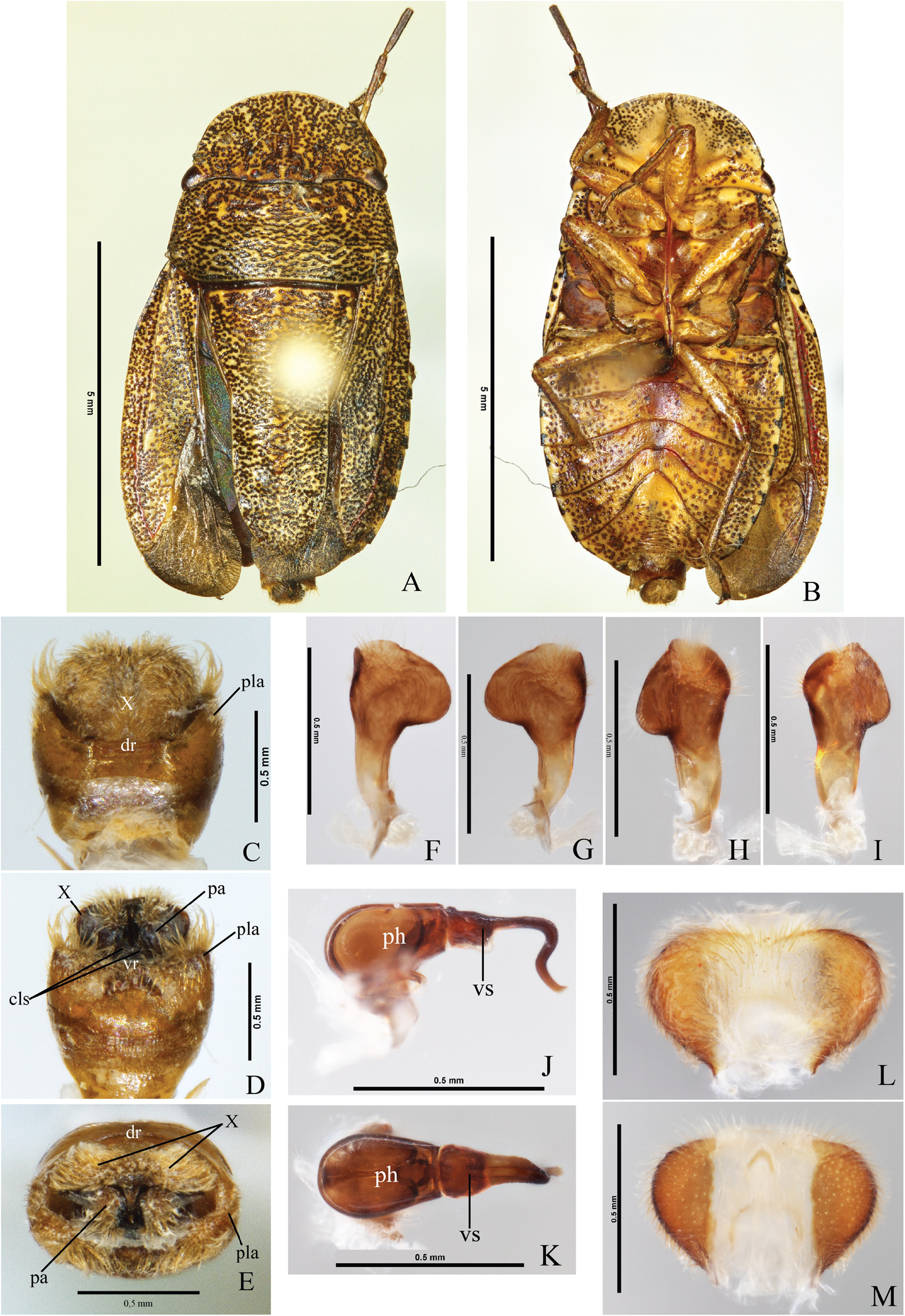
Ischnopelta vellozia Rosso & Campos , sp. n. ( Figs. 4B View FIGURE 4 , 5M View FIGURE 5 , 46–47 View FIGURE 46 View FIGURE 47 )
Etymology. The epithet refers to the Reserva Particular Vellozia, located in Serra do Cipó, component of the geological province Serra do Espinhaço, Southeast of Minas Gerais, Brazil, where most of the specimens used for the description of this species were collected.
Type Locality. BRAZIL, Minas Gerais, Santana do Riacho [-19,279444, -43,59].
Holoype. Male. Brazil, Minas Gerais, Santana do Riacho (RPPN Fazenda Vellozia), 29.III.2008, C. F. Schwerthner Col. Deposited at Museu de Zoologia da Universidade de São Paulo ( MZSP), São Paulo (SP), Brazil .
Paratypes. 5 males and 4 females. BRAZIL, Minas Gerais, Diamantina (20 km NE, Rod. BR 367 ) , 1 male, 8. I .1997, T. J . Henry & A . Paula, [-18.15, -43.502777], ( USNM); Santana do Riacho ( RPPN Fazenda Vellozia) , 2 males and 4 females, 29. III .2008, C. F . Schwertner Col., [-19,279444, -43,59], ( UFRG); Jaboticatubas ( Serra do Cipó ) , 2 males, 30. IV .1973, Montouchet Col. , [-19.5000, -43.7500], ( MZSP) GoogleMaps .
Description. The overall somatic morphology is as describedfor I.scutellata ,exceptforthefollowingfeatures. Body densely punctured, brownish.Head.Labrum inserted anteriorly to half the distance between the anterior margin of the eyes and the apex of mandibular plates. Antennae: segment I and dorsal surface of segments II and III dark yellowish with minute brown blotches, ventral surface of segments II and III light-brown, segments IV and V dark brown; segments ratio: I>II<III<IV<V.
Thorax. Hemelytra: corium as long as scutellum; conspicuous spot at apex of the radial vein blotch. Pro-, meso- and metapleura dark yellowish.. Setae on posterodorsal margin of protibiae as long as the others.
Abdomen. Dark spots at the lateral of urosternites irregularly shaped narrow; male urosternite VII unarmed.
Male. Apical margin of membrane of hemelytra convex; median portion of posterior margin of urosternite VII subrectilinear; urosternite VII not reaching anteriorly the imaginary line connecting the spiracles of urosternite V.
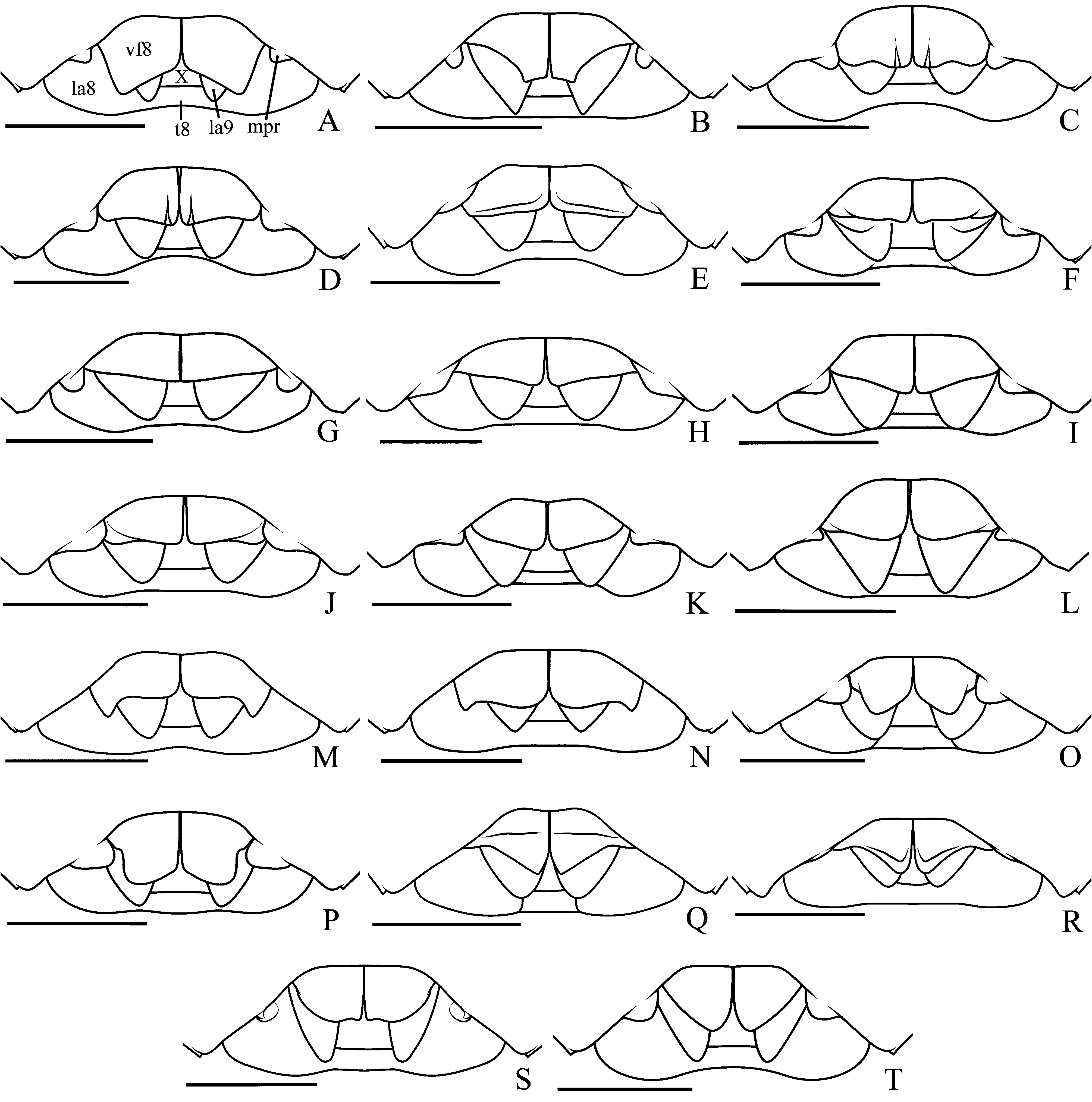
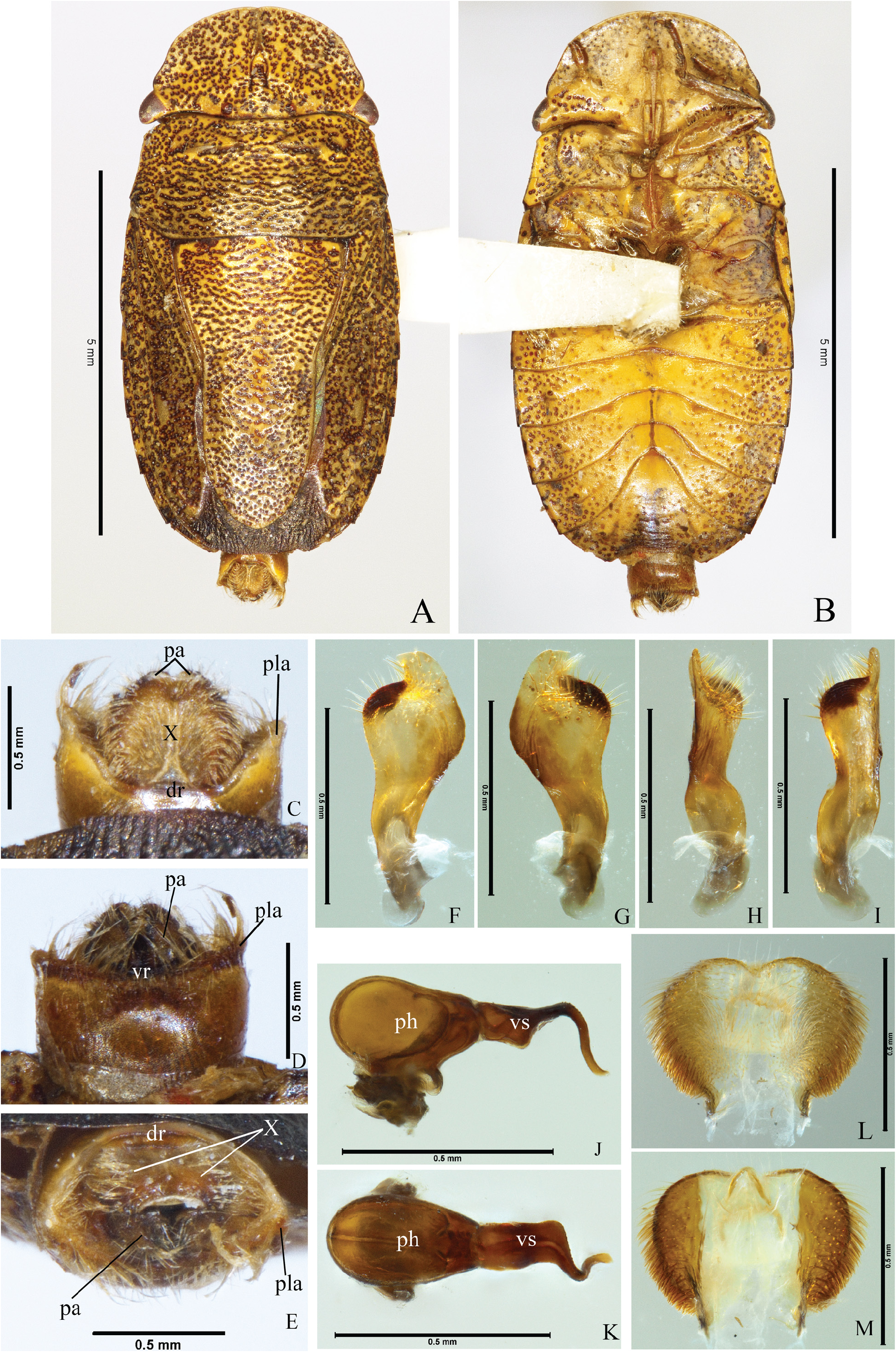
Genitalia. Pygophore with dorsal rim concave ( Fig. 46C View FIGURE 46 , dr); ventral rim sinuous with wide and shallow median depression ( Fig. 46D View FIGURE 46 , vr). Posterolateral angles 0.5 times shorter than the rest of the pygophore, perpendicular to the frontal plane and subparallel ( Fig. 46C–E View FIGURE 46 , pla). Setae in a narrow band on the ventral rim, on the median depression of ventral rim and outer surface of posterolateral angles; longer and denser on the laterals of ventral rim and apex of posterolateral angles. Segment X wider than long, surpassing the apex of posterolateral angles and dorsally covering the parameres; cordiform; apical margin sclerotized and emarginated; lateral margins sclerotized, densely covered by long setae; mid-longitudinal region membranous covered by setae, short and sparse on basal portion and longer and denser on distal portion Figs. 46C–E, X; 46L–M View FIGURE 46 ). Parameres claviform, flat, oblique to the frontal plane; outer margin sinuous, distal portion strongly convex; inner margin sinuous; setae covering the distal portion of ventral surface of the head ( Figs. 46D View FIGURE 46 , pa; 46F–I). Cup-like sclerites externally visible, apices rounded and subparallel ( Fig. 46D View FIGURE 46 , cls). Phallus: vesica dorsally flat, ventrally expanded; distal portion of vesica sinuous, ventrally directed; secondary gonopore beveled ( Fig. 46J–K View FIGURE 46 ).
Female. Membrane of hemelytra not reaching the posterior margin of mediotergite VIII, posterior margin convex; median portion of posterior margin of mediotergite VIII concave; median portion of posterior margin of urosternite VII subrectilinear; projections of urosternite VII thicke and slightly oblique to the surface of urosternite VII ( Fig. 47C View FIGURE 47 , mpr). Genitalia. Valvifers VIII wider than long; posterior margin sinuous, median portion subrectilinear, lateral portion forming a subtriangular projection; sutural margins subrectilinear and folded dorsally; surface longitudinally convex, dark yellowish with brown punctures, setae on the distal portion of sutural margins and on median portion of posterior margin; longitudinal grooves narrow and shallow on basal portion ( Figs. 5M View FIGURE 5 ; 47C View FIGURE 47 , vf8). Valvifers IX partially covered by the valvifers VIII, lateral margin subrectilinear, setae on mid-basal portion of ventral surface ( Fig. 47C–D View FIGURE 47 , vf9). Laterotergites IX not reaching the posterior margin of mediotergite VIII; lateral margin convex; setae on mid-basal portion of lateral margin and ventral surface ( Fig. 47C–D View FIGURE 47 , la9). Thickening of vaginal intima slightly wider than long; proximal margin subrectilinear; distal margin convex, emarginated, and sclerotized; lateral margins convex; surface membranous on the proximal margin and on mid-longitudinal subtriangular area ( Fig. 47D View FIGURE 47 , vi). Vesicular area anterior to the collar 1/5 of the posterior portion; median duct anterior and posterior to the collar with proximal widening ( Fig. 47D View FIGURE 47 , md, mdp); inner duct coiled in the proximal widening ( Fig. 47D View FIGURE 47 , id). Distal ductus receptaculi of same caliber as the proximal one ( Fig. 47D View FIGURE 47 , drd, drp). Pars intermedialis broader distally ( Fig. 47D View FIGURE 47 , pi); proximal annular crest directed to the ductus receptaculi; distal annular crest perpendicular to the pars intermedialis and almost twice the diameter of the proximal one ( Fig. 47D View FIGURE 47 , dac, pac). Capsula seminalis globose; laterobasal projection sinuous, long; lateral projection minute ( Fig. 47D View FIGURE 47 , cs, pr).
Measurements: Table 21.
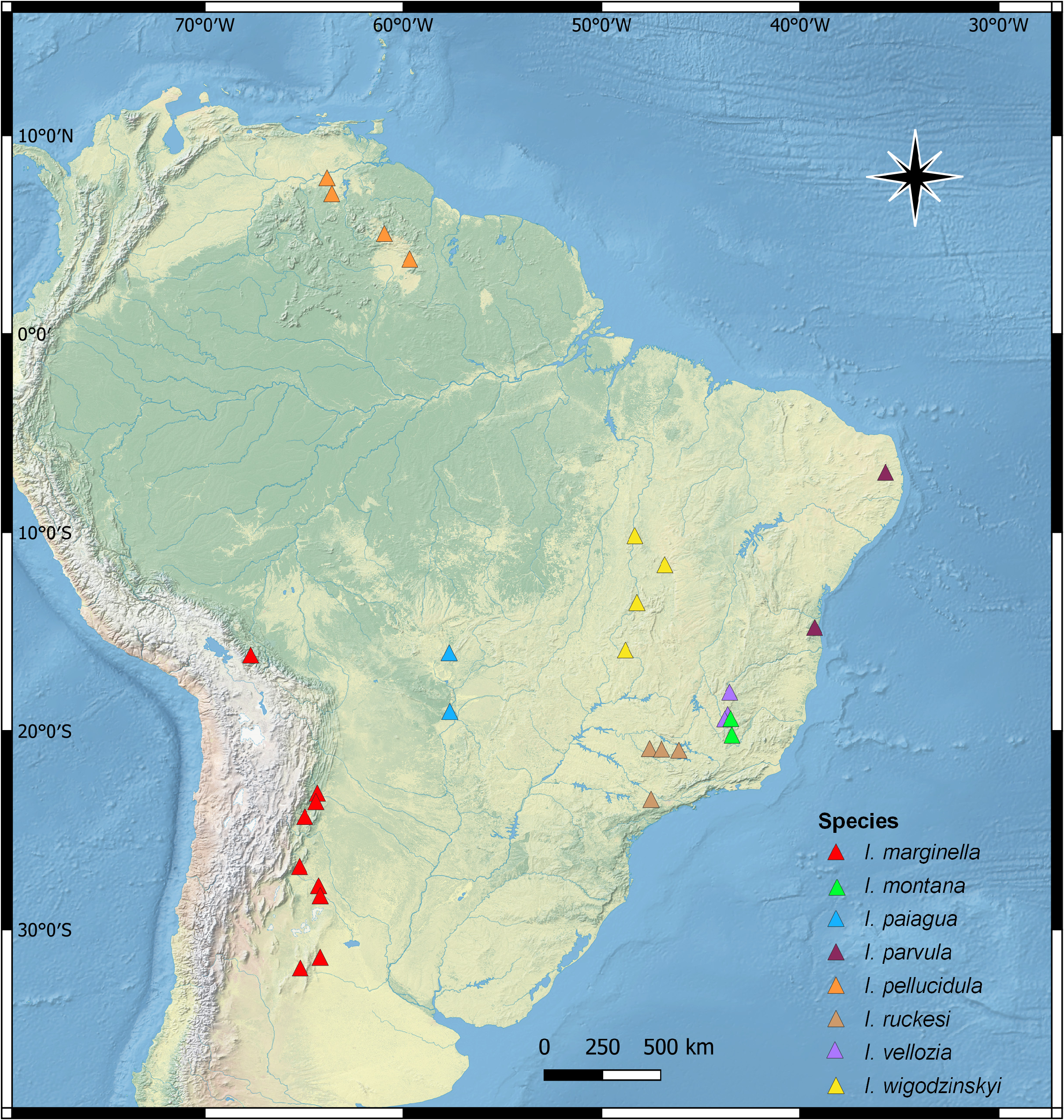
Distribution. Brazil (Minas Gerais) ( Fig. 8 View FIGURE 8 ).
Comments. The males of Ischnopelta vellozia sp. n. have a cordiform segment X ( Fig. 46L–M View FIGURE 46 ), as in I. anangulata sp.n. ( Fig. 13C, X View FIGURE 13 ), I. cordiformis sp. n. ( Fig. 20L–M View FIGURE 20 ), and I. wigodzinskyi sp. n. ( Fig. 48L– M View FIGURE 48 ). However, in I. anangulata the posterlateral angles of the pygophore are not developed ( Fig. 13C–D View FIGURE 13 ), in I. wigodzinskyi they are 0.3 times the length of the pygophore ( Fig.48C View FIGURE 48 , pla), whereas about 0.5 times in I. vellozia ( Fig. 46C–D View FIGURE 46 , pla) and I. cordiformis ( Fig. 20C–D View FIGURE 20 , pla). Precisely defining the identity of males on the last two species requires a more detailed analysis of the genital structures. For the females, the triangular projection on the lateral angle of valvifers VIII is larger in I. vellozia ( Figs. 5M View FIGURE 5 ; 47C View FIGURE 47 , vf8) than in I. wigodzinskyi ( Fig. 5N View FIGURE 5 , 49C View FIGURE 49 , vf8).


| MZSP |
Brazil, Sao Paulo, Sao Paulo, Museu de Zoologia da Universidade de Sao Paulo |
| NE |
University of New England |
| I |
"Alexandru Ioan Cuza" University |
| T |
Tavera, Department of Geology and Geophysics |
| J |
University of the Witwatersrand |
| A |
Harvard University - Arnold Arboretum |
| USNM |
Smithsonian Institution, National Museum of Natural History |
| C |
University of Copenhagen |
| F |
Field Museum of Natural History, Botany Department |
| UFRG |
Instituto de Biologia |
No known copyright restrictions apply. See Agosti, D., Egloff, W., 2009. Taxonomic information exchange and copyright: the Plazi approach. BMC Research Notes 2009, 2:53 for further explanation.
|
Kingdom |
|
|
Phylum |
|
|
Class |
|
|
Order |
|
|
Family |
|
|
Genus |