
Iheringichthys syi, Azpelicueta & Britski, 2012
|
publication ID |
https://doi.org/ 10.1590/S1679-62252012000100004 |
|
persistent identifier |
https://treatment.plazi.org/id/039987E2-FFF7-D476-A833-FF6BFCBCF837 |
|
treatment provided by |
Felipe |
|
scientific name |
Iheringichthys syi |
| status |
sp. nov. |
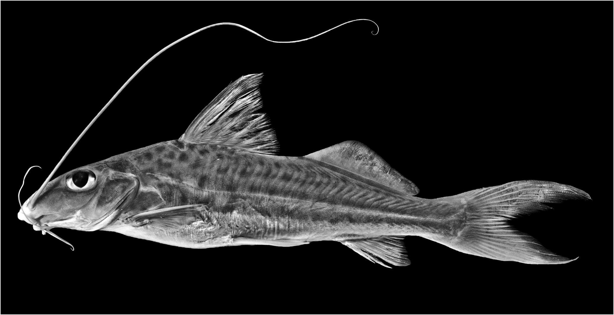
Iheringichthys syi View in CoL , new species
Figs. 1 View Fig , 2a View Fig , 3-5 View Fig
Holotype. MZUSP 108404, 149.1 mm SL ( Fig. 1 View Fig ), Brazil, Mato Grosso do Sul, Três Lagoas Municipality, rio Paraná basin, rio Paraná in Jupiá, 20°45’04’’S 51°40’42’’W, 15 Oct 1962, Excursão Departamento de Zoologia. GoogleMaps
Paratypes. Brazil. Mato Grosso do Sul. AI 279, 3 (1 C&S), 144.0- 171.8 mm SL and MZUSP 22507 View Materials , 15 View Materials , 109.9 View Materials - 160.5 mm SL, collected with the holotype. MZUSP 22626 View Materials , 5 View Materials , 91.2-130.2 mm SL, rio Paraná basin, rio Paraná in front of Jupiá , 20°45’14’’S 51°40’42’’W, 11 Dec 1960, P.E. Vanzolini, S. Saiar & J.H. Vanzolini GoogleMaps . São Paulo. MZUSP 23082 View Materials , 3 View Materials , 133.4 View Materials -185.0 mm SL, rio Paraná basin, rio Paraná in Ilha Solteira (20°25’45’’S 51°20’00’’W), Sep 1965, Excursão Departamento de Zoologia GoogleMaps .
Diagnosis. Iheringichthys syi is most readily distinguished from I. labrosus and I. megalops by the presence of only a fine serration on anterior pectoral spine margin, very small serrae on the posterior pectoral-spine margin, and a shorter pectoral fin (16.3-20.1% SL vs. 20.1-23.2% in I. labrosus and 20.3-22.3% in I. megalops ). In addition, I. syi has a shorter snout (42.0-51.0% HL vs. 56.6-62.0 HL) and a larger eye (23.0- 31.2% HL vs. 22.4-26.7% HL) than I. labrosus . Furthermore, I. syi has a narrow interorbital (16.2-23.0% HL), a relatively long postorbital length (30.6-34.0% HL), low adipose fin (4.8-7.8% SL), ratio between eye diameter by of interorbital length 97.0- 140.0%, adpressed dorsal remote from adipose-fin origin, tip of pectoral fin far from pelvic-fin origin, upper lip curved upward with a very small cleft, and body with numerous, small dots irregularly scattered on flanks, especially marked on anterior half of flank.
Description. Morphometric data for holotype and 26 paratypes presented in Table 1. Medium-sized pimelodid with maximum size of 220 mm in total length. Dorsal profile of body from snout tip to eye strongly convex, slightly concave across eye and supraoccipital bone, slightly convex from that point to dorsal-fin origin; straight or slightly convex from origin of dorsal fin to origin of adipose fin; slanted ventrally from that point to caudal peduncle, and concave along caudal peduncle. Ventral profile of body straight, slanted dorsally from anal-fin origin to caudal peduncle; caudal peduncle convex.
Maximum body width across cleithra in front of pectoral-spine insertion 7.1-9.4 times in SL. Maximum body depth at dorsal-fin origin 4-5 times in SL.
Dorsal fin somewhat triangular, distal margin straight or slightly concave in some specimens. Dorsal-fin origin located at vertical through mid-length of pectoral fin. Tip of adpressed dorsal fin remote from adipose-fin origin, even in small specimens. Dorsal fin lepidotrichia II,6; spinelet short, broad, triangular, with few longitudinal striae, covered by skin. Dorsal-fin spine strong, with longitudinal striae; its osseous portion shorter than first branched dorsal-fin ray. Serrae absent on anterior margin, only weak serration present at midlength.Serrae developed posteriorly on distal third of spine very small. Adipose-fin origin located at vertical through posterior fourth of pelvic-fin length.Adipose-fin low (its depth 13.8-17.4 times in its length), long (its base 3.3-4.2 times in SL), almost equal in size than head. Caudal fin deeply forked, dorsal lobe elongated, longer than ventral lobe; lower lobe rounded, little wider than upper lobe. Principal caudal-fin rays i,7-8,i. Anal-fin origin located at vertical through first third of adipose fin. Anal fin short, with posterior margin slightly concave; second branched ray longest (one specimen with first branched ray longest), first three branched rays forming small lobe. Anal-fin iv,7 (2), iv,8 (15), iv,9 (2), or v,8* (7) rays.
Pectoral fin with I,9 (2), I,10* (20), or I,11 (4) rays. First branched pectoral-fin ray longer than osseous spine plus its soft portion. Longitudinal striae covering dorsal and ventral surfaces of pectoral spine, less notable on ventral one. Retrorse serrae on posterior margin of spine very fine, small (about 30-32 regularly distributed in spines of 30 mm, less in small specimens), posterior serrae absent in last third of spine in some specimens. Serration along anterior margin of spine fine, always covered by skin, inapparent ( Fig. 2a View Fig ). First branched pectoral-fin ray longer than bony spine plus soft portion. Pectoral axillary gland pore, above posterior portion of pectoral fin, very close to ventral margin of cleithral process. Cleithral surface covered by very fine tubercles; posterior cleithral process broad, scarcely pointed forming almost straight angle. Pelvic-fin origin located at vertical through last dorsal-fin ray, fin tip reaching posterior third of distance between pelvic and anal fin origins. Pelvic fin with i,5 rays. Inner fin rays covering anus and urogenital papillae.
Head triangular in dorsal view, of moderate length (contained 3.1-3.5 times in SL), relatively deep (its depth at base of supraoccipital process equal to body width or scarcely less). Head covered by thin skin; roof of skull, especially posterior part and supraoccipital posterior process ornamented with tubercles and striae. Numerous branches of laterosensory canal system expanded between eye and posterior tip of supraoccipital process. Anterior orbital ridge very low, formed by frontal bone. Snout long, pointed (contained 1.9-2.2 times in HL). Anterior cranial fontanelle narrow, oval, anterior margin nearer to posterior nostril than eye and finished before posterior margin of eye. Posterior fontanelle absent. Supraoccipital posterior process prominent, pointed (contained 1.9-2.2 times in HL), longer than wide reaching nuchal plate ( Fig. 3 View Fig ); its dorsal surface strongly angular in cross section, sharply rounded. Anterior nostril with short tubular rim, located at level of maxillary barbel insertion; posterior nostril preceded by large anterior membrane covering almost completely oblique opening. Distance between snout and anterior nostril longer than distance between nostrils ( Fig. 3 View Fig ).
Eye large, placed laterodorsally, located little behind midpoint of head. Interorbital space flat, scarcely concave behind this area; interobital narrow (0.7-0.9) times eye diameter.
Mouth very small, ventral ( Fig. 4). Upper jaw projecting. Fleshy lips prominent (width of lower lip about 0.5 times in width of upper lip); upper lip curved upward with small median cleft. Premaxillary teeth completely or partially exposed with mouth closed. Premaxillary tooth band slender, its transverse axis short; its posterolateral corner bluntly rounded; its anterior margin slightly rounded. Premaxillary teeth conical, fine. Dentary tooth band wider at symphysis, with teeth of similar shape.
Maxillary barbel originated at low sulcus between anterior nostril and eye; maxillary barbel long, usually reaching procurrent caudal-fin rays; sometimes longer, ending about midlength of caudal lobes. Lateral mental barbel surpassing pectoral-fin origin. Tip of medial mental barbel scarcely reaching branchiostegal membrane. Branchiostegal membranes diverging without overlap.
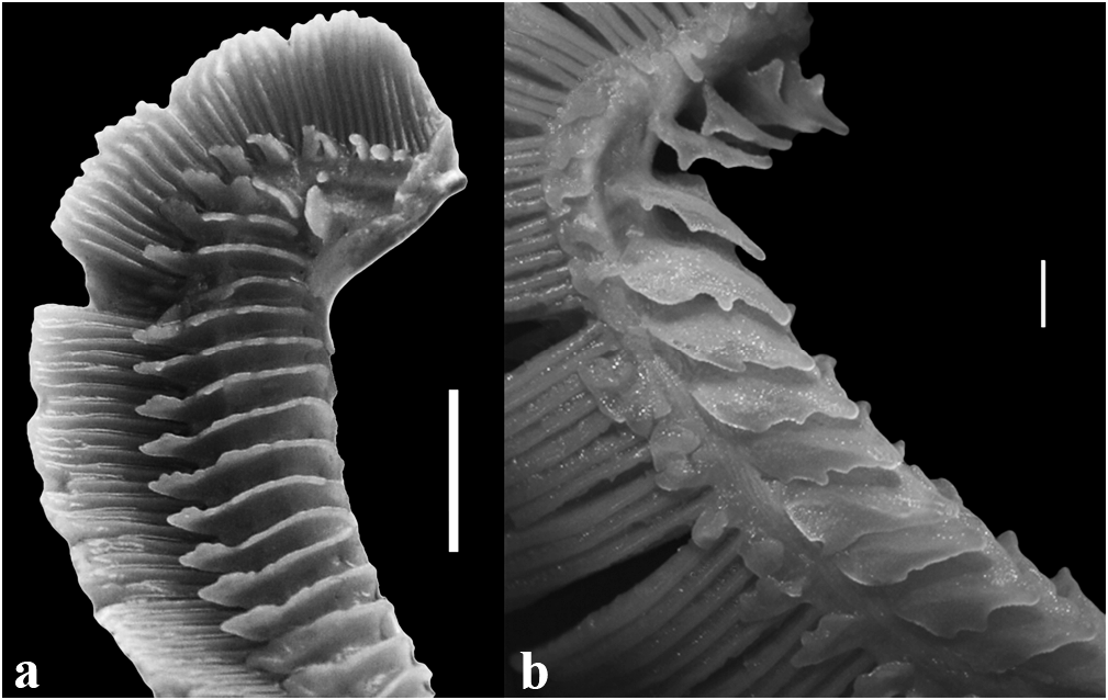
Inner surface of bucal cavity with rounded or filamentous papillae, larger and more abundant in large specimens. Skin of anterior and posterior surfaces of all branchial arches covered with papillae. Size of papillae smaller on anterior face of first and reduced fifth arches, and posterior face of fourth arch. Papillae developed on anterior surface of first arch and posterior surface of fourth arch rounded or conical, close to branchial filaments. Anterior surfaces of second to fifth arches, and posterior surfaces of first to third arches covered by broad papillae located transverse to longitudinal axis of arch ( Fig. 5a View Fig ). Larger, broad papillae with crenate margins. Skin around pharyngeal tooth plates covered by numerous papillae.
Gill rakers lobate ( Fig. 5b View Fig ), distributed as follows on first gill arch: 4 (1), 5* (20), or 6 (6) on upper limb; 1 on cartilage, and 14 (1), 15 (6), 16* (10), 17 (4), or 18 (2) on lower limb (n = 23). Gill rakers absent on second to fifth arches.
Lateral line complete; side-branch tubules of laterosensory canals alternatively directed horizontally and posteroventrally,
2a. Horizontal orbital diameter 27.3-32.0 % of HL; snout length 44.3-46.7 % of HL; adipose-fin length 92.5-100.7 % of distance between dorsal-fin origin and adipose-fin origin ........................................................... Iheringichthys megalops
2b. Horizontal orbital diameter 22.4-26.7 % of HL; snout length 47.0-51.7 % of HL; adipose-fin length 79.7-88.9 % of distance between dorsal-fin origin and adipose-fin origin ......................................................... Iheringichthys labrosus from head to vertical between pelvic-fin origin and short distance after last dorsal-fin ray insertion.
Urogenital papilla with numerous small ephitelial folds, forming furrows, placed between anus and urogenital papillae. Also small folds developed laterally to anus in large specimens.
Margins of swimbladder straight; swimbladder heartshaped, with three incomplete, large chambers. Sonic muscles inserted ventrolaterally on anterior third.
Color in alcohol. Background brownish, scarcely darker on dorsum; lower third of flanks cream, without dots. Many brown spots irregularly distributed on flanks. Spots more numerous on anterior half or two-thirds of body; spots very faint or absent on posterior half or third of body. Spots of anterior part of body, close to head, smaller. Very narrow horizontal line pale whitish, crossing flanks at level of pores of lateral line sensory canals in most specimens. Dorsal surface of head and cheeks light brown, without dots. Black chomatophores at dorsal-fin origin and around nuchal plates.
Distribution. The species is found in Brazil from the upper rio Paraná, in Jupiá (Três Lagoas Municipality, Mato Grosso do Sul State) and Ilha Solteira Municipality (São Paulo State).All specimens of Iheringichthys examined from those areas belong to I. syi . Most -and possibly all- records of Iheringichthys labrosus from those areas likely correspond to I. syi .
Etymology. The Guaraní word syi means straight, here alluding to the fine serration present on anterior margin of the pectoral-fin spine. The name is applied as an adjective.
Artificial key for Iheringichthys species:
1a. Anterior margin of pectoral-fin spine with fine serration, inapparent ................................................... Iheringichthys syi View in CoL
1b. Anterior margin of pectoral-fin spine with well developed serrae ....................................................................................... 2


| MZUSP |
Museu de Zoologia da Universidade de Sao Paulo |
No known copyright restrictions apply. See Agosti, D., Egloff, W., 2009. Taxonomic information exchange and copyright: the Plazi approach. BMC Research Notes 2009, 2:53 for further explanation.