
Heteropachylus gracilis, Mendes, 2011
|
publication ID |
https://doi.org/ 10.1111/j.1096-3642.2011.00706.x |
|
DOI |
https://doi.org/10.5281/zenodo.5492059 |
|
persistent identifier |
https://treatment.plazi.org/id/A60A87F2-FFBA-FFA2-D995-EA465D9440C8 |
|
treatment provided by |
Valdenar |
|
scientific name |
Heteropachylus gracilis |
| status |
sp. nov. |
HETEROPACHYLUS GRACILIS View in CoL SP. NOV.
( FIGS 19 View Figure 19 , 20 View Figure 20 , 25G, H View Figure 25 , 28 View Figure 28 )
Heteropachylus View in CoL sp. nov. 2: Sigrist & Carvalho, 2008: 39.
Distribution ( Fig. 28 View Figure 28 ): BRAZIL. Espírito Santo (Linhares and São Matheus). WWF Ecoregion: NT0103, Bahia coastal forests.
Type material: m holotype. ( IBSP440 View Materials ), BRAZIL, Espírito Santo, São Matheus, Reserva Florestal do Rio Doce , i.1998, Brescovit et al . Paratypes. 2m ( IBSP347 View Materials ), m ( IBSP450 View Materials ), m, 3f, and 1juv ( IBSP393 View Materials ) same data as holotype; 2m ( MNRJ4428 View Materials ), Linhares. 27.i.1973. Jim J, Caramaschi U & Carvalho C .
Etymology: Species name comes from the Latin gracilis , which means thin, slight, in reference to the comparatively slender aspect and weak armature of dorsal scutum.
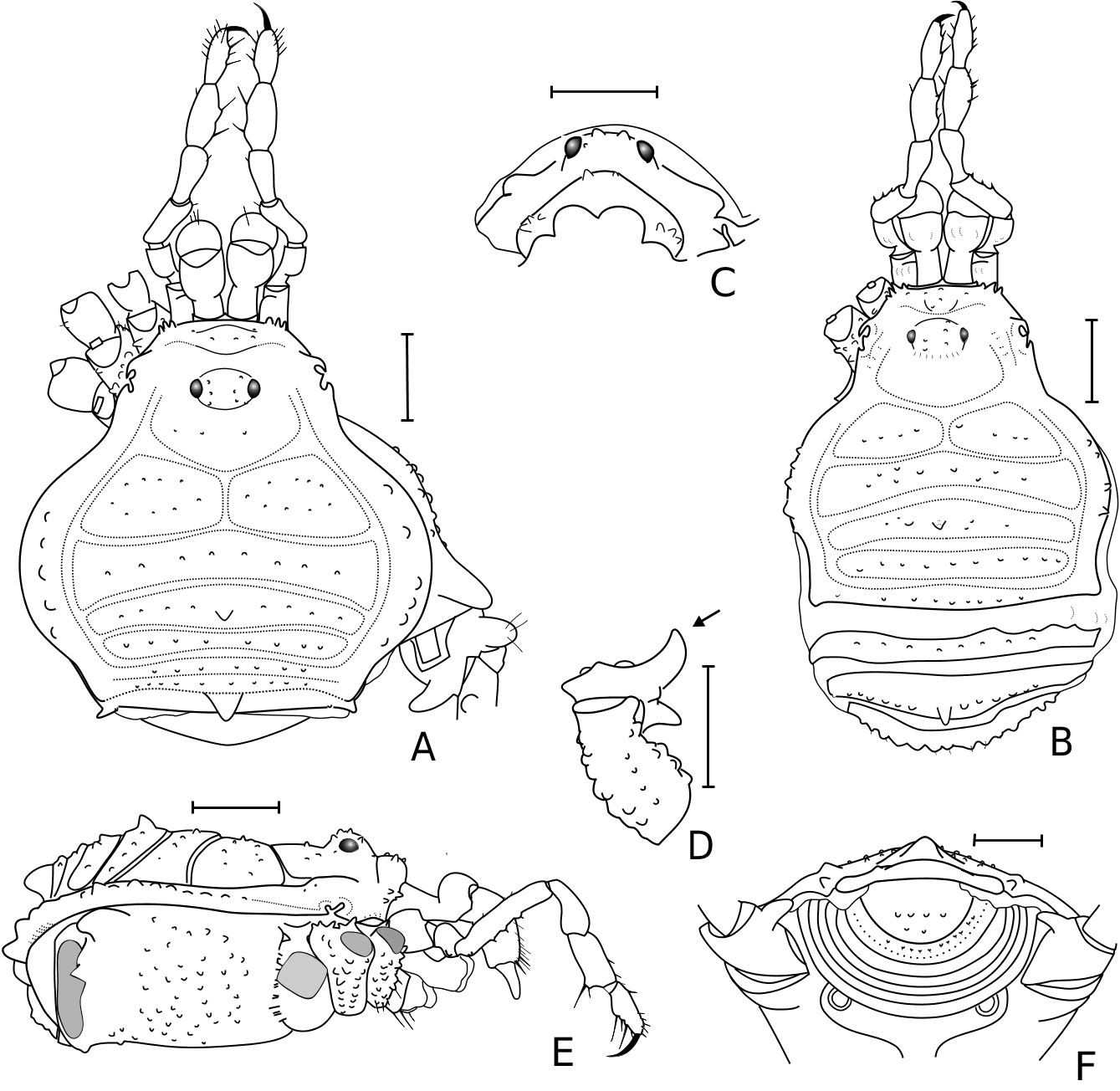
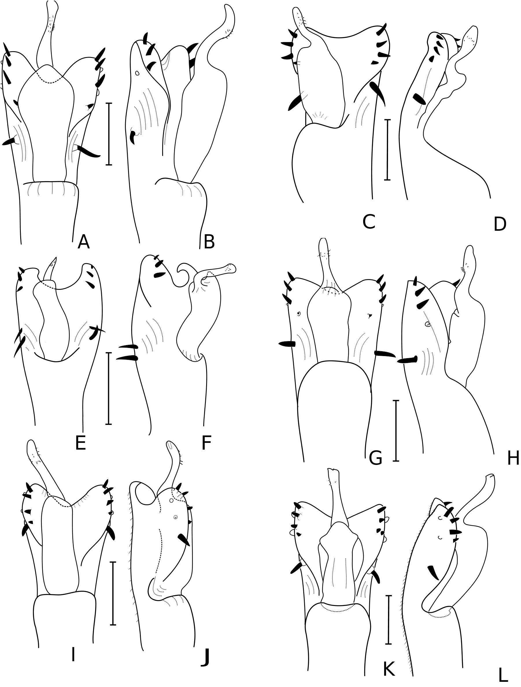
Diagnosis: Four mesotergal areas ( Fig. 19A View Figure 19 ). Mesotergal area I divided by longitudinal division; groove between mesotergal areas II and III complete ( Fig. 19A View Figure 19 ); mesotergal area III bearing unpaired armature and at the same level as the others; mesotergal area IV entire, not divided by longitudinal groove ( Fig. 19A, E View Figure 19 ). Free tergite I with unarmed corners; free tergite II with a tubercle on each corner and a conical median apophysis ( Fig. 19A View Figure 19 ); free tergite III unarmed, without keel and not fused to the dorsal scutum ( Fig. 19A View Figure 19 ). Dorsal anal operculum with granules ( Fig. 19F View Figure 19 ). Femur IV ( Fig. 20 View Figure 20 ) bearing conical dorsobasal apophysis and lacking dorsodistal one. Male genitalia ( Fig. 25G, H View Figure 25 ): fourth pair of setae dislocated to the centre of the ventral plate, distal group formed by only the first three setae.
Measurements: Male holotype. CL: 1.8; MCW: 2.5; ASL: 2.9; MASW: 4.8. FL: 3.6. Males (N = 5). CL: 1.7 (1.6–1.8); MCW: 2.3 (2.1–2.4); ASL: 3.0 (2.9–3.3); MASW: 4.5 (4.0–4.8). FL: 3.3 (2.9–3.6) Females (N = 3): CL: 1.7 (1.6–1.8); MCW: 2.2 (2.2–2.3); ASL: 2.4 (2.3–2.6); MASW: 3.9 (3.6–4.3). FL: 3.3 (3.1–3.4).
Description
Male holotype: Dorsum ( Fig. 19A View Figure 19 ). Scutum outline pyriform, widest at mesotergal area II. Anterior margin of carapace with three granules at each side. Frontal hump covered with granules. Eye mound with small granules near the eyes, without conspicuous armature. Carapace with sparse minute granules. Mesotergum divided into four areas; mesotergal area I divided into left and right halves by median longitudinal groove, with two rows of granules, the paramedian pair larger than the others; mesotergal areas II–IV with a row of granules each; mesotergal area III bearing median tubercle; mesotergal area IV entire. Posterior margin with transverse row of granules. Free tergites I–II fused to the dorsal scutum ( Fig. 19A View Figure 19 ); free tergite I with a transversal row of granules; free tergite II bearing small blunt median apophysis pointed upwards and one tubercle on each corner ( Fig. 19A, F View Figure 19 ); free tergite III smooth, with posterior border slightly convex and protuberant corners ( Fig. 19A, F View Figure 19 ). Lateral areas of mesotergum each with row of tubercles reaching area III, larger at area II ( Fig. 19A, E View Figure 19 ). Dorsal anal operculum with row of granules ( Fig. 19E View Figure 19 ). Appendages. Pedipalps ( Fig. 19A, E View Figure 19 ) – trochanter with two ventral setiferous granules. Femur with ventral row of setiferous granules and with subapical mesal spine. Tibia with three mesal (I+Ii) and two ectal ([Ii]) spines. Tarsus with four mesal (IiIi) and four ectal (IiIi) spines. Legs – calcaneus I about half length of metatarsus I, lighter in colour, and thicker than astragalus. Coxa IV ( Fig. 19A View Figure 19 ) armed with apical prolateral blunt apophysis. Trochanter IV ( Fig. 19A View Figure 19 ) armed with apical prolateral blunt apophysis and distal retrolateral curved apophysis. Femur IV ( Fig. 20 View Figure 20 ) short, distinctly curved in dorsal view and with two curvatures (‘S-shaped’) in lateral view, bearing rows of tubercles; with conical dorsobasal apophysis preceded by basal rounded tubercle and granules; dorsal surface with thre rows of rounded tubercles equal sized; retrolateral surface with row of rounded tubercles, with three to four conspicuous apophyses at distal portion, one to two smaller between two larger ones; prolateral surface with row of blunt apophyses located at the distal half, larger at the median portion of the row; apex ( Fig. 19D View Figure 19 ) with retrolateral conical apophysis, pointed upwards in apical view and prolateral rounded apophysis of acuminate apex. Tarsal counts. 6/9–10/6/6. Male genitalia ( Fig. 25G, H View Figure 25 ). Ventral plate subrectangular, narrower in basal portion, with distal border distinctly concave; dorsolateral surface excavated, seeming to be divided into ventral and dorsal layers. Group of three distal short setae on dorsum and two blunt setae on venter of ventral plate; fourth seta dislocated to the centre of ventral plate and much smaller; followed by one large basal seta. Glans without dorsal process. Stylus with apical setae of subequal size. Colour (in alcohol). Background colour of body and legs yellowish-amber to reddish-yellow, carapace with reddish-brown spots. Chelicerae and pedipalps yellow, reticulated with dark brown. Tarsi pale yellow. Female ( Fig. 19B View Figure 19 ). Free tergites not fused to the dorsal scutum, free tergite II bearing a small median spine. Apical apophysis of coxa IV reduced in relation to the male and spiniform. Trochanter IV narrow without armature. Femur IV without conspicuous armature.
Variation: Less developed exemplars have body less convex, with ratio between the maximum width of abdominal scutum and carapace smaller. The body granulation is more evident and the armature of the coxa, trochanter, and femur IV are not very developed.


No known copyright restrictions apply. See Agosti, D., Egloff, W., 2009. Taxonomic information exchange and copyright: the Plazi approach. BMC Research Notes 2009, 2:53 for further explanation.
|
Kingdom |
|
|
Phylum |
|
|
Class |
|
|
Order |
|
|
Family |
|
|
Genus |
Heteropachylus gracilis
| Mendes, Amanda Cruz 2011 |
Heteropachylus
| Sigrist MS & Carvalho CJB 2008: 39 |