
Repenomamus
|
publication ID |
https://doi.org/ 10.1046/j.1096-3642.2003.00064.x |
|
persistent identifier |
https://treatment.plazi.org/id/03D087D6-3261-5B71-FC5A-FCAB8184FDED |
|
treatment provided by |
Carolina |
|
scientific name |
Repenomamus |
| status |
|
IMPLICATIONS OF REPENOMAMUS
Developmental studies of living mammals have revealed that the posterior portion of Meckel’s cartilage forms the anlage of the malleus and that the middle portion of the cartilage degenerates in the later stages of ontogeny. The sheath of the middle portion of the cartilage becomes the sphenomandibular ligament (pterygomandibular in monotremes) or the anterior ligament of the malleus ( Gaupp, 1913; Goodrich, 1930; Allin & Hopson, 1992). By its relationship with the ear region, the OMC in Repenomamus and Gobiconodon provides evidence for a relationship of Meckel’s cartilage with the definitive mammalian middle ear in early mammals, which is otherwise only inferred from embryological evidence of living mammals. The Repenomamus specimens showed that, while the anlage of the malleus is reduced, or posteriorly shifted, to form a small malleus, a significant middle segment of Meckel’s cartilage persists and is even ossified in adults of some early mammals and close relatives. This fact supports the assumption that a persisting and possibly ossified Meckel’s cartilage connecting the malleus to the lower jaw was present in the common ancestor of mammals ( Zeller, 1993). The OMC in Repenomamus probably remains in its early ontogenetic position, as in extant mammals in which the cartilage extends from the dentary to the ventral side of the ear region. A similar condition is possibly present in other early mammals, such as triconodontids and symmetrodontids, in which an internal groove similar to the meckelian groove of Repenomamus is present.
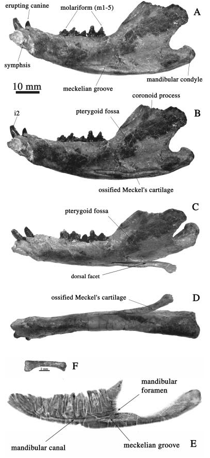
The function of the ossified Meckel’s cartilage in adults of Repenomamus and Gobiconodon is unclear. Because a facet exists on the dorsal side of the ossified Meckel’s cartilage in Repenomamus ( Fig. 1F View Figure 1 ), we have speculated that a portion of the medial pterygoid muscle originates on the pterygoid region of the skull and inserts to the ossified Meckel’s cartilage. There is enough space between the pterygoid area of the skull and the dorsal facet of the OMC to allow the muscle to work. This speculation was based on a comparison with marsupials and multituberculates, in which the medial pterygoid inserts to the inflected angular process in marsupials ( Sánchez-Villagra & Smith, 1997), or to the pterygoid shelf in multituberculates ( Gambaryan & Kielan-Jaworowska, 1995). As in other triconodonts and in symmetrodonts, the mandible of Repenomamus lacks the angular process or pterygoid shelf. Although a ridge defining the ventral border of the pterygoid fossa is present in the mandible of Repenomamus , it is a much weaker structure than is the inflected angular process in marsupials or the pterygoid shelf in multituberculates. The position and shape of the OMC in relation to the dentary and the pterygoid fossa in Repenomamus resembles the inflected angular process or the pterygoid shelf; it may have functioned as an additional site for the partial insertion of the medial pterygoid muscle. This condition may represent an intermediate condition during the evolutionary shift of the jaw-elevating muscle insertion from the postdentary bones to the dentary, a trend documented in the transition from cynodonts to mammals ( Crompton, 1963; Barghusen, 1968; Barghusen & Hopson, 1970).
Rugosities on the posterior end of the OMC in Repenomamus suggest a connection by ligament to the lateral flange in life. This contact functions as a hinge that operates in concert with that of the dentary-squamosal. Although the hinge is anterior to that of the dentary-squamosal, and therefore not coaxial, the ossified cartilage can still rotate with the dentary as the latter was depressed and elevated because of the possible mobility at the contact of the dentary and the OMC.
The ear ossicles in early mammals other than therians are poorly documented in the fossil record. The presence of these elements are known only in multituberculates ( Miao & Lillegraven, 1986; Miao, 1988; Meng & Wyss, 1995; Rougier et al., 1996), but are inferred in other groups, such as triconodonts ( Allin & Hopson, 1992), based on jaw and cranial morphologies. For most Mesozoic mammals and their close relatives, however, jaw and skull materials are often fragmentary, which obstructs inference of the shape and size of the ear ossicles and their relationships with surrounding structures.
Although actual ear ossicles are not preserved in Repenomamus , well preserved skulls and mandibles provide a reliable basis for inference of the ear ossicles. Identification of the OMC leads to the conclusion that Repenomamus and Gobiconodon have a definitive mammalian middle ear because the dentary of the two taxa lacks other scars for the postdentary bones. In addition, lack of the angular process, and therefore absence of the angular gap, suggests a separation of the middle ear from the dentary. For the same reason, the definitive mammalian middle ear is probably present in Zhangheotherium . A similar conclusion has been reached for triconodontids ( Allin & Hopson, 1992; Rowe, 1996a,b), in which the ear ossicles have not been recovered. Given the phylogenetic relationship and other ear structures, such as the fossa incudis and fenestra vestibuli, which are comparable to those of other species that have the ear ossicles (or their equivalents), we have no reason to believe that ear ossicles are phylogenetically absent in Repenomamus and Gobiconodon .
Compared to those of close relatives of mammals, such as Morganucodon , Probainognathus and Pachygenelus ( Hopson, 1966; Allin, 1975; Allin & Hopson, 1992; Luo & Crompton, 1994), the fossa incudis and fenestra vestibuli, and distance between these structures, as well as the general size of the tympanic region of Repenomamus are relatively small. In addition, the ectotympanic and malleus of Repenomamus were probably inclined, similarly to those of monotremes ( Kuhn, 1971; Zeller, 1987, 1993), multituberculates ( Meng & Wyss, 1995; Rougier et al., 1996) and those in early ontogenetic stage of therians ( Starck, 1967). The inclined orientation of these elements is also suggested by the tilted fenestra vestibuli of Repenomamus , based on a roughly parallel relationship between the fenestra vestibuli and ectotympanic in living mammals ( Rougier et al., 1996). Moreover, there is no supporting structure for a vertically orientated ectotympanic. The lateral flange, crista parotica and anteromedial ridge of the petrosal can only support an inclined ectotympanic and malleus.
The fossa incudis is immediately medial to the secondary craniomandibular joint (dentary-squamosal) in Repenomamus and Zhangheotherium . This relationship shows that in early mammals the secondary craniomandibular joint is lateral to the primary joint (malleo-incudal = quadrate-articular), not anterior to the primary joint as was shown in the ontogenesis of living mammals ( Reichert, 1837; Gaupp, 1913; Zeller, 1989, 1993; Maier, 1990). It also shows that in early mammals the ear ossicles are medial, not posterior, as in extant mammals ( Rowe, 1996a,b), to the secondary craniomandibular joint. The ear structure of Repenomamus probably represents an intermediate condition between the mandibular ear of nonmammalian synapsids, such as Morganucodon ( Kermack et al., 1981) , and the definitive mammalian middle ear of mammals in which the ear ossicles are widely separated from the secondary craniomandibular joint and lie behind intervening secondary auditory structures ( Rowe, 1996a,b).
Coexisting with the detached ear ossicles in Repenomamus are other auditory and feeding features. These include an elongated promontorium, a distinct external auditory meatus, an expanded glenoid fossa and mandibular condyle, enlarged pterygoid fossa, and broad masseteric fossa. These features can be attributed to the more efficient hearing of air-borne sound, and to more powerful mastication, respectively.
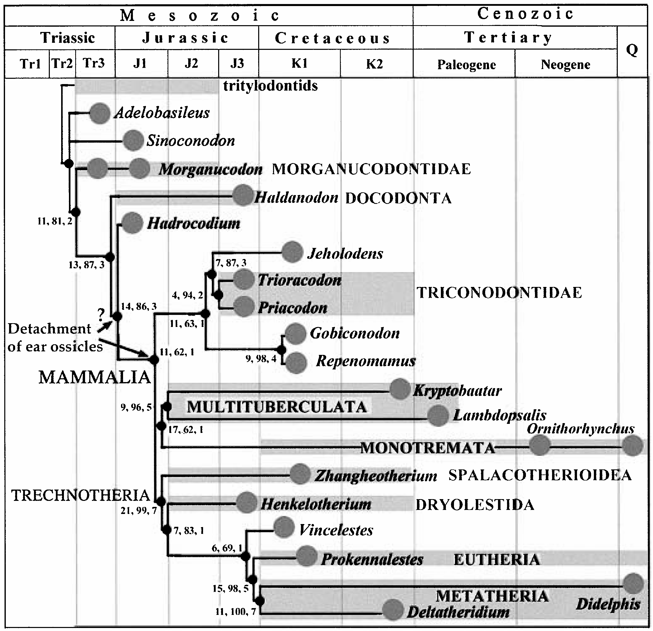
With the new data from Repenomamus and the new Gobiconodon , a phylogenetic analysis has been attempted ( Wang et al., 2001). The cladogram ( Fig. 6 View Figure 6 ) is the consensus (tree length = 275; CI = 0.589; RI = 0.706) of four equally most parsimonious trees that are obtained by branch-and-bound searches using PAUP* 4.0 b8 ( Swofford, 2000) based on a data set consisting of 112 craniodental characters across 20 taxa. The phylogeny is largely in keeping with other recent phylogenetic hypotheses of mammals and their relatives ( Rowe, 1988; Rougier et al., 1996; Hu et al., 1997; Ji et al., 1999; Luo, Crompton & Sun, 2001). Within the phylogeny, acquisition of the definitive mammalian middle ear in Repenomamus and Gobiconodon is consistent with the prediction that triconodontids have fully detached ear ossicles ( Allin & Hopson, 1992). Whether the definitive mammalian middle ear is a synapomorphy for Mammalia, which probably occurred in the middle Jurassic, based on fossil records, or it is shared by Mammalia and Hadrocodium and thus evolved in the early Jurassic ( Luo et al., 2001), depends on the interpretation of Hadrocodium ( Wang et al., 2001; see below).
No known copyright restrictions apply. See Agosti, D., Egloff, W., 2009. Taxonomic information exchange and copyright: the Plazi approach. BMC Research Notes 2009, 2:53 for further explanation.