
Exechonella pumicosa Canu & Bassler, 1928
|
publication ID |
https://doi.org/ 10.11646/zootaxa.4305.1.1 |
|
publication LSID |
lsid:zoobank.org:pub:1192C3A0-5CCB-4A86-903C-A2B82906A5F9 |
|
DOI |
https://doi.org/10.5281/zenodo.6017320 |
|
persistent identifier |
https://treatment.plazi.org/id/CF0AB852-FFE6-E933-FF03-F9C294DBE5E1 |
|
treatment provided by |
Plazi |
|
scientific name |
Exechonella pumicosa Canu & Bassler, 1928 |
| status |
|
Exechonella pumicosa Canu & Bassler, 1928
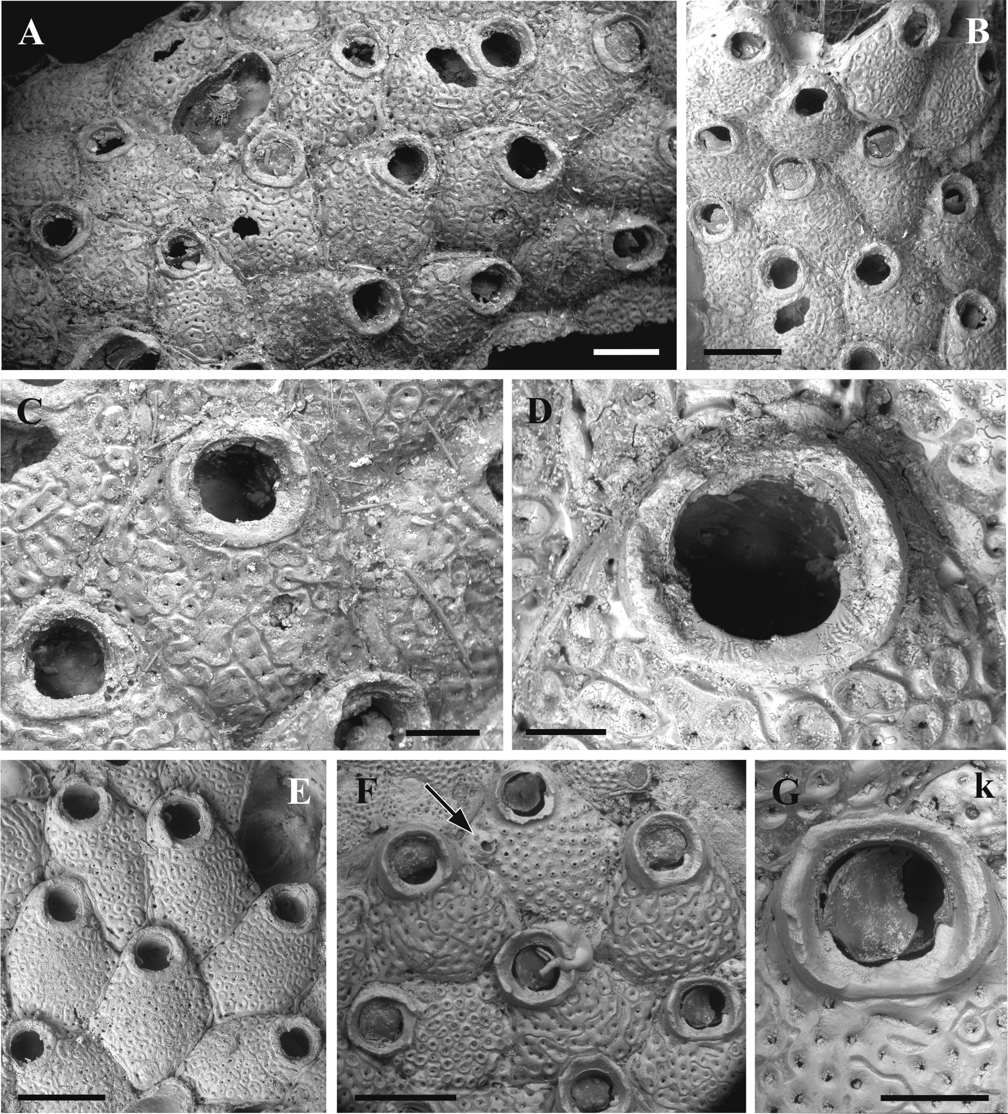
( Fig. 9 View FIGURE 9 , Table 8)
Exechonella pumicosa: Canu & Bassler 1928 , p.70–71, pl. 14, fig 1, text fig. 11a. Phylactella labrosa: Osburn 1914 View in CoL , p. 213.
? Exechonella antillea: Winston 1982 View in CoL , p. 136, fig. 60.
Material examined. Holotype: USNM 7838 View Materials , encrusting on worm tube. Atlantic Ocean , Florida, Fowey Light , 15 miles south of Miami , depth 40 fathoms, November 1914, coll. by J.B. Henderson. Additional material examined: USNM 10127 View Materials , Atlantic Ocean, Florida, Fowey Light, 15 miles south of Miami, depth 40 fathoms, November 1914, coll. by J. B. Henderson; USNM 545922 View Materials , [Dry] Tortugas, depth 22 fathoms.
Description. Colony encrusting, unilaminar, multiserial. Autozooids pentagonal in shape, longer than wide, separated by narrow grooves. Zooidal periphery surrounded by a narrow gymnocystal rim that is seen in most zooids. Primary orifice wide, pear-shaped, almost as long as wide. Poster (one-third) narrower than anter (twothirds), mostly of rounded outline, but tending to be more quadrate (angular) in some zooids. The anter wall is underlain by an inner lamina, the ends of which form well-defined mostly triangular condyles, with their points directed to the midline and extending beyond the edge of the step-like curve below. The collar-like peristome is thick-walled, oval or parallel-sided laterally, having the various width proximally, sometimes with low blunt processes. In most zooids the collar proximal edge is wide and flat, with a smooth medial area, but slightly wrinkled either side. In some zooids a tiny central (blunt or pointed) projection occurs on it. Frontal shield with 40– 62 well-separated, rounded or oval foramina, each with a wide, smooth gymnocystal rim, the walls raised slightly above the frontal shield and sloping towards the small lumen. Rims of most foramina fuse into ‘chains’ of 2–8, sometimes forming radiating pore rows known in cribrimorph cheilostomes. With age the foraminal rims occupy most of the frontal surface, leaving only narrow furrows between each other. Small marginal pores are predominantly oval and elongated. Avicularia not observed. Kenozooids with 3–5 pores were sometimes met on autozooidal margins. Vertical zooidal walls wide, represented by multiporous mural septula with 1–2 rows of communication pores. Ancestrula not observed.
Florida, Atlantic Ocean
m±sd r n AzL 798±61 714–900 8 AzW 639±73 529–757 8 OrL 194±18 171–214 8 OrW 192±14 171–214 8 FoN 47±7 40–62 8 FoD 49±8.4 25–65 34 OD 9±2.5 5–14 34 Remarks. While being very similar, Exechonella pumicosa differs from E. antillea by thicker peristomes many of which have wide and flat proximal edge normally without a blunt projection. Also foraminal rims occupy most of the frontal surface in the former species, leaving only narrow furrows between each other. Many of them fuse forming ‘chains’ (linear or branching) of 3–7 foramina. In the latter species the frontal surface between foraminal rims is well seen and ‘fused’ foramina are not so numerous and ‘chains’ much shorter (2–3 foramina). In the studied specimens the number of frontal foramina ranges from 40 to 62 in E. pumicosa and from 34 to 52 in E. antillea . In addition, avicularia are not seen in the studied specimens of E. pumicosa (while a presence of these polymorphs is not a stable character). On the other hand, most of zooids are surrounded by a narrow raised ‘gymnocystal’ rim in this species whereas it is only rarely seen in E. antillea . It should be noted that different specimens from the Fowey Light show a varying degree of foraminal rim fusion, with most zooids predominantly having short chains of 2–3 zooids that are similar to E. antillea .
Exechonella pumicosa was originally described by Canu and Bassler (1928b) from Miami , Florida, but subsequently it has been synonymized under E. antillea View in CoL (e.g. Osburn 1940, 1950; Shier 1964; Winston 1982 and Fransen 1986). However , examination of the holotype of E. pumicosa using the SEM allows for the distinction between these two closely related species.
Examination of the specimen USNM 545922 described as Phylactella labrosa View in CoL by Osburn (1914) from Dry Tortugas, Florida, (and later synonymized by him with E. antillea View in CoL , see above) showed numerous chains of the fused foramina in its zooids that points to E. pumicosa . The maximal number of foramina was 68 that also correspond more to this species than to E. antillea View in CoL . Also, while kenozooids are present in this specimen, avicularia are missing. This specimen also shows the early developmental stages of the frontal shield with foramina being round elevated projections without a rim. In older zooids foraminal rims were aquired. With time becoming wider they eventually fuse.
Winston (1982), in her study of the Indian River area , Florida, described and illustrated a specimen (as Exechonella antillea ) that in our opinion belongs to E. pumicosa although the foraminal luminae are larger than those in the holotype . The original description of E. pumicosa made by Canu and Bassler (1928b) mentions the presence of avicularia, but we did not observe them in the holotype. Winston’s (1982) figure 60 does not show avicularia either, although these polymorphs were mentioned in her description.
In addition to three specimens from Florida we checked a non-numbered specimen from Bocas del Toro, Panama (collected by K.J. Tilbrook and J.E. Winston in 2004, and currently kept in the Virginia Museum of Natural History , USA; authors possess four SEM-images) that strongly reminiscent a holotype of E. pumicosa . However, in comparison with the holotype it has a smaller zooidal size and larger foramina. It also possesses avicularia on the wall of two lateralmost foramina, which are larger than the rest of the foramina. It should be noticed that avicularia were predominantly seen in the peripheral zooids in this specimen that might explain their absence in the holotype of E. pumicosa . More material from Panama is required to make a definite conclusion if this specimen could belong to the new species or not.
Distribution. Exechonella pumicosa is currently known only from Dry Tortugas, Gulf of Mexico, and Atlantic side of Florida, USA.
| USNM |
Smithsonian Institution, National Museum of Natural History |
No known copyright restrictions apply. See Agosti, D., Egloff, W., 2009. Taxonomic information exchange and copyright: the Plazi approach. BMC Research Notes 2009, 2:53 for further explanation.
|
Kingdom |
|
|
Phylum |
|
|
Class |
|
|
Order |
|
|
Family |
|
|
Genus |
Exechonella pumicosa Canu & Bassler, 1928
| Cáceres-Chamizo, Julia P., Sanner, Joann, Tilbrook, Kevin J. & Ostrovsky, Andrew N. 2017 |
Exechonella antillea
| : Winston 1982 |
E. antillea
| : Winston 1982 |
E. antillea
| : Winston 1982 |
E. antillea
| : Winston 1982 |
Exechonella pumicosa
| : Canu & Bassler 1928 |
Exechonella pumicosa
| : Canu & Bassler 1928 |
E. pumicosa
| : Canu & Bassler 1928 |
E. pumicosa
| : Canu & Bassler 1928 |
Phylactella labrosa
| : Osburn 1914 |
Phylactella labrosa
| : Osburn 1914 |