
Etmopterus lailae, Ebert, David A., Papastamatiou, Yannis P., Kajiura, Stephen M. & Wetherbee, Bradley M., 2017
|
publication ID |
https://doi.org/ 10.11646/zootaxa.4237.2.10 |
|
publication LSID |
lsid:zoobank.org:pub:43FE1ADD-00F5-4A65-B878-3EF231A03A2B |
|
DOI |
https://doi.org/10.5281/zenodo.5625248 |
|
persistent identifier |
https://treatment.plazi.org/id/03AC87E3-764F-FFD3-FF2D-C082FC9481E9 |
|
treatment provided by |
Plazi |
|
scientific name |
Etmopterus lailae |
| status |
sp. nov. |
Etmopterus lailae View in CoL , new species
Laila’s Lanternshark
( Figures 1–3 View FIGURE 1 View FIGURE 2 View FIGURE 3 a, 4–5, Table 1 View TABLE 1 )
Holotype. BPBM 40183, 368 mm TL, immature male, R/V Townsend Cromwell cruise 8805, leg 2, station 216, Koko Seamount, 35° 16.48´N 171° 17.13´E to 35° 16.55´N 171° 17.20´E, 314–358 m, 13 August 1988.
Paratypes. BPBM 40174, 303 mm TL, immature male, R/ V Townsend Cromwell cruise 8805, leg 2, station 167, South Kanmu Seamount , 32° 03´N 173° 04´E to 32° 02´N 173° 06´E, 336–338 m, 6 August 1988 GoogleMaps ; BPBM 40182, 265 mm TL, immature male, R/V Townsend Cromwell cruise 8805, leg 2, station 218, Koko Seamount, 35° 17.05´N 171° 22.01´E to 35° 17.05´N 171° 21.54´E, 368–384 m, 14 August 1988.
Diagnosis. Etmopterus lailae is a moderately large, slender species of linear–denticled Etmopterus that can be separated from most of its congeners within the E. lucifer clade by the length of its anterior flank marking branch being much longer relative to its posterior branch; all other members of this genus, except for two species, have a posterior branch that is equal to or longer than the anterior branch. The only two species with an anterior branch relatively longer than the posterior branch, E. lucifer Jordan & Snyder, 1902 and E. sculptus Ebert, Compagno , & De Vries, 2011, can be separated from the new species by a lower spiral valve count (8–9 vs 14–16 for E. lailae ), a slightly higher precaudal vertebral count (55–64 vs 53–57), and a higher number of teeth on the lower jaw (30–43 vs 26–28). Etmopterus lailae lacks dermal denticles between the nostrils on the ventral snout surface and on the dorsal fins, while the other two species have denticles present on the snout and dorsal fins.
Description. Proportional measurements expressed as a percentage of total length (TL) are given for the holotype and the range for two paratypes ( Table 1 View TABLE 1 ).
Body fusiform, trunk sub-cylindrical, width 1.2 (0.7–1.0) in trunk height; head sub-conical, moderate-sized, length 14.9 (13.9–16.2)% TL, slightly depressed, height 1.6 (1.3–1.6) times width. Snout moderately long, triangular–shaped becoming rounded at snout–tip in dorsal view, slightly depressed in lateral view, head width 11.1 (9.2–10.9)% TL. Eyes oval-shape, large, length 2.9 (2.6–3.3) in head, 1.9 (1.6–1.8) times width of eye; orbits with anterior and posterior notches; inter–orbital space 1.6 (1.3–1.5) in width of head; eye length 1.3 (1.4–1.5) times in inter–orbital distance. Spiracles small, greatest diameter 1.9 (1.3–1.5)% TL, 2.7 (3.3–4.0) times into length of eye, distance to eye 2.4 (2.6)% TL, eye–spiracle length 0.9 (0.9) times into height of eye. Nostrils large, oblique, length 1.4 (1.5–1.6) times into internarial width, 2.7 (2.6–2.7) times eye diameter; anterior nasal flap well developed, triangular, anterior tip extending across nasal opening, length 0.6 (0.5) times into spiracle length. Gill openings small, slightly oblique, in horizontal series, height decreasing progressively posteriorly, first two openings noticeably larger than last three openings, fifth opening just in front of pectoral fin origin; height of first gill slit 1.8 (1.3–1.8) in height of fifth gill opening; inter-gill length about equal, 1.1 (1.2–1.3) times, to length of eye. Mouth broad, length 4.0 (3.0–3.3) times width, slightly arched, width 0.7 (0.6) in preoral length.
......continued on the next page Teeth dissimilar in upper and lower jaw ( Fig. 2 View FIGURE 2 ); upper jaw teeth with strong central cusp flanked on each side by two smaller lateral cusplets, less than one-half the height of median cusp, and decreasing in size distally; lower jaw teeth unicuspid, blade-like, oblique, fused into a single row. Tooth count in first row of upper jaw 24 (22–24) and in first row of lower jaw 28 (26–26).
First dorsal fin small, length of first dorsal fin 9.0 (7.9–8.6)% TL, anterior margin slightly curved, rounded at apex, origin slightly behind pectoral fin free rear tip; fin base insertion well forward of pelvic-fin origin; pre–first dorsal length 1.6 (1.7–1.8) in inter–dorsal distance; first dorsal–fin spine nearly straight, short, (0.4–0.9) times into height of first dorsal fin, located posterior to pectoral fin posterior margin. Second dorsal fin noticeably larger than first dorsal fin, length of first dorsal fin 0.7 (0.6–0.7) into second dorsal fin, height of first dorsal fin (0.5–0.6) into second dorsal fin; apex broadly rounded, posterior margin concave, free rear tip elongated, length 13.0 (12.5– 13.2)% TL, pre–second dorsal length 2.8 (2.9–3.1) in inter–dorsal distance; second dorsal–fin spine large, height about equal to or slightly higher than fin height, slightly curved near tip towards fin apex; origin behind over or slightly behind pelvic fin free rear tips. Interspace between first and second dorsal fins 2.8 (2.9–3.1) times into pre– pectoral length.
Pectoral fin length 9.0 (8.3)% TL, broadly rounded at free rear tips, base into anterior margin length ratio 1.7 (1.7–2.0), posterior margin nearly straight edged. Caudal peduncle rounded, relatively short, 12.0 (10.6–10.9)% TL, and tapering posteriorly; height slightly greater than width, 1.3 (1.0–1.4) times width; distance less than upper caudal fin length. Caudal fin elongated, slightly greater than head length, sub–terminal notch conspicuous; preventral caudal fin margin 2.4 (3.2–3.0) into dorsal caudal fin margin.
Dermal denticles on dorsal body surface erect, thorn-like, curved rearwards, in distinct longitudinal rows extending from dorsal head surface to caudal fin; distance between rows appear to decrease behind pelvic fin insertions to caudal fin. Ventral snout surface with prominent pores (ampullae of Lorenzini) surrounded by dermal denticles, except for bare patch between nostrils and extending just behind posterior nostril edges; area above upper lip of jaw without dermal denticles ( Fig. 3 View FIGURE 3 a). Dorsal fins mostly naked, without dermal denticles extending on fin base or ceratotrichia.
Luminescent markings distinct, covering ventral head surface, extending to level of nostrils and at about orbital anterior notch, descending below eye level to mouth, then extending upward over mouth corners, but not encircling mouth; discontinuous with belly marking posterior to mouth at about first gill openings, demarcated by a faint band of transverse dermal folds across throat extending from below lower edges of first and second gill openings on either side.
Belly marking originates behind mouth on posterior portion of transverse dermal folds and extends ventrally along pectoral fin bases slightly extending upwards to about level with fin base insertion, about level with ventral edges of gill openings, and posteriorly to pelvic fin bases; ventral surface of pectoral fin with two very dark lobeshaped markings, one at fin origin and another at insertion, each extending from fin base to origin of ceratotrichia bisected by distinct lighter band forming a two–prong fork pattern; margin on pectoral–pelvic space clearly demarcated, line extending from rear margin of pectoral–fin insertion nearly to pelvic–fin origin except for lighter space at pelvic–fin origin; dark ventral belly surface terminates under free rear inner margin of second dorsal fin; ventral caudal peduncle surface with darker marking just behind cloaca and extending about halfway to lower caudal origin ( Fig. 4 View FIGURE 4 ).
Flank markings distinctive, with anterior and posterior branches present; anterior branch slender, curving slightly over pelvic fins with a downward concavity, thin proximally near marking base, thickening medially before narrowing to acute tip distally; length relatively long, 11.1 (9.6–10.4)% TL as measured from marking base to tip, extending beyond origin of pelvic–fin; posterior branch nearly straight, relatively thick, length less than anterior branch, 6.3 (6.3–7.9)% TL, extending just beyond second dorsal fin base insertion, terminating below and before midpoint of inner fin margin; base of flank marking relatively narrow, origin posterior to pelvic fin insertion.
Caudal central marking distinct, thick, ovoid-shaped, length 6.0 (6.3–6.8)% TL, longer than base width of flank marking. Caudal fin upper marking very narrow, its length 1.2 (1.0–1.1) times central caudal marking.
Vertebral counts: total vertebral counts 86 (79–82); monospondylous 40 (40); diplospondylous 17 (13–15); total precaudal 57 (53–55); caudal 29 (26–27). Spiral valve count is 16 (14–16).
Coloration. After preservation dorsal and lateral surface a light to medium brown, except for prominent dark brown lateral and caudal markings, ventral surfaces also a dark brown; transition between lateral and ventral surfaces sharply demarcated. Body with 1 to 3 rows of prominent dark photophores extending from head posteriorly along flanks to about upper caudal origin; an irregular row of dark photophores also extending between pectoral and pelvic fins. Ventral surface a dark brown to blackish around mouth, belly, between pelvic fin origins and lower caudal origin; area around mouth distinctly dark brownish, sharply contrasting the lighter brown snout; area across throat slightly lighter than area anterior and posterior to this region. Gills and area just below darker than lighter brown area above. Pectoral and pelvic fin bases light brown above, darker below, and with blackish posterior and inner margins; remainder of fins becoming translucent. Dorsal fins light brown at base, becoming lighter to translucent. Lateral and caudal flank markings prominent, sharply demarcated, but without lighter colored lateral flanks. Caudal fin after preservation lacks vertical dark bar or any noticeable markings except for upper caudal marking.
Size. Maximum length is at least 370 mm TL (holotype: BPBM 40183) for an immature male, both paratypes are immature males; females were not available for examination.
Distribution. The new species presently is known only from the Koko and South Kanmu seamounts, Northwestern Hawaiian Islands, and at a depth range of 314–384 m ( Fig. 5 View FIGURE 5 ).
Etymology. The new species is named after Laila Mostello-Wetherbee, shark enthusiast and daughter of coauthor Brad Wetherbee. The proposed common name is Laila’s Lanternshark.
Comparisons. Etmopterus lailae can be assigned to the “ E. lucifer clade” as defined by Straube et al. (2010) with its predominant lateral flank markings displaying conspicuous anterior and posterior branches. The members of this group are also referred to as linear denticle etmopterids due to the characteristic arrangement of distinct linear rows of denticles on the dorsal head surface that also extends to the flanks, caudal peduncle and caudal base. The “ E. lucifer clade” can further be subdivided into three distinct subgroups based on the relative lengths of the lateral flank marking branches ( Ebert et al., 2016); anterior flank marking branch longer than posterior, anterior branch shorter than posterior, and anterior and posterior branches relatively equal in length. Eleven Etmopterus species are currently recognized to fall within the “ E. lucifer clade”, of which six species ( E. alphus Ebert, Straube, Leslie & Weigmann, 2016 , E. brachyurus Smith & Radcliffe, 1912 , E. bullisi Bigelow & Schroeder, 1957 , E. decacuspidatus Chan, 1966 , E. dislineatus Last, Burgess , & Séret, 2002, and E. molleri Whitley, 1939 ) have an anterior flank branch that is shorter than the posterior, and three species ( E. burgessi Schaaf-DaSilva & Ebert, 2006, E. evansi Last, Burgess , & Séret, 2002, and E. pycnolepis, Kotlyar, 1990 ) have anterior and posterior branch lengths that are about equal in length ( Ebert et al., 2011, 2016). The remaining two species, E. lucifer and E. sculptus Ebert, Compagno , & De Vries, 2011, each has an anterior branch length that is longer than the posterior, and appear to be closest to E. lailae based on the relative branch lengths of flank markings ( Ebert et al. 2011).
Etmopterus lailae can be separated from its two closest congeners by a combination of external morphological and internal meristic characteristics. Externally, E. lailae can be separated from E. sculptus by a shorter posterior flank branch marking (6.3–7.9 vs 7.5–8.6)% TL; a longer central caudal marking (6.0–6.8 vs 3.4–5.0)% TL; central caudal marking length 3.3–3.6 vs 1.4–1.9 times flank base marking width; and a shorter upper caudal marking (1.1–1.2 vs 2.0–2.5) relative to the central caudal marking. The ventral snout surface of E. lailae between the nostrils is bare, but posteriorly has dermal denticles surrounding tiny pores (ampullae of Lorenzini), while E. sculptus is rather uniformly covered with dermal denticles, with small naked patches along the upper lips, and around and between the nostrils. Internally, E. lailae compared to E. sculptus has a higher spiral valve turn count (14–16 vs 8–9), a lower precaudal vertebral count (53–57 vs 60–64), and a lower tooth count on the lower jaw (26– 28 vs 36–43). The biogeography is also informative since E. lailae is only known from the north central Pacific Ocean, while E. sculptus occurs from off Namibia to southern Mozambique in the southern hemisphere ( Ebert et al., 2011).
The closest congener geographically and morphologically to E. lailae is the Japanese E. lucifer , but these two species can be separated by a combination of meristic and external morphological characteristics. Etmopterus lailae can be separated meristically from Japanese E. lucifer by having a higher spiral valve turn count (14–16 vs 8–9), a lower tooth count on the lower jaw (26–28 vs 30–39), and an overlapping although slightly lower precaudal vertebral count (53–57 vs 55–63) and a lower total vertebral count (79–86 vs 85–90), although it should be noted that precaudal and total vertebral counts vary widely depending on the region and within some regions ( Last & Stevens, 2009; Last & Stewart, 2015). For example, Yamakawa et al. (1986) based on 65 Japanese E. lucifer specimens reported a range of 55–62 precaudal vertebral, while we found the range for five Japanese E. lucifer to be 59–63. Although Yamakawa et al. (1986) did not report on the number of total vertebrae from their study, we found this number to range from 85–90 based on five Japanese E. lucifer specimens, while E. lailae had a range of 79–86.
Etmopterus lailae can be separated from E. lucifer by the following body ratios: mouth width shorter (6.6–7.6 vs 7.4–10.1)% TL; pre-pectoral length slightly shorter (21.8–22.6 vs 22.6–25.3)% TL; pectoral pelvic space shorter (21.5–24.4 vs 24.2–33.2)% TL; anterior pectoral fin length shorter (7.9–8.6 vs 8.8–11.0)% TL; first dorsal fin length longer (7.9–9.0 vs 5.4–8.0)% TL; second dorsal fin height relatively low, its height 26.3–34.3 vs 34.5– 41.9% TL of its overall length. The flank markings are also informative in separating these two species: E. lailae when compared to E. lucifer has a shorter anterior flank marking branch (9.8–12.6% vs 12.7–14.0)%; a lateral flank marking base width that is slightly narrower (1.8–2.0% vs 2.1–2.5%); a slightly narrower flank base width 9.2–9.3 vs 9.6–13.9; a longer central caudal marking (6.0–6.8% vs 2.8–5.1%); central caudal marking length 3.3– 3.6 vs 2.5–2.7 times flank base marking width.
The arrangement of the dermal denticles, including their presence or absence, is informative when separating E. lailae from E. lucifer . The ventral snout surface of E. lailae has a naked patch between, and extending just posterior to, the space between the inner nostrils ( Fig. 2 View FIGURE 2 a). Also, beginning just posterior to the inner nostril opening is a bare patch, absent dermal denticles, extending rearwards to the upper lip except for small patch of denticles bisecting it about one-third the distance from the inner nostril opening to the upper lip; this area has several large pore openings. The area just anterior to the upper lip of the jaw, and traversing its length, also has a bare area with a few large scattered pores. Distinct pores cover the remainder of the snout ventral surface. In contrast, the snout ventral surface of E. lucifer is densely and relatively evenly covered by dermal denticles, but has no bare patches, except along the upper lip ( Fig. 2 View FIGURE 2 b); some small pores are apparent, but scattered. The dorsal fins of E. lailae are mostly naked, without dermal denticles extending onto the dorsal fins, while E. lucifer has linear rows of denticles present on the bases and extending onto the dorsal fins, including the ceratotrichia. The lateral dermal denticles extending the length of the body on E. lailae appear more sculpted than observed in E. lucifer .
In addition to the above parameters these two species differ significantly in the size at maturity. All three known specimens of E. lailae are immature males, with the largest measuring 368 mm TL after preservation (370 mm TL before preservation), while E. lucifer from Japanese waters is a much smaller species with males maturing (before preservation) at a minimum length of 278 mm TL, with a maximum of 310 mm TL, based on the individuals examined in this study.
The separation of “ E. lucifer clade” etmopterids, including the new species E. lailae , is complicated due to the inadequate original description of E. lucifer from Japan and the convoluted taxonomic history of this species from Japanese waters and elsewhere. The original description and type series of E. lucifer was based on a combination of two different etmopterid species, one form with an anterior branch longer than the posterior, and the other with the posterior branch being longer than the anterior. Examination of several of the syntypes by DAE and J.A. Schaaf-Da Silva (California Department Fish & Wildlife) confirms that two different species are among the type series. Furthermore, a lectotype has never been designated for the species, thus leaving open the question of what species exactly constitutes true E. lucifer , and leaves unresolved the status of the other species comprising the type series. Resolution to, and a description of, true E. lucifer with a lectotype designation are currently under investigation by DAE and N. Straube (Bavarian State Collection of Zoology).


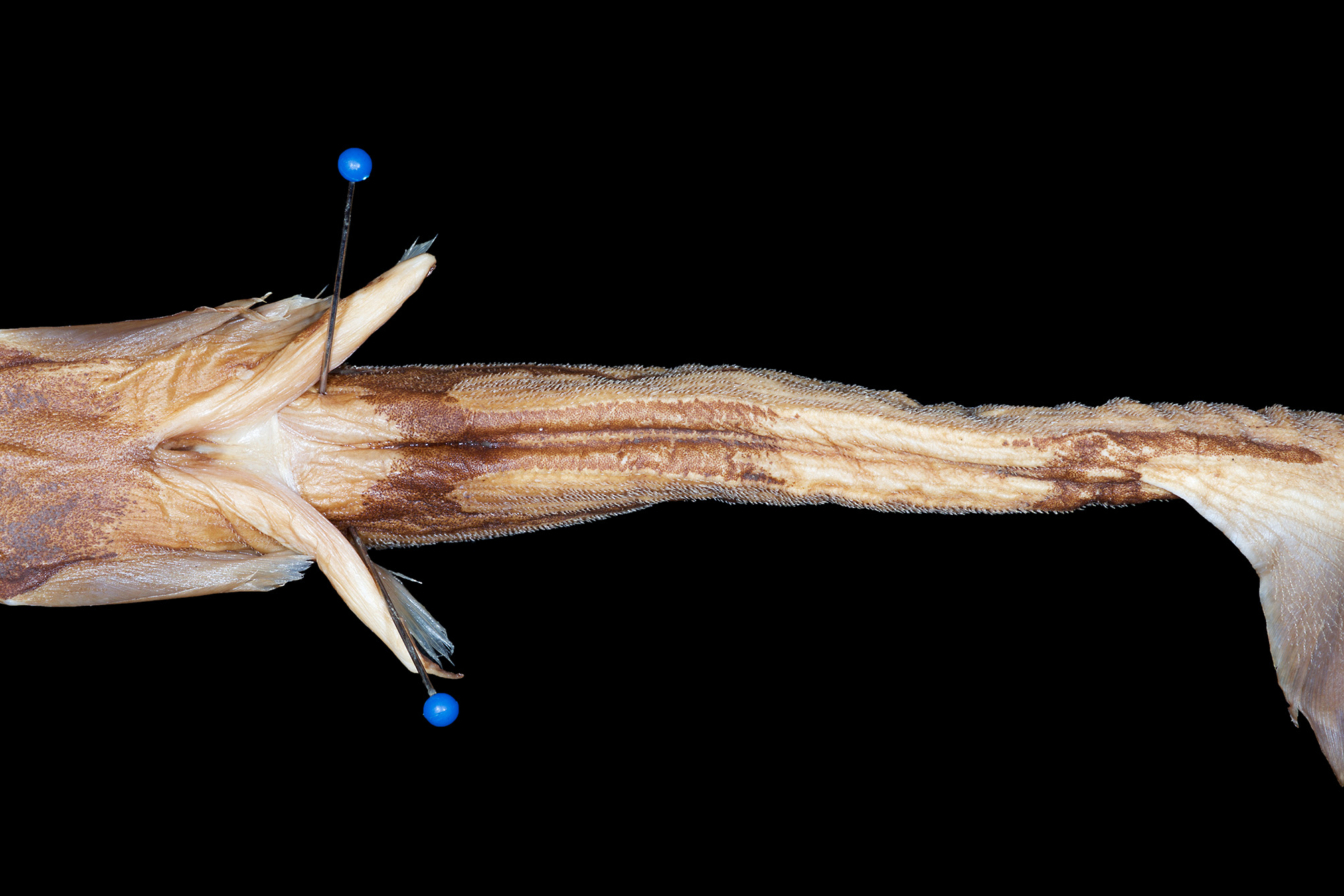
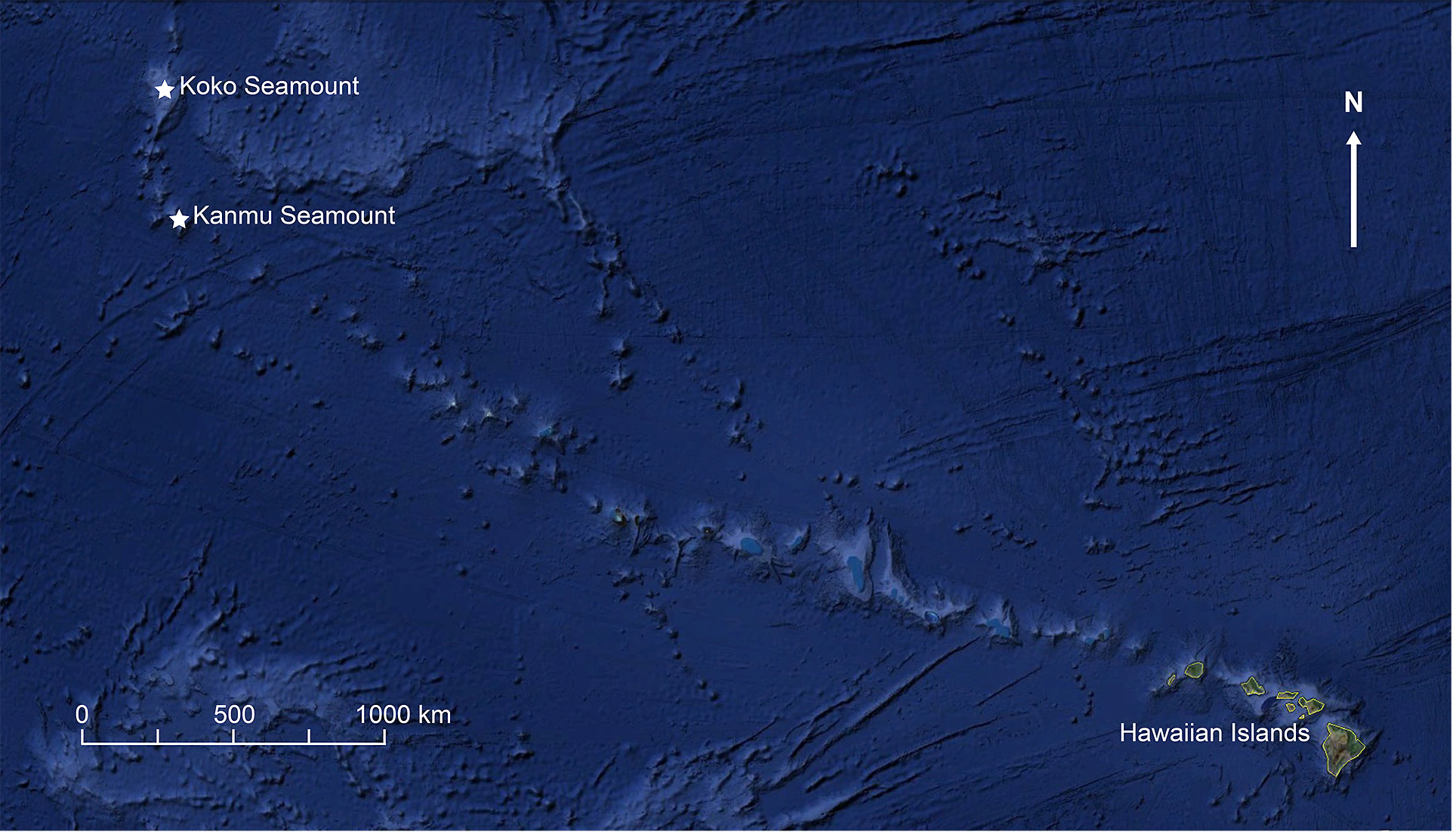
TABLE 1. Morphometrics and meristics of Etmopterus lailae holotype (BPBM 40183) and two paratypes (BPBM 40174; BPBM 40182). Values expressed as percent TL.
| Measurement | Holotype | Paratypes |
|---|---|---|
| Total length (mm) | 368 | 265–303 |
| Precaudal length | 80.4 | 75.5–78.5 |
| Prenarial length | 2.4 | 2.3–2.3 |
| Preoral length | 10.6 | 10.9–11.2 |
| Preorbital length | 6.5 | 5.3–5.9 |
| Prespiracle length | 13.0 | 12.8–13.9 |
| Pregill length | 19.0 | 18.1–18.8 |
| Head length | 14.9 | 13.9–16.2 |
| Prepectoral length | 23.9 | 21.8–22.6 |
| Prepelvic length | 53.3 | 48.3–50.8 |
| Snout–vent length | 57.6 | 52.1–54.5 |
| Pre–first dorsal fin length | 35.1 | 32.8–33.3 |
| Pre–second dorsal fin length | 61.4 | 56.6–57.4 |
| Interdorsal fin length | 22.0 | 18.1–20.1 |
| Dorsal–caudal length | 12.0 | 10.6–10.9 |
| Pectoral–pelvic length | 22.3 | 21.5–24.4 |
| Pelvic–caudal length | 17.1 | 16.2–19.2 |
| Eye length | 5.2 | 4.9–5.3 |
| Eye height | 2.7 | 3.0–3.0 |
| Interorbital length | 6.8 | 7.2–7.3 |
| Nostril width | 1.9 | 1.9–2.0 |
| Internarial length | 2.7 | 3.0–3.0 |
| Anterior nasal flap length | 1.1 | 0.7–0.8 |
| Spiracle length | 1.9 | 1.3–1.5 |
| Eye–spiracle length | 2.4 | 2.6–2.6 |
| Mouth length | 1.9 | 2.0–2.3 |
| Mouth width | 7.6 | 6.6–6.8 |
| Upper labial furrow | 1.6 | 2.3–2.3 |
| Lower labial furrow | 1.6 | 1.3–1.9 |
| First gill height | 1.9 | 1.9–2.3 |
| Second gill height | 1.6 | 1.5–2.0 |
| Third gill height | 1.1 | 1.5–1.7 |
| Fourth gill height | 1.1 | 1.3–1.5 |
| Fifth gill height | 1.1 | 1.3–1.5 |
| Head height | 7.1 | 6.8–6.9 |
| Head width | 11.1 | 9.2–10.9 |
| Pectoral fin length | 9.0 | 8.3–8.3 |
| Pectoral fin anterior margin length | 7.9 | 8.3–8.6 |
| Pectoral fin base length | 4.6 | 4.3–4.9 |
| Pectoral fin height | 6.8 | 4.6–7.2 |
| Pectoral fin inner margin length | 5.2 | 4.5–5.0 |
| Pectoral fin posterior margin length | 6.5 | 4.6–7.2 |
| Pelvic fin length | 11.1 | 8.6–10.6 |
| Pelvic fin anterior margin length | 4.9 | 5.3–6.0 |
| Pelvic fin base length | 6.5 | 5.3–6.8 |
| Pelvic fin height | 1.9 | 2.0–2.6 |
| Pelvic fin inner margin length | 5.4 | 3.3–4.2 |
| Pelvic fin posterior margin length | 6.0 | 4.0–6.4 |
| First dorsal fin length | 9.0 | 7.9–8.6 |
| First dorsal fin spine length | Broken | 3.0–3.4 |
| First dorsal fin spine exposed length | Broken | 1.7–1.9 |
| First dorsal fin anterior margin length | 3.8 | 4.5–4.6 |
| First dorsal fin base length | 5.2 | 5.3–5.3 |
| First dorsal fin height | 2.4 | 1.5–2.6 |
| BPBM |
Bishop Museum |
No known copyright restrictions apply. See Agosti, D., Egloff, W., 2009. Taxonomic information exchange and copyright: the Plazi approach. BMC Research Notes 2009, 2:53 for further explanation.
|
Kingdom |
|
|
Phylum |
|
|
Class |
|
|
Order |
|
|
Family |
|
|
Genus |