
Drepanoneura
|
publication ID |
https://doi.org/ 10.5281/zenodo.183222 |
|
DOI |
https://doi.org/10.5281/zenodo.6231934 |
|
persistent identifier |
https://treatment.plazi.org/id/038087FC-FFDC-4907-FF66-D37976C3F830 |
|
treatment provided by |
Plazi |
|
scientific name |
Drepanoneura |
| status |
|
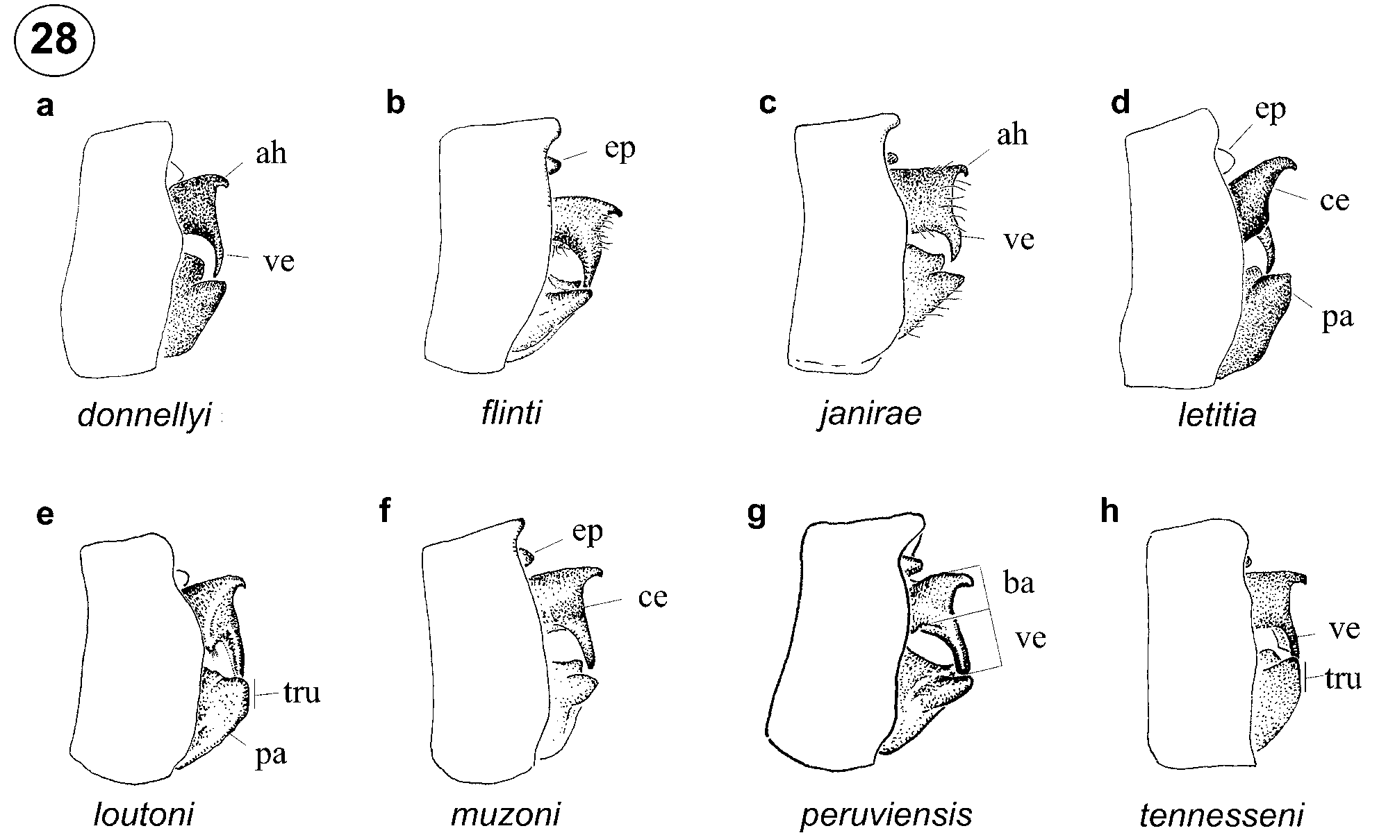
Key to males of Drepanoneura View in CoL
Caution should be used in using this key. Identification relies on subtle differences of the cerci, paraprocts, and apex of the genital ligula. Extrusion and cleaning of anal appendages with a paint brush following relaxation of specimens will more easily allow examination of the ventral branch of the cercus in lateral view. Some species pairs, D. flinti and D. peruviensis , and D. donnellyi and D. letitia , can be separated with assurance only by characters of the genital ligula. We recommend comparison with illustrations and diagnoses before determinations are considered final.
1. Ventral branch of cercus in posterior view arising at mid-width of cercus, slightly convergent to branch of opposite cercus at tip ( Figs. 30 View FIGURE 30 b, f–g)..........................................................................................................2
1'. Ventral branch of cercus in posterior view aligned with inner margin of cercus at base, slightly divergent to branch of opposite cercus at tip ( Figs. 30 View FIGURE 30 a, c–e, h).................................................................................4
2. Ventral branch of cercus in lateral view as long as base of cercus ( Fig. 28 View FIGURE 28 f); in posterior view narrowing gradually to tip, with inner side approximately linear ( Fig. 30 View FIGURE 30 f); Orellana Prov. in Ecuador to Madre de Dios Dept. in Peru ( Fig. 37 View FIGURE 37 )......................................................................................................... D. muzoni View in CoL
2'. Ventral branch of cercus in lateral view longer than base of cercus ( Figs. 28 View FIGURE 28 b, g); in posterior view narrowing abruptly at base, with inner side concave ( Figs. 30 View FIGURE 30 b, g).................................................................3
3. Apex of genital ligula with a shallow v-shaped cleft (14a); Amazonas Dept. in Colombia ( Fig. 37 View FIGURE 37 ) .......... .......................................................................................................................................................... D. flinti View in CoL
3'. Apex of genital ligula with a deep u-shaped cleft (19a); Loreto Dept. in Peru ( Fig. 37 View FIGURE 37 )...... D. peruviensis View in CoL
4. In lateral view postero-dorsal margin of S10 projected posteriorly and ventral branch of cercus shorter than base of cercus ( Fig. 28 View FIGURE 28 c); N Bolivia and W Brazil ( Fig. 37 View FIGURE 37 ) .............................................. D. janirae View in CoL
4'. In lateral view postero-dorsal margin of S10 not or only slightly projected posteriorly and ventral branch of cercus as long as or longer than base of cercus ( Figs. 28 View FIGURE 28 a, d–e, h).........................................................5
5. Apex of paraproct truncate in lateral view ( Figs. 28 View FIGURE 28 e, h)............................................................................6
5'. Apex of paraproct truncate pointed in lateral view ( Figs. 28 View FIGURE 28 a, d)...............................................................7
6. Ventral branch of cercus in lateral view cylindrical, gradually narrowing distally ( Fig. 28 View FIGURE 28 e); usually a long yellow stripe along ventral margin of humeral suture ( Fig. 2 View FIGURE 2 h); Morona-Santiago Prov. in Ecuador to Madre de Dios Dept. in Peru ( Fig. 37 View FIGURE 37 ) ........................................................................................ D. loutoni View in CoL
6'. Ventral branch of cercus in lateral view wide at base, laminar, and becoming abruptly truncate apically ( Fig. 28 View FIGURE 28 h); usually a short yellow stripe or spot at posterior end of humeral suture ( Fig. 2 View FIGURE 2 n); Sucumbíos to Napo Provs. in Ecuador ( Fig. 37 View FIGURE 37 ) ........................................................................................... D. tennesseni View in CoL
7. Apex of genital ligula approximately linear ( Fig. 13 View FIGURES 13 – 20 a) and latero-apical lobes long, narrow, and directed externally ( Fig. 13 View FIGURES 13 – 20 c); Antioquia to Tolima Depts. in Colombia ( Fig. 37 View FIGURE 37 )................................ D. donnellyi View in CoL
7'. Apex of genital ligula with a shallow v-shaped cleft ( Fig. 16 View FIGURES 13 – 20 a) and latero-distal lobes short, broad, and curved medially ( Fig. 16 View FIGURES 13 – 20 c); Panamá Prov. in Panama ( Fig. 37 View FIGURE 37 ).................................................... D. letitia


No known copyright restrictions apply. See Agosti, D., Egloff, W., 2009. Taxonomic information exchange and copyright: the Plazi approach. BMC Research Notes 2009, 2:53 for further explanation.