
Electrogena gridellii ( Grandi, 1953 )
|
publication ID |
https://doi.org/ 10.11646/zootaxa.4362.3.3 |
|
publication LSID |
lsid:zoobank.org:pub:8530FD3C-A0FC-4092-A7C3-E50BC5030F48 |
|
DOI |
https://doi.org/10.5281/zenodo.6051313 |
|
persistent identifier |
https://treatment.plazi.org/id/03CDF150-FFC2-9611-75A5-466CFBDAF9BE |
|
treatment provided by |
Plazi |
|
scientific name |
Electrogena gridellii ( Grandi, 1953 ) |
| status |
|
Electrogena gridellii ( Grandi, 1953) View in CoL
Heptagenia gridellii: Grandi 1953 male and female imagines Ecdyonurus gridellii: Belfiore 1981 lectotype designation Ecdyonurus gridellii: Belfiore 1982 nymph description
nec Ecdyonurus gridellii sensu Zurwerra & Tomka 1984
nec Ecdyonurus gridellii sensu Sartori & Dethier 1985
Electrogena gridellii: Zurwerra & Tomka 1985 View in CoL new combination nec Electrogena gridellii sensu Sartori 1987 View in CoL
Electrogena gridellii: Gaino et al. 1987 View in CoL egg description
Electrogena vipavensis: Zurwerra & Tomka 1986 View in CoL female imago, male subimago, nymph exuvia, egg NEW SYNONYM
Nymph description. Length: Male: body 8.5– 12 mm, cerci: 11–16 mm. Female: body 12–14 mm, cerci: 13–17 mm. General colouration dull, little contrasted.
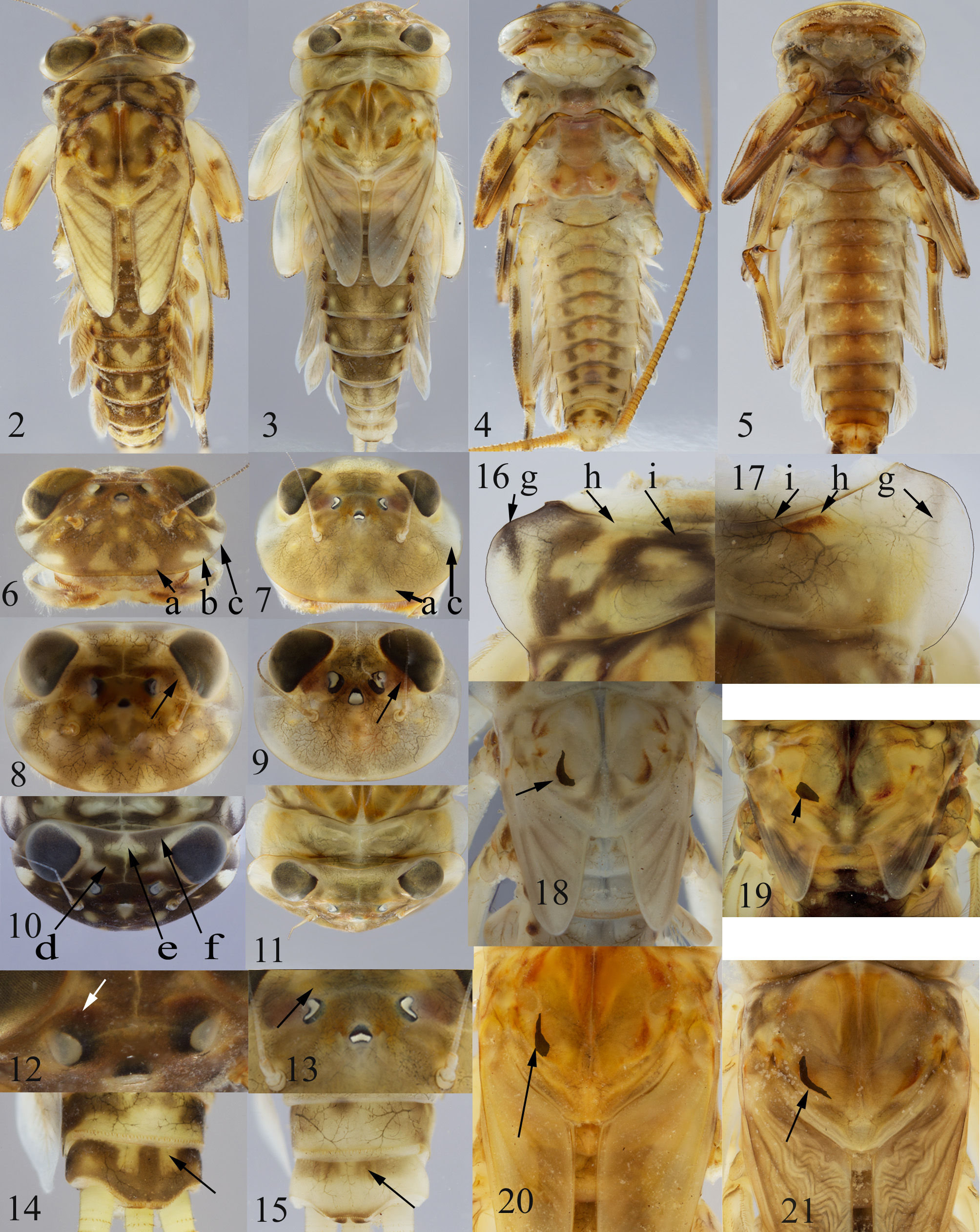
Head: surface little contrasted ( Figs 7, 9, 11, 13 View FIGURES 2–21 ); noteworthy the presence of a double sagittal faint (sometimes lacking) triangular marking on the anterior margin of head (a) and a lateral light zone ahead the compound eyes (c) ( Fig. 7 View FIGURES 2–21 ); no contrasted pattern on the posterior part ( Fig. 11 View FIGURES 2–21 ), only a greyish band on the posterior margin; a dark marking touching the compound eyes, no colouration behind the ocelli ( Figs 9, 13 View FIGURES 2–21 ). Labrum rather slender R_LBR = 4.2–4.6. Mandible with a prostheca bearing 9–20 bristles (N_PRO), the first bristles thick, then the size of them tapering, often ending with very tiny hairy bristles (not counted) ( Fig. 43 View FIGURES40–51 ). Addition of right and left N_PRO: 22–39 bristles. Maxillae with a row of 19–26 comb-shaped setae on anterior margin (N_CBS), the 5th (starting from inner side) with 10–15 teeth (N_TCB), outer margin of galea-lacinia bearing 11–43 setae (N_OUT), inner margin and ventral face of the first segment of palpus bearing 24–46 thin setae (N_PLP). Hypopharynx with laterally expanded superlinguae, densely covered with long thin setae, apex of lobes with long setae ( Fig. 49 View FIGURES40–51 ), sometimes missing when worn or very difficult to see because obscured by dirt as E. brulini in Fig. 48 View FIGURES40–51 . Labium, ventral face, bristles reaching about half of the width of the paraglossae ( Fig. 47 View FIGURES40–51 ), inner distal margin can be concave, R_GLA = 3.1–5.2, R_GLB = 2.5–3.0.
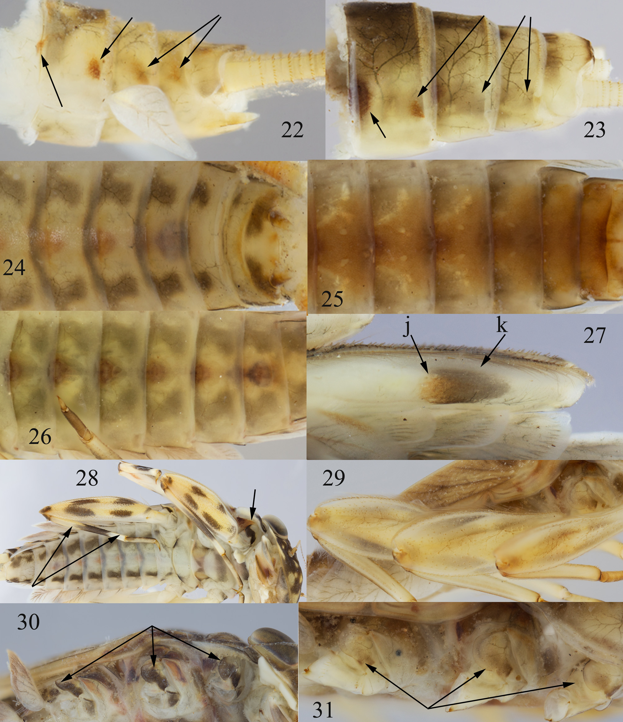
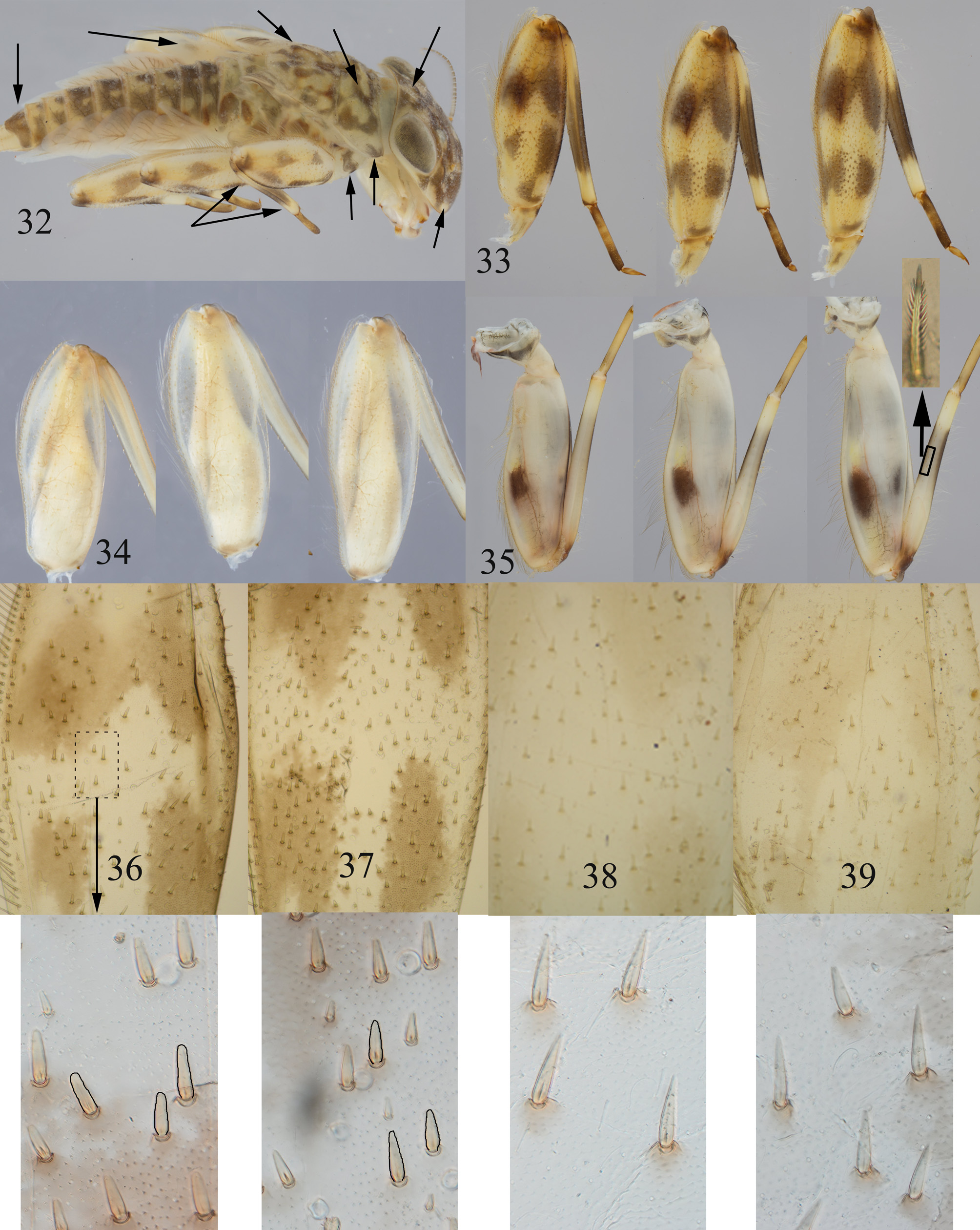
Thorax: Pronotum without a pattern, faint, but presence of a reddish-brown spot (h), usually hidden by the head, the lateral margins regularly rounded with a faint greyish line (g) ( Fig. 17 View FIGURES 2–21 ). Mesonotum with several light brown markings and an oval-shaped spot in young instars (arrow in Fig. 19 View FIGURES 2–21 ), becoming an oblique semilunar marking (arrow in Fig. 21 View FIGURES 2–21 ). Coxa little contrasted: proximal half white, distal part greyish ( Fig. 31 View FIGURES 22–31 ). Thoracic sterna: sclerites light, imaginal colouration becoming brown during nymph development. Legs: faint ornamentation rather similar between fore-, mid and hind legs ( Figs 29 View FIGURES 22–31 , 34 View FIGURES 32–39 ). Femora dorsal face with four (some can be joined) less contrasted brownish spots delimiting an often incomplete light cross, the weak contrast disappearing in the last instars ( Fig. 29 View FIGURES 22–31 ); ventral face of femora very rarely with a greyish cuticular suffusion in last instars; all femora with long, narrow and pointed bristles on the dorsal face ( Figs 38, 39 View FIGURES 32–39 ); only one (can miss) strong spine in the middle of the ventral face of all femora, near the outer margins (N_BVF). Tibia faint, when contrasted, only the apex lighter; pectinate setae scarce on foretibia, numerous on mid and hind tibia (as E. brulini Fig. 35 View FIGURES 32–39 , inset). Tarsi distal quarter slightly darkened (mainly on dorsal face), exceptionally forming a less or more visible ring on distal part, colouration not strengthening toward the proximal part; claws with 2 teeth, rarely 1 or 3, exceptionally 0 or 4 (N_CLW).
Abdomen: Terga with little contrasted markings: terga (III) IV–VI (VII–VIII) anterior margin: grey slender triangle in the middle ( Fig. 3 View FIGURES 2–21 ); terga (IV–VI and VIII–X) posterior margin: grey line, not visible when fused with entirely dark terga; terga II–VIII anterolateral epidermal brown spot larger than the posterolateral one ( Fig. 23 View FIGURES 22–31 ), anterolateral spot hidden under the preceding terga in small nymphs, growing up to last instar ( Figs 23 View FIGURES 22–31 ); terga II– VII posterolaterally with a reddish-brown spot ( Fig. 23 View FIGURES 22–31 ); in the last instars, tergum VII (darker) contrasting with terga VIII–X ( Fig. 3 View FIGURES 2–21 ), tergum X anterior half brownish with a light area in the middle, posterior half white with only a slight line on the posterior margin ( Fig. 15 View FIGURES 2–21 ); posterior margin of each tergum with thin and elongated spines with microdenticulations at their bases. Sterna: no cuticular colouration except a light greyish stain, epidermis light in young nymphs, becoming brown in last instars ( Fig. 25 View FIGURES 22–31 ); ganglionic chain brownish pigmented, thoracic ganglia a bit darker, in last instars, ganglia hardly visible due to the dark epidermal colouration ( Fig. 25 View FIGURES 22–31 ). In the male last instar nymphs, styliger processes and titillator already visible through sternum 9, but titillators not enough formed to be diagnostic. Gill I tongue-shaped, gills II–VI broad and asymmetrical, gill VII elongated, narrow at apex and slightly pointed ( Fig. 51 View FIGURES40–51 ). Paracercus and cerci light, becoming darker only in the last instars, colouration fading apically.
Male imago: Length: body 11–13 mm, forewing 12–14 mm, cerci 31–36 mm.
Head: Eyes clearly separated, sclerite joining the compound eyes W-shaped, generally well-marked ( Fig. 85 View FIGURES68–85 ).
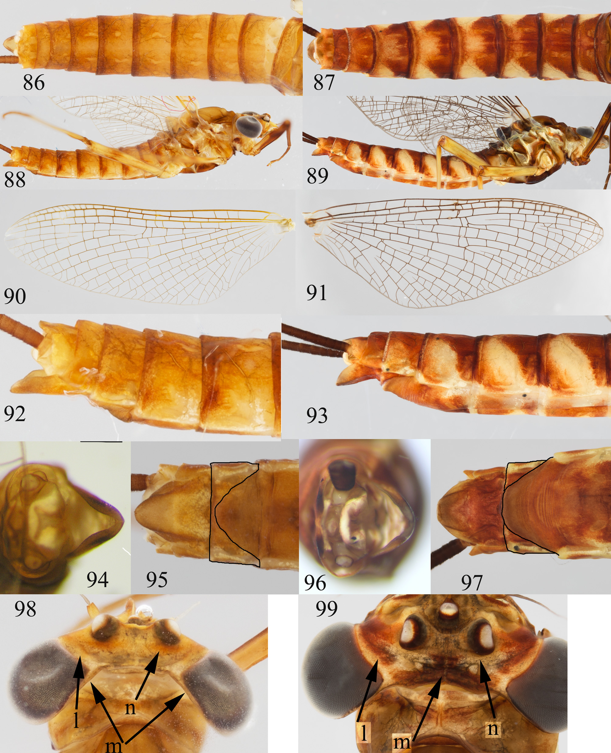
Thorax: dark brown ( Figs 53, 55 View FIGURES52–55 ). Wings veins all dark brown ( Fig. 65 View FIGURES 56–67 ). Central part or entire forefemora darkened ( Figs 62–63 View FIGURES 56–67 ); mid and hind femora dorsally with a very light transversal brown band ( Fig. 62 View FIGURES 56–67 ); ventral face at most with a barely visible band ( Fig. 63 View FIGURES 56–67 ). Abdomen: Terga II–VII (VIII) anterior margin brown with a pair of large subtriangular markings pointed backwards reaching or not the posterior margin and a dark marking between them, posterior margin darkened (as female in Fig. 87 View FIGURES 86–99 ), these markings can be fused, covering the dorsal surface ( Fig. 53 View FIGURES52–55 ); in lateral view, the subtriangular markings are fused with the anterolateral and the posterolateral markings of the preceding tergum building a large oblique band ( Fig. 55 View FIGURES52–55 ). Sterna pale with a large subtriangular light orange-brown marking covering more or less the entire face, ganglia violet ( Fig. 59 View FIGURES 56–67 ). Cerci brown, colouration fading to the apex. Penis lobes subrhomboidal, narrowed towards the apex, slightly bent inwards ( Fig. 69 View FIGURES68–85 ), only a few small dorsal spines basolaterally on penis lobes ( Fig. 73 View FIGURES68–85 ). Titillator convex before the unique tooth, than concave until a more or less visible basal tip ( Figs 79–81 View FIGURES68–85 ). Styliger almost straight with lateral humps (as E. brulini on Fig. 66 View FIGURES 56–67 ), but humps can be very inconspicuous as in Fig. 67 View FIGURES 56–67 .
Female imago: Length: body: 11–14 mm, forewing: 14–17 mm, cerci: 25–32 mm.
Same general colouration as in male; only thorax somewhat lighter ( Figs 87, 89 View FIGURES 86–99 ). Wings veins all medium brown ( Fig. 91 View FIGURES 86–99 ). Light area behind the pair of ocelli (n), area between the pair of ocelli and the compound eyes dark (l), sclerite joining the compound eyes with an extended dark brown area ( Fig. 99 View FIGURES 86–99 , m). Subgenital plate covering ¾ of the sternum VIII surface, subanal plate in ventral and apical view rounded ( Figs 96, 97 View FIGURES 86–99 ).
Male and female subimago: Same colouration as imago, but paler. Wings without pattern, entirely grey.
Egg: Dimensions: 155/120 µm; micropyle: 11/6.5 µm.
Chorion entirely and regularly covered with relatively dense microgranules, KCT’s evenly distributed over the surface, more concentrated only on a very restricted area of one pole ( Fig. 102 View FIGURES 100–103 ). Micropyle bordered with the same microgranules as surface ( Fig. 103 View FIGURES 100–103 ).
Diagnosis. Nymph: general colouration faded, femora without spot on the ventral face, two teeth on tarsal claws; (0) 1 bristle on ventral face of femora, near the outer margin; bristles of femora long, thin and pointed; prostheca consisting in 9–20 bristles ( Fig. 43 View FIGURES40–51 ), terga anterolateral spot smaller than the posterolateral one ( Fig. 23 View FIGURES 22–31 ).
Imago: dorsal face of mid and hind femora with an inconspicuous band ( Fig. 62 View FIGURES 56–67 ); ventral face of mid and hind femora without colouration ( Fig. 63 View FIGURES 56–67 ); wings veins dark brown in male ( Fig. 65 View FIGURES 56–67 ), medium brown in female ( Fig. 91 View FIGURES 86–99 ); terga well contrasted, white and dark brown, posterolateral and anterolateral spots well visible forming an oblique streak ( Figs 55 View FIGURES52–55 , 57 View FIGURES 56–67 , 89 View FIGURES 86–99 ); titillators with 1 apico-lateral tooth ( Figs 79–81 View FIGURES68–85 ).
Although many differences in colorations make it possible to easily separate E. brulini from E. gridellii , the two species have always been confused because the value of colouration characters is underrated (or because of the use of old faded specimens); therefore those characters are generally omitted. Among colouration characters are true patterns, but also simple colourations which seem to have even less taxonomic importance. However, some of these are conservative characters, visible both in nymphs and adults, and have been positively tested as to their discriminating value to separate E. brulini from E. gridellii .
Only the discriminating characters between E. gridellii and E. brulini tested and easily usable in the vast majority of cases are presented here.
At the nymphal stage:
- all colourations of E. brulini contrasted, those of E. gridellii very dull.
- pattern and different markings contrasted on the head in E. brulini ( Figs 6,8,10 View FIGURES 2–21 , a–f), no pattern and only some pale markings (notice the absence of spot (b) ) in E. gridellii ( Figs 7, 9, 11 View FIGURES 2–21 ).
- each prostheca consisting in 5-8 bristles (all bristles evenly thick, at the most the basal one a little thinner) in E. brulini ( Fig. 42 View FIGURES40–51 ), 9–20 bristles (the first bristles thick, then the size of them tapering, often ending with very tiny hairy bristles) in E. gridellii ( Fig. 43 View FIGURES40–51 ).
- pronotum with a contrasted pattern but no reddish-brown spot (h), the lateral margins with a dark line (g) in E. brulini ( Fig. 16 View FIGURES 2–21 ), faint, but presence of a reddish-brown spot (h), usually hidden by the head, the lateral margin with a faint greyish line (g) in E. gridellii ( Fig. 17 View FIGURES 2–21 ).
- mesonotum with brown semilunar vertical markings in all instars in E. brulini (arrow in Figs 18, 20 View FIGURES 2–21 ), ovalshaped spot becoming an oblique semilunar marking in E. gridellii ( Figs 19, 21 View FIGURES 2–21 ).
- femora ventral face always with a visible dark (generally double) spot in E. brulini ( Fig. 27 View FIGURES 22–31 ), at most with a greyish cuticular suffusion in the last instars in E. gridellii .
- bristles of dorsal face medium sized blunt pointed on forefemora, more peaked in mid and hind femora in E. brulini ( Figs 36, 37 View FIGURES 32–39 ), thin, long and pointed on all femora long in E. gridellii ( Figs. 38, 39 View FIGURES 32–39 ).
- coxa bicoloured, proximal half white, distal half dark grey in E. brulini ( Figs 28, 30 View FIGURES 22–31 , 32 View FIGURES 32–39 ), little contrasted in E. gridellii ( Fig. 31 View FIGURES 22–31 ).
- tibia contrasted, distal quarter and proximal third light, middle darker in E. brulini ( Fig. 28 View FIGURES 22–31 , 32, 33 View FIGURES 32–39 ), entirely faint, when contrasted, only the apex lighter in E. gridellii .
- tergum X dark with a light marking on each side in E. brulini ( Fig. 14 View FIGURES 2–21 ), anterior half brownish with a light area in the middle, posterior half white with only a slight line on the posterior margin in E. gridellii ( Fig. 15 View FIGURES 2–21 ).
- sterna with an arch-shaped cuticular pattern, epidermis always entirely light in E. brulini ( Fig. 24 View FIGURES 22–31 ), without cuticular pattern, epidermis light in young nymphs, becoming brown in last instars in E. gridellii ( Fig. 25 View FIGURES 22–31 ).
At imaginal stages:
- general colouration of abdomen uniform in E. brulini ( Figs 56 View FIGURES 56–67 , 86 View FIGURES 86–99 ), contrasted in E. gridellii ( Figs 57 View FIGURES 56–67 , 87 View FIGURES 86–99 ).
- wings veins yellowish-brown; costa, subcosta and radius anterior brighter yellow in E. brulini ( Figs. 64 View FIGURES 56–67 , 90 View FIGURES 86–99 ), regularly dark brown in male, medium brown in female in E. gridellii ( Figs 65 View FIGURES 56–67 , 91 View FIGURES 86–99 ).
- mid and hind femora dorsally with a brown spot at the distal third and a second spot, variably visible, in the proximal third in E. brulini ( Fig. 60 View FIGURES 56–67 ), with a very light transversal brown band in E. gridellii ( Fig. 62 View FIGURES 56–67 ).
- mid and hind femora ventrally with a brown spot at the distal third in E. brulini ( Fig. 61 View FIGURES 56–67 ), at most with a barely visible band in E. gridellii ( Fig. 63 View FIGURES 56–67 ).
- male: sclerite joining the compound eyes weakly rounded forming a wide V in E. brulini ( Fig. 84 View FIGURES68–85 ), W-shaped in E. gridellii ( Fig. 85 View FIGURES68–85 ).
- male: titillator massive, straight or concave before the two teeth (rarely one or three), but no basal tip in E. brulini ( Figs 75–78 View FIGURES68–85 ), convex before the unique tooth, than concave until a more or less visible basal tip ( Figs 79–81 View FIGURES68–85 ).
- female: subgenital plate covering about half of sternum VIII surface in E. brulini ( Fig. 95 View FIGURES 86–99 ), covering ¾ of the sternum in E. gridellii ( Fig. 97 View FIGURES 86–99 ).
- female: subanal plate tapering to apex, in apical view narrow and lateral margins rolled up towards the inside in E. brulini ( Figs 94, 95 View FIGURES 86–99 ), subanal plate in ventral and apical view rounded in E. gridellii ( Figs 96, 97 View FIGURES 86–99 ).
At both stages:
- nymph pattern ( Fig. 10 View FIGURES 2–21 , d - f) disappearing, only narrow coloured lines remaining laterally on posterior margin in female imagines in E. brulini ( Fig. 98 View FIGURES 86–99 , m), nymph posterior part of the head with only a greyish band (so faint that it is not visible on photos) on the posterior margin, becoming a large centered brown band in female imagines in E. gridellii ( Fig. 99 View FIGURES 86–99 , m).
- a light line along the inner margins of compound eyes, a dark colouration behind the ocelli ( Figs 8, 12 View FIGURES 2–21 ); characters still present in female imagines in E. brulini ( Fig. 98 View FIGURES 86–99 , 1 View FIGURE 1 , n), a dark marking touching the compound eyes, no colouration behind the ocelli ( Figs 9, 13 View FIGURES 2–21 ); characters still present in female imagines in E. gridellii ( Fig. 99 View FIGURES 86–99 , 1 View FIGURE 1 , n).
- terga anterolateral spot small and hidden, posterolateral bigger in E. brulini ( Figs 22 View FIGURES 22–31 , 54 View FIGURES52–55 , 88, 92 View FIGURES 86–99 ), anterolateral spot large, posterolateral spot little developed in nymphs, grows and merges with dorsal coloration in E. gridellii imagines ( Figs 23 View FIGURES 22–31 , 55 View FIGURES52–55 , 89, 93 View FIGURES 86–99 ).
Rationales for the proposed synonymy. The type material of E. vipavensis currently housed in MZL is composed of the following items:
- the holotype, a male subimago (N° 4155) with corresponding exuvia on slide (N° 4155_D_a_male). Subimago: sclerite joining the compound eyes W-shaped, styliger straight with lateral humps, all the colouration faded, but the drawings in the original description fits with our contrasted E. gridellii subimagines: anterior margin of terga III–V with a pair of subtriangular markings pointed backwards and a spot between them, posterior margin darkened, posterolateral part of terga III–V with a dark marking near the border fused with the anterior margin marking of next tergum, an oval white area oblique from the anterolateral corner. Exuvia: all femora with long, narrow and pointed bristles on the dorsal face, N_BVF: 0, N_CLW: 2, N_PRO: 11 / 11. According to all observed characters we found no difference between the holotype and E. gridellii .
- paratype female abdominal segments VII–X (N° 4175) with corresponding exuvia on slide (N° 4175_a_fem) (assumed to be the paratype drawn in the original description because of the precision « fem » on the exuvia slide) and the forewings on slide (N° 4175_b). According to the original drawing, the pattern of the terga III–V in lateral view is composed by the following elements: a large oval white area oblique from anterolateral corner, an anterolateral spot, posterolateral spots, a lateral dark streak at base of terga, central part of the subanal plate tapering towards apex. Exuvia: all femora with long, narrow parallel and truncate bristles on the dorsal face, N_BVF: numerous, N_CLW: 2, N_PRO: 9 / 9. According to all observed characters we found no difference between this paratype and E. ujhelyii except that the 2–3 posterolateral spots of the original drawing are usually fused in E. ujhelyii .
- paratype female abdominal segments VII–X (N°4291) with corresponding exuvia on slide (slide 4291_a). Exuvia: all femora with long, narrow and pointed bristles on the dorsal face, N_BVF: 0, N_CLW:2, N_PRO: 9. New identification: E.gridellii .
- paratype female abdominal segments VII–X (N° 4223). Central part of the subanal plate tapering towards apex, but not sufficient for a secure identification: E. cf ujhelyii .
- paratype female abdominal segments VII–X (N° 4198) plus a second sample with 5 legs and 1 forewing (same number N° 4198). Not sufficient for identification: Electrogena sp.
Together with the type material and from the same sampling data: one exuvia named E. ozrensis on slide (N° 4167_a). All femora with long, narrow and pointed bristles on the dorsal face, N_BVF: 0, N_CLW:2, N_PRO: 14. New identification: E. gridellii .
Our examination of the type material of E. vipavensis indicates that the holotype, male subimago (N° 4155) and its exuvia on slide (N° 4155_D_a_male) and the female paratype exuvia on slide (N° 4291_a) are conspecific with E. gridellii . Therefore, we propose that Electrogena vipavensis Zurwerra & Tomka, 1986 should be considered as a subjective junior synonym of Electrogena gridellii ( Grandi, 1953) syn. nov.
After comparison of the original drawing with our material and re-investigation of the female paratype exuvia slide (N° 4175_a_fem), we consider that this specimen is conspecific with E. ujhelyii .
Material examined: 97 male imagines, 45 female imagines, 8 nymphs, 140 nymph exuviae, 31 male subimagines, 39 female subimagines: IT / FV, San Dorligo della Valle, Rosandra , Bagnoli Superiore , 45.6195°N 13.8654°E 91 m, 9.IV.2015 (emerged 9.IV–23.V.2015); 75 nymphs: same locality, 7.IV.2015; 11 nymphs: IT / FV, Povoletto , Via Reclusane , 46.2658°N 13.3126°E 158 m, 6.IV.2015; 1 nymph: IT / FV, Attimis , Rio Valle tributary, 46.1748°N 13.3211°E 177 m, 6.IV.2015; 6 nymphs: IT / FV, Attimis , Torrente Racchiusano , 46.1733°N 13.3330°E 230 m, 6.IV.2015; 1 nymph: IT / FV, Stregna , Torrente Erbezzo , 46.1287°N 13.5699°E 232 m, 6.IV.2015; 1 nymph: IT / FV, Prepotto , Fiume Judrio , 46.0826°N 13.5434°E 145 m, 6.IV.2015; 15 nymphs: IT / FV, Prepotto , Fiume Judrio tributary, 46.0827°N 13.5427°E 147 m, 6.IV.2015; 1 nymph: SL / IS, Koper, Rizana , 45.5328°N 13.8729°E 66 m, 7.IV.2015, A. Wagner leg. [ MZL]; 1 nymph: IT / FV, Manzano, Fiume Natisone basin, Oleis, 46.02°N 13.38°E 75 m, F. Desio leg, E GoogleMaps . Bauernfeind det. [NHMW]
E. vipavensis : one male subimago holotype (N° 4155 with exuvia on slide), 4 female paratypes, SL / GO, Ajdovscina, Stream Vipava, 16.IV.1983, Hefti, Tomka & Zurwerra leg. [ MZL]


| MZL |
Musee Zoologique |
No known copyright restrictions apply. See Agosti, D., Egloff, W., 2009. Taxonomic information exchange and copyright: the Plazi approach. BMC Research Notes 2009, 2:53 for further explanation.
|
Kingdom |
|
|
Phylum |
|
|
Class |
|
|
Order |
|
|
Family |
|
|
Genus |
Electrogena gridellii ( Grandi, 1953 )
| Wagner, Andre, Vuataz, Laurent & Sartori, Michel 2017 |
Electrogena gridellii sensu
| Sartori 1987 |
Electrogena gridellii:
| Gaino et al. 1987 |
Electrogena vipavensis
| : Zurwerra & Tomka 1986 |
Ecdyonurus gridellii sensu
| Sartori & Dethier 1985 |
Electrogena gridellii:
| Zurwerra & Tomka 1985 |
Ecdyonurus gridellii sensu
| Zurwerra & Tomka 1984 |
Ecdyonurus gridellii:
| Belfiore 1982 |
Ecdyonurus gridellii:
| Belfiore 1981 |
Heptagenia gridellii:
| Grandi 1953 |