
Dyscophus insularis, Grandidier, 1872
|
publication ID |
https://doi.org/ 10.1111/j.1096-3642.2007.00329.x |
|
DOI |
https://doi.org/10.5281/zenodo.10544919 |
|
persistent identifier |
https://treatment.plazi.org/id/039D87F8-FFB8-FFFA-76D0-FC457EB2CD96 |
|
treatment provided by |
Felipe |
|
scientific name |
Dyscophus insularis |
| status |
|
DYSCOPHUS INSULARIS View in CoL GRANDIDIER, 1872
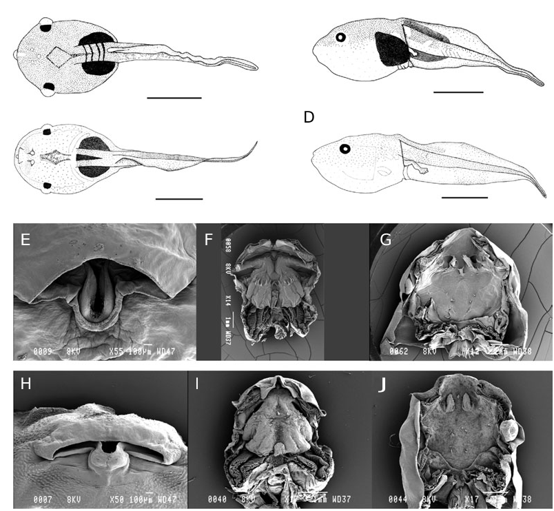
Specimens were collected from an ephemeral breeding pool in the Kirindy forest. This pool was large (> 1000 m 2), with a depth of 80 cm and clear water. A large proportion of the pond area was covered with standing, floating and submerged aquatic vegetation. The external morphological description is based on a specimen at stage 34 (TL and BL are 26.9 and 9.5 mm, respectively) included in batch ZSM 402/2004. Buccopharyngeal features are described on the basis of a tadpole at stage 38 from the same batch.
External morphology: In dorsal view ( Fig. 3A View Figure 3 ), body roughly ovoid, widest at the level of eyes, snout truncate. In profile ( Fig. 3B View Figure 3 ), body depressed, BW 118% of BH, snout very small, round and almost acute. Eyes moderately sized, ED 13% of BL, bulging, visible in ventral view, positioned dorsolaterally and directed almost laterally. Nares not open, positioned dorsally, closer to snout than to pupils, RN 67% of NP, very close to each other, NN 21% of PP. Spiracle ventral, a large fold of skin free at the rear of the spiracular tube in the form of a half-circle, not attached to body wall, orientated posteriorly, very close to ventral tube, SS 89% of BL. Tail musculature moderately weak in the proximal third to weak in the distal two-thirds, TMH 32% of BH and 39% of MTH, the proximal quarter parallel then slightly tapering, almost reaching tail tip. Upper fin moderately high, UF 41% of MTH, convex, extending slightly onto body, SU 84% of BL, lower fin shallow, LF 34% of MTH, straight on the most part; point of maximum height of tail located just after the ventral tube opening, MTH 83% of BH, tail tip a flagellum. Ventral tube small, tubular, medial, directed posteroventrally, its anterior part linked to body wall, its posterior part linked to ventral tail fin, opening medial. Neither lateral line organs nor glands visible.
Oral disc ( Fig. 3E View Figure 3 ) moderately large, in position and orientation terminal, not emarginated, ODW 28% of BL and 36% of BW. Upper labium as a large flap of skin hanging down the lower labium, concave medially; lower labium flat with a large U-shaped extension medially covered in great part by upper labium, only the extremity of U going beyond it. No papillae, no denticulate papillae. No keratodonts. No jaw sheaths.
Coloration in preservative: Upper side uniformly punctuated with small dark brown spots. Upper labium coloured as the back. Upper part of flanks coloured as the back except an unpigmented area dorsolaterally at the back of body. Lower part of flanks and ventral side immaculate. Upper part of caudal muscle coloured as the back but less regularly, some rare spots on the lower part. Fins immaculate except the external half of the proximal part of the upper fin. A bright white area encompassing the caudal muscle and half of fins in the proximal third. Hindlimbs immaculate.
Variation: The ratios taken on seven tadpoles at stages 33–38 vary in the following proportions: BW 120–141% of BH; RN 36–89% of NP; NN 12–18% of PP; SS 87–94% of BL; TMH 29–43% of BH; TMH 24– 46% of MTH; UF 28–42% of MTH; LF 29–40% of MTH; SU 84–96% of BL; MTH 86–141% of BH; ODW 23– 37% of BL; ODW 29–41% of BW.
Buccal floor ( Fig. 3F View Figure 3 ): Buccal floor wider than long, its maximum width at the level of the buccal pockets. Prelingual arena very short and narrow; two pairs of small pustules anterolaterally and two pairs of small prelingual papillae posterolaterally; part immediately posterior to prelingual arena vertical, forming a deep depression. Tongue anlage prominent, elongate, without lingual papillae, lying at the bottom of the depression. Buccal floor arena round, delimited by a papilla medially to buccal pocket and posteriorly by a transversal row of about 15–20 large papillae of equal size; interior of arena smooth except a few small papillae just anterior to the row of buccal floor arena papillae; a transversal row of two or three papillae between buccal pocket and tongue anlage. Buccal pockets wide, deep, almost transversely to obliquely orientated, unperforated; four prepocket papillae, one on the edge of the anterior wall of the buccal pocket, orientated posteriorly and above the buccal pocket; about ten small postpocket papillae. Glottis just posterior to the
A
C
B
row of buccal floor arena papillae, anterodorsal in orientation, far ahead of the end of the ventral velum. Ventral velum wide with spicular support, bearing an indistinct projection above each filter cavity (the most medial being the better defined); velum interrupted by the laryngeal anlage; secretory pits not visible. Branchial baskets almost straight, anteroposteriorly directed, longer than wide, with three filter cavities, filter plates almost vertically arranged, filter mesh very dense with tertiary folds.
Buccal roof ( Fig. 3G View Figure 3 ): Prenarial arena wide and pentagonal, with 4–5 pustules. Choanae large, round; anterior wall slightly elevated, pustulate, without papilla; narial valve greatly enlarged into an anteromedially large and elongate structure with three projections distally, anteromedially and dorsally directed, covering the posterior end of the choana. Postnarial arena small, without ornamentation. Median ridge depressed anteroposteriorly, long and thin bearing three digitations on top, directed ventrally. One pair of elongate and smooth lateral ridge papillae, posterolateral to narial valves. Buccal roof arena non-existent, a very few and small pustulations scattered within, three pairs of very small buccal roof arena papillae. Posterolateral ridges few prominent, present through the buccal roof, their lateral ends laying relatively far anteriorly. Glandular zone present at least laterally; secretory pits not visible. Dorsal velum continuous, medial part straight, directed ventrally. Two pressure cushions on each side.
No known copyright restrictions apply. See Agosti, D., Egloff, W., 2009. Taxonomic information exchange and copyright: the Plazi approach. BMC Research Notes 2009, 2:53 for further explanation.