
Dugesia sinensis Chen & Wang, 2015
|
publication ID |
https://doi.org/ 10.11865/zs.20150301 |
|
publication LSID |
lsid:zoobank.org:pub:33F3BEB5-B79A-45DF-9681-AA5FCB90A3CC |
|
DOI |
https://doi.org/10.5281/zenodo.5542197 |
|
persistent identifier |
https://treatment.plazi.org/id/038887A8-330D-C454-FF28-23AE7B43FC6B |
|
treatment provided by |
Carolina |
|
scientific name |
Dugesia sinensis Chen & Wang |
| status |
sp. nov. |
Dugesia sinensis Chen & Wang , sp. nov.
Locality. Specimens were collected from the ventral side of the pebbles in the rivulet (0.4 m in maximum depth, 25 m in width) beside the Caoxi spring resort, Shaoguan, Guandong Province, China (24°39′42″N, 113°36 ′ 50 ″ E; elev. 75 m). There are plenty of pebbles in the bottom of the rivulet. Wheel animalcule and different insect’s larvae can be observed in the water samples GoogleMaps .
Material examined. Holotype PLA-0090-1–21, serial section vertically, rivulet (0.4 m in maximum depth, 25 m in width) beside Caoxi Spring Resort , Shaoguan, Guandong Province, China (24°39 ′ 42 ″ N, 113°36 ′ 50 ″ E; elev. 75 m) GoogleMaps . Paratypes: PLA -0091-1–9, PLA-0092-1–5, PLA-0093-1–4, serial section horizontally, same data as holotype. Other materials: PLA- 0094–0098, the whole body is fixed in 10% formaldehyde, same data as holotype. All specimens are deposited in IZCAS, Beijing, China GoogleMaps .
Etymology. The species is referring to its type locality, China.
Diagnosis. The new species is characterized by the following features: triangle-shaped head, two hyperplastic ovaries, asymmetric opening of oviducts, bursal canal to the left of the copulatory apparatus, a distance between copulatory bursa and the mouth, developed diagram in the male atrium.
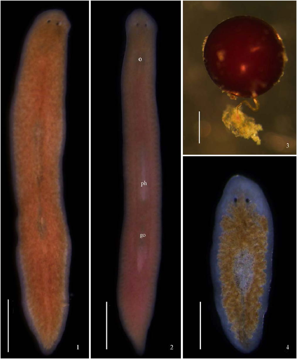
Description. Body size of the living mature individuals ranges from 12.0–15.0 mm in length and 2.48 mm in width. Dorsal side of body uniformly light brown while ventral side pale. Margin of body and areas of copulatory apparatus appear gray in color. Mobilizable triangle head has two blunt-pointed auricles. Bar-shape unpigmented auricular grooves locate posteriorly to two kidney-shaped eyes. Fan-shaped unpigmented area placed just at the lateral part of eyes ( Figs 1–2 View Figs 1–4 ). Mouth opens at 1/3 rear part of body and distance between mouth and gonopore 3.0– 3.3 mm in living individuals (1.6–2.0 in fixed samples).
Habits. Newly collected individuals cannot adapt to the environment in the laboratory and refuse to eat either the pork liver slices or cooked egg yolk, resulting in disintegration. After using the mineral water to rear, the animals start to ingest the pork livers. In the daytime, they like to flock under the ventral side of the granite while moving slowly along the wall of the glass bowl during the night. When preying, they stretch out the pharynx and swallow the pork liver tissue, then the intestine turns into dark red in the next few hours. The whole length of the body in stationary state is 2/3 of the length in the state of movement.
Reproduction. The animals were collected in August 2011. They started the state of sexual reproduction in June 2013, and appeared fissiparous states in November 2013. Until May 2014, the individuals presented the sexual reproduction mode again. The area copulatory apparatus on the dorsal side of the ex- fissiparous individuals is clearly unpigmented ( Fig. 1 View Figs 1–4 ). The sphere-shape eggs are dark red in color and the egg stalk is twisted ( Fig. 3 View Figs 1–4 ). The eggs scattered in the ventral side of the granite or the back of aquatic plants’ leaves. The larva is 1.9 mm in length and 0.6 mm in width without evident pigmentation throughout the dorsal side of the body, and the intestine is clear under the microscope ( Fig. 4 View Figs 1–4 ).
Digestive system. Digestive system is comprised of mouth, pharynx and intestine. The cylinder-shape pharynx is located in the central part of body, extending longitudinally to the rear. The surface of pharynx is generally pale without visible pigment. The inner and outer pharynxgeal musculature is bilayered. The base of pharynx was connected to 3 intestinal branches. While the middle one reaches to the anterior part of eyes, the other two extend to the end of the body ( Fig. 4 View Figs 1–4 ).
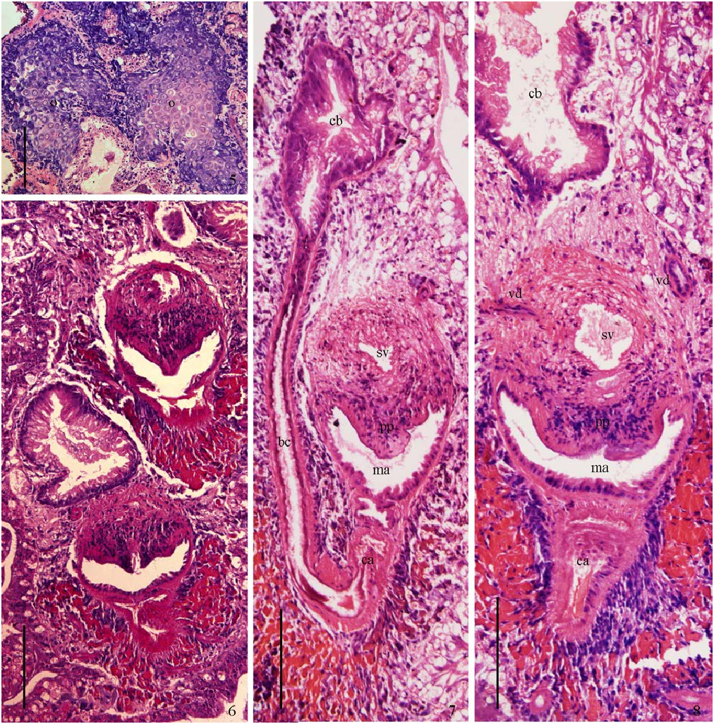
Reproductive system. Female reproductive system consists of ovaries, bursa canal, copulatory bursa and common atrium. A pair of hyperplasic ovaries are closely next to each other, showing an irregular shape ( Fig. 5 View Figs 5–8 ). The distance between ovaries and the brain is 760 μ m. Each ovary has an oviduct elongating ventrally along the outer side of the ventral nerve cord and opening asymmetrically into the common atrium. While the right oviduct opens at the ventral side of the common atrium, the left oviduct opens more dorsally. There is a significant distance (1.0 mm) from the anterior wall of copulatory bursa to the mouth ( Fig. 12 View Figs 9–12 ). The irregular copulatory bursa is lined by glandular epithelium, surrounded by a layer of longitudinal muscle ( Figs 7–11 View Figs 5–8 View Figs 9–12 ). Copulatory bursa connects the common atrium by bursal canal. Pipe-shaped bursal canal lies dorsally to the left of the penis ( Figs 7 View Figs 5–8 , 13–14 View Figs 13–14 ). The area around the opening of oviducts is covered by abundant shell glands and the gonopore receives the a few cement glands ( Figs 7–11 View Figs 5–8 View Figs 9–12 ). A few samples present 2–3 duplicated copulatory bursas ( Fig. 6 View Figs 5–8 ).
Male reproductive system is made up of tests, vas deferens, penis, male atrium, common atrium. The vas deferentia extend ventrally to the base of the penis bulb, and then go up anterior-dorsally before penetrating the penis bulb, and finally open at the proximal anterior part of the seminal vesicle ( Fig. 14 View Figs 13–14 ). The penis consists of penis bulb, seminal vesicle and penis papilla. The penis bulb is comprised of interwoven circular and longitudinal muscle fibers with an oval-shape seminal vesicle inside ( Figs 9–10 View Figs 9–12 , 14 View Figs 13–14 ). A small diaphragm, receiving abundant penis glands, separates the seminal vesicle and ejaculatory duct ( Fig. 14 View Figs 13–14 ). The ejaculatory duct runs ventrally and opens at the blunt tip of the penis papilla, making penis papilla in an asymmetric shape. The irregular lumen in the ejaculatory duct receives numerous penis glands. The conical penis papilla has a rather blunt tip and the margin of the papilla is folded, without distinguishing penis folds. There is a developed male diaphragm separating the male atrium and common atrium. The male diaphragm is lined by nucleated epithelium which is underlain by thick circular muscle fibers ( Figs 9–11 View Figs 9–12 , 14 View Figs 13–14 ). The specimen with 3 duplicated penises has one ejaculatory duct opening opposite to the mouth and the others are arranged like the normal individuals. All size of the duplicated penis is relatively small than that in normal individuals ( Fig. 6 View Figs 5–8 ).
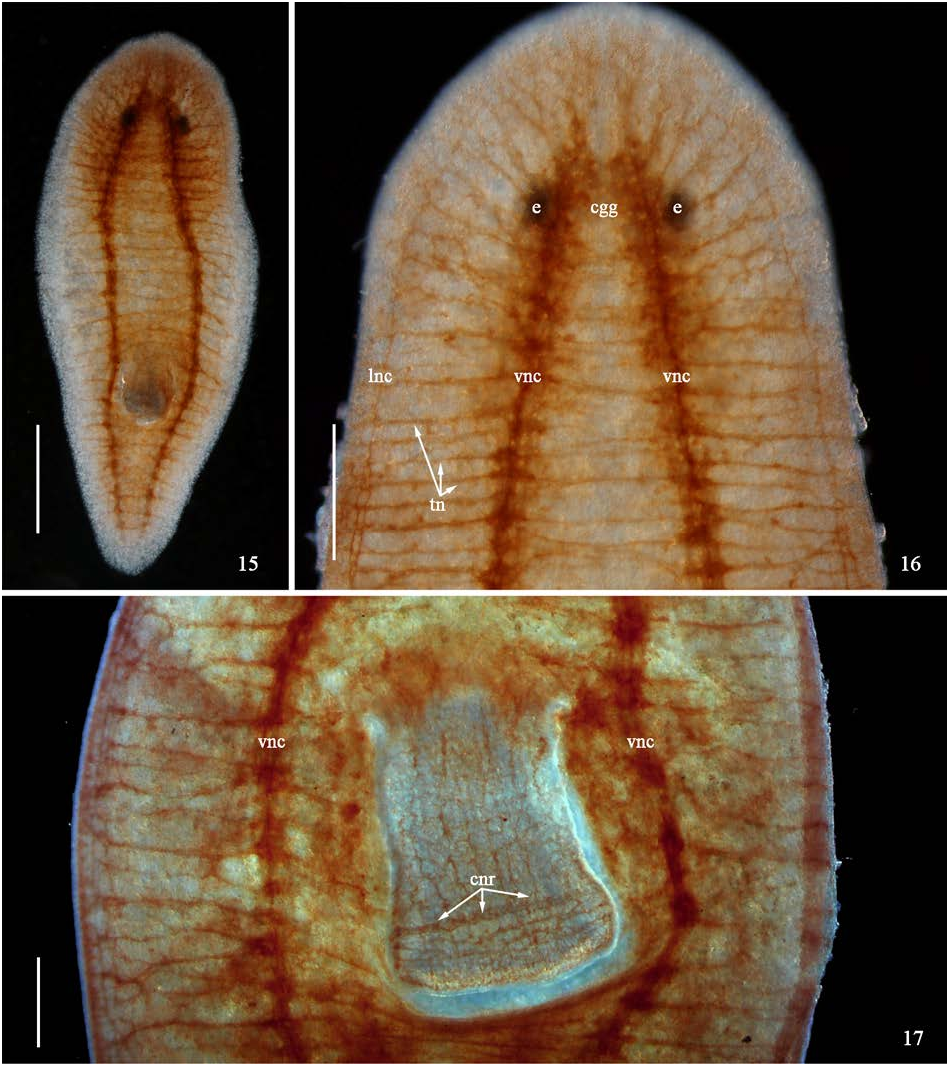
AChE + nerve structure. After histochemical localization, the AChE + nerve structure appears copper- red ladder structure ( Figs 15–16 View Figs 15–17 ). The AChE + nerve structure is composed of cerebral ganglion, ventral nerve cord, lateral nerve cord, circular nerve ring and transverse nerves. The control specimens present a negative reaction.
The reverse "U" shape cerebral ganglions are filled with numerous nerve cells. Each side of the cerebral ganglions extends backward, narrows gradually and forms the ventral nerve cord ( Fig. 16 View Figs 15–17 ). The sites where the ventral nerve cord sent out the transverse nerves always bulge and the transverse nerves end at the lateral nerve cord ( Fig. 17 View Figs 15–17 ). The lateral nerve cords originate from the cerebral ganglions, terminate at the end of the tail and are linked by a crescent-shaped transverse nerve ( Figs 15–16 View Figs 15–17 ). Transverse nerves rarely send out branch nerves. A thick circular nerve ring locates near the mouth and connects the pharynx nerve by pharyngal nerve net ( Fig. 17 View Figs 15–17 ). The boundary between cerebral ganglion and ventral nerve cord is hard to define.
Molecular phylogenetic analysis. The NJ and ML trees based on COI sequences are closely similar ( Figs 18–19 View Fig View Fig ). Three individuals of Duegsia sinensis Chen & Wang , sp. nov. comprise a sister group to the other Duegsia group with 100% bootstrap in both trees. All the COI sequences of genus Duegsia used in analysis form a clade with 100% bootstrap in both trees, suggesting the genus Dugesia is likely to be a sister group of the genus Bipalium , Schmidtea , Girardia from the family Geoplanoidea .


| IZCAS |
Institute of Zoology, Chinese Academy of Sciences |
| ML |
Musee de Lectoure |
No known copyright restrictions apply. See Agosti, D., Egloff, W., 2009. Taxonomic information exchange and copyright: the Plazi approach. BMC Research Notes 2009, 2:53 for further explanation.
|
Kingdom |
|
|
Phylum |
|
|
Class |
|
|
Order |
|
|
Family |
|
|
Genus |