
Dragmacidon sanguineum ( Burton, 1933 )
|
publication ID |
https://doi.org/ 10.11646/zootaxa.4587.1.1 |
|
publication LSID |
lsid:zoobank.org:pub:CC6CDA5A-E283-49AD-9F31-CE95C123A379 |
|
persistent identifier |
https://treatment.plazi.org/id/224C879C-2B5B-FFD9-FF08-89D5FDD46473 |
|
treatment provided by |
Plazi |
|
scientific name |
Dragmacidon sanguineum ( Burton, 1933 ) |
| status |
|
Dragmacidon sanguineum ( Burton, 1933)
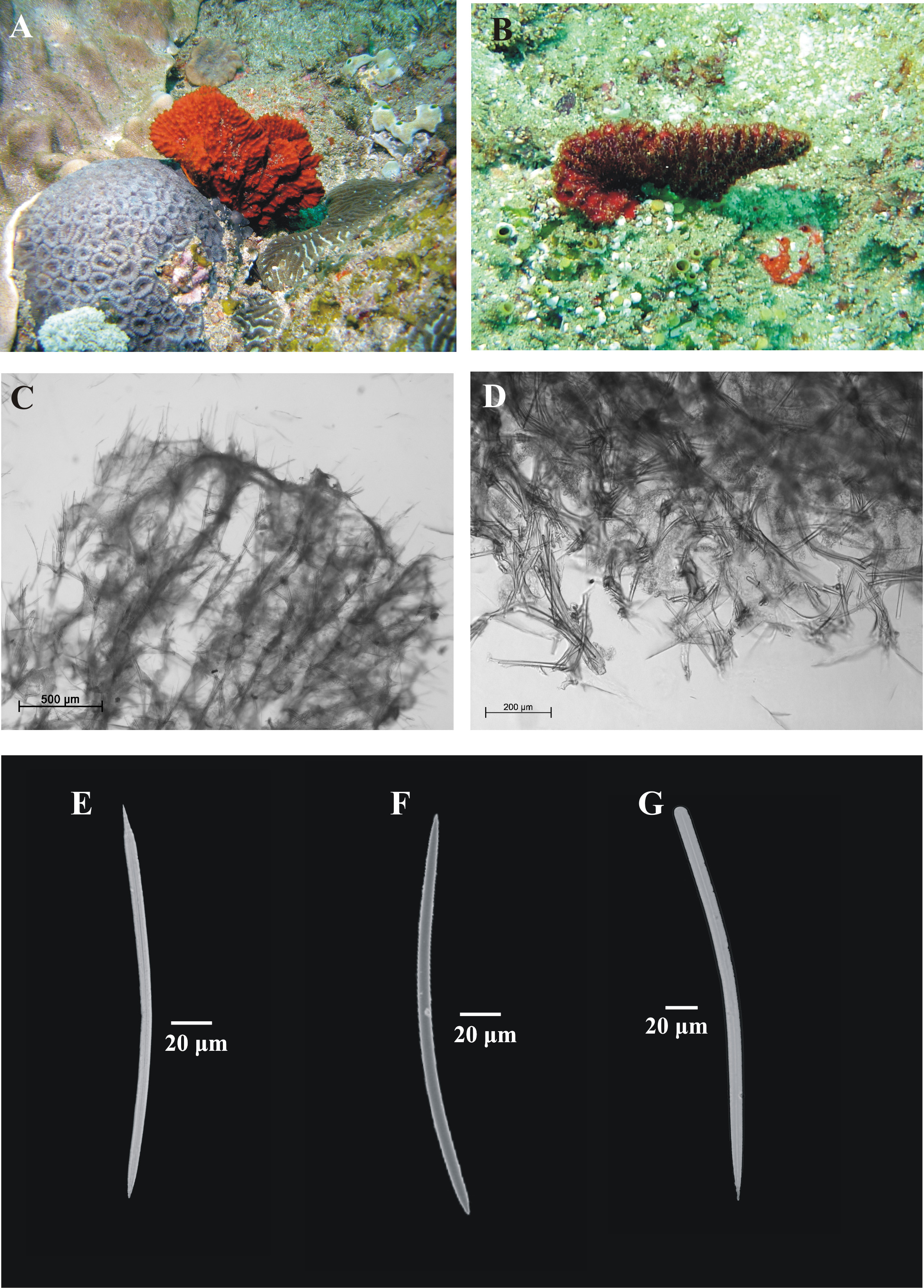
( Fig. 18 View FIGURE 18 A–G)
Synonomy
Axinella sanguinea Burton, 1933, p 27 , Fig. 4 View FIGURE 4
Material examined. SAMC–A24764 (cross-reference TS 841 & Saf 03-Sod 64), Two Mile reef, Sodwana Bay (27.5167°S, 32.6834°E), South Africa, 0 5 November 2003, collected by T. Samaai, depth 20 m. GoogleMaps SAMC–A24765 (cross-reference TS 890 & Saf 03-Sod 14), Ramsay reef, Sodwana Bay (27.4466°S, 32.7152°E), South Africa, 0 3 November 2003, collected by T. Samaai, depth 18 m. GoogleMaps
Description. Massive, flabellate sponge, with irregular thick, flattened planar lamellae, with irregular margins, 120 × 90 × 40 mm diameter, growing from a common base ( Fig. 18A, B View FIGURE 18 ). Sponge attached to substrate directly, up to 100 mm diameter or by small basal stalk, 30 mm long. Surface finely hispid, fleshy and conulose, with fine ridges running vertically down blades. Conules are irregular, 1 cm high, and linked to form meandering surface ridges. Oscules inside of ridges along axis of blade, 2 mm in diameter. Texture soft and spongy, very compressible. Colour in life dark brick-red; in preservative, beige.
Skeleton ( Fig. 18C, D View FIGURE 18 ). Choanosomal skeleton differentiated into axial and extra-axial region. Well-developed spongin enclosed primary fibres; fibres reticulated in the axial region and cored by primary choanosomal styles or oxeas (rare). Primary fibres connected by secondary unenclosed spongin and formed by styles, 30–65 µm in diameter, containing 3–7 spicules at any point. The mesohyl is augmented with long auxiliary oxeas. The extraaxial skeleton consists of several multispicular tracts (usually 2–3 megascleres) of ectosomal styles cemented by spongin, but not enclosed in sponging, running longitudinally diverging into a plumoreticulation towards the ectosome. The ectosomal skeleton has distinct brushes of ectosomal styles.
Spicules. Megascleres ( Fig. 18 View FIGURE 18 E–G). Styles, in two size classes: I) sharply pointed, fusiform with evenly rounded base, usually curved centrally, 244 (229–262) × 8 (8) µm, n = 10, II) long, slender and fusiform, 421 (395– 438) × 6 (6) µm, n = 10. Oxeas, in two size classes I) shorter, fairly thick, with one end fusiform and the other end hastate, 242 (222–263) × 8 (8) µm, n = 10, II) long and very slender, fusiform, 262 (254– 273) × 2 (2) µm, n = 10.
Microscleres. Absent
Substratum, depth range and ecology. Found on rocky ledges at a depth of 20 m.
Geographic distribution. Sodwana Bay, east coast of South Africa.
Remarks. Our specimens agree well with the original published description of Dragmacidon sanguineum ( Burton, 1933) from Umkomaas, KwaZulu-Natal, South Africa. Differences concern mainly variations in spicule size. The spicule dimensions recorded for the specimens described here are larger than recorded for the holotype (styles 140 µm; oxea 211 µm).
Key diagnostic characters.
• Sponge massive and flabellate.
• Long slender styles.
• Blood-red colour.
• No microscleres present.

No known copyright restrictions apply. See Agosti, D., Egloff, W., 2009. Taxonomic information exchange and copyright: the Plazi approach. BMC Research Notes 2009, 2:53 for further explanation.
|
Kingdom |
|
|
Phylum |
|
|
Class |
|
|
Order |
|
|
Family |
|
|
Genus |
Dragmacidon sanguineum ( Burton, 1933 )
| Samaai, Toufiek, Pillay, Ruwen & Janson, Liesl 2019 |
Axinella sanguinea
| Burton 1933: 27 |