
Discothyrea chimera Hita Garcia & Lieberman, 2019
|
publication ID |
https://doi.org/ 10.1093/isd/ixz015 |
|
publication LSID |
lsid:zoobank.org:pub:F01A07B1-90C0-41A9-AFF8-1722885CE35C |
|
DOI |
https://doi.org/10.5281/zenodo.5922596 |
|
persistent identifier |
https://treatment.plazi.org/id/03D9AC4A-E574-FF9F-FCC4-F95EBF9C0508 |
|
treatment provided by |
Plazi |
|
scientific name |
Discothyrea chimera Hita Garcia & Lieberman |
| status |
sp. nov. |
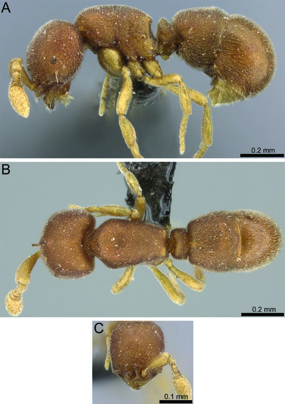
Discothyrea chimera Hita Garcia & Lieberman sp. n.
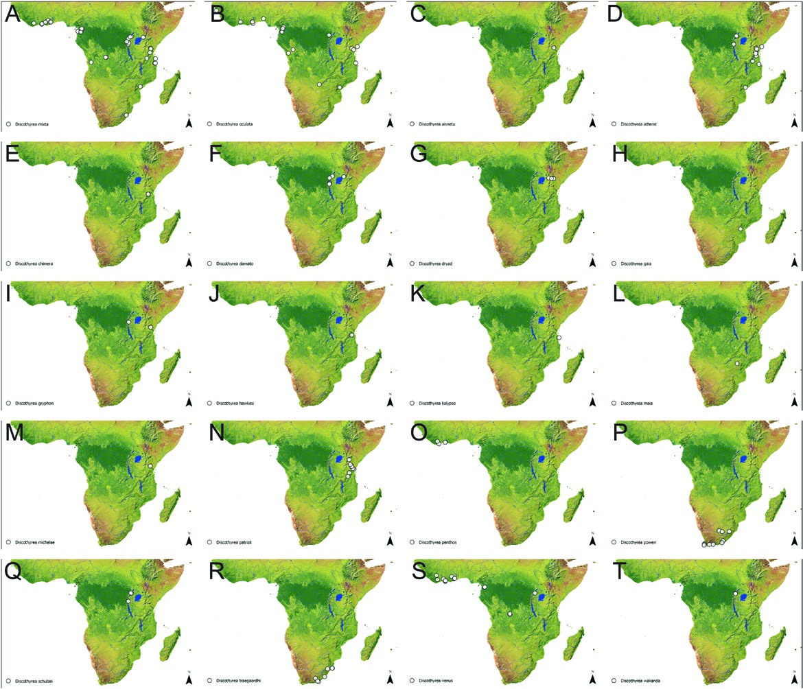
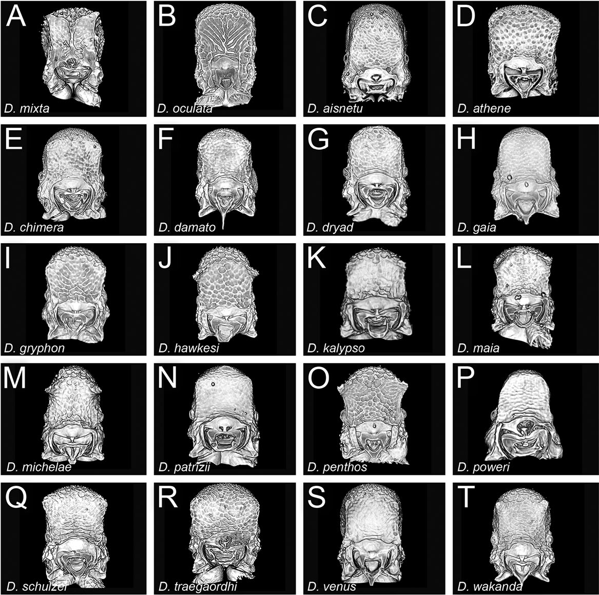
( Figs. 4E View Fig , 6E View Fig , 7E View Fig , 8E View Fig , 9E View Fig , 10E View Fig , 11E View Fig , 12E View Fig , 14E View Fig , 27 View Fig , 28 View Fig ; Supp Video S5 [online only])
Type Material
HOLOTYPE, pinned worker, TANZANIA, Morogoro Region, Kilosa, Mamiwa-Kisara Forest Reserve , −6.3753, 36.93711, 1989 m, primary forest, leaf litter, Winkler, collection code CEPF-TZ-1.2, GoogleMaps
16.–21.VIII.2005 (P. Hawkes, J. Makwati & R. Mtana) ( SAMC: CASENT0235471).
Cybertype. Volumetric raw data (in DICOM format), 3D rotation video, still images of surface volume rendering, and 3D surface (in PLY format) of the physical holotype (CASENT0235471) in addition to stacked digital color images illustrating head in full-face view, profile and dorsal views of the body. The data are deposited at Dryad (Hita Garcia et al. 2019, http://doi.org/10.5061/ dryad.3qm4183) and can be freely accessed as virtual representation of the type. In addition to the cybertype data at Dryad, we also provide a freely accessible 3D surface model of the holotype at Sketchfab (Model 5).
Diagnosis
The following character combination distinguishes D. chimera from the remainder of the complex: masticatory margin of mandible with a conspicuous, curved subapical tooth; subquadrate head with anterolateral genal angles sharply defined; anterior clypeal margin with long erect setae; propodeum not dentate; mesotibia without apicoventral spur; petiolar node strongly attenuated dorsally and squamiform in profile (LPeI 329); AT4 around 1.2 times longer than AT3; erect setae absent on dorsal surfaces.
Worker Measurements and Indices (n = 1)
EL 0.03; HL 0.45; HW 0.35; SL 0.23; PH 0.19; PW 0.31; DML
0.33; PrH 0.34; WL 0.54; HFL 0.31; PeL 0.07; PeW 0.20; PeH 0.23;
LT3 0.32; LT4 0.39; OI 6; CI 90; SI 60; LMI 52; DMI 57; DMI2 93;
ASI 124; HFI 57; DPeI 286; LPeI 329.
Worker Description
Head very broad (CI 90), appearing subquadrate posterad antennal sockets posterior head margin straight; posterodorsal corners of head rounded; in frontal view, sides of head slightly convex; eyes present, relatively long (OI 6), oval, situated slightly less than halfway between anterolateral corner of gena and posterior head margin; eyes just visible in frontal view; anterolateral corner of gena squared, nearly right-angled; frontal lamella bladelike in profile, apex acute, lamella more or less evenly translucent across its disc, without a clearly defined fenestra; medial clypeus broad and sharply transverse, lateral clypeus curving moderately strongly between antennal sockets and anterolateral corners of head, bearing row of long erect setae. Antenna with moderately long scape (SI 60), scape very strongly incrassate, gently bent; pedicel subcylindrical, longer than broad; apparent antennomere count eight, flagellomeres basad apical club highly compressed, taken together only about as long as apical club. Ventral head with short horizontal postoccipital ridge with very short, triangular anteromedian carina; medial region of hypostooma somewhat truncate, arms narrowed; palpal formula not examined. Mandible with prominent, curved subapical tooth and small, sharp prebasal denticle; prebasal denticle located nearly at basal angle; ectal face with longitudinal carina running from preapical tooth to basal angle, leaving short depressed prebasal region on masticatory margin.
Mesosoma gently convex in profile, pronotum distinctly higher than propodeum; in dorsal view mesosoma moderately thick (DMI 57; DMI2 93) and somewhat narrowed posteriorly; pronotal humeri moderately rounded; posterior propodeal margin weakly concave; posterodorsal corners of propodeum subangulate, without teeth; declivitous face of propodeum very shallowly concave in profile and oblique posterior view; propodeal spiracle fairly large and round but inconspicuous, directed posteriorly; propodeal lobes poorly developed, very short and bluntly truncated.
Legs short (HFI 57) and relatively robust; mesotibia without apicoventral spur, with small but distinct seta inserted in apical pit; mesobasitarsus relatively short, about as long as tarsomeres II–IV taken together.
Petiolar node strongly attenuated dorsally and squamiform in profile, about 3.3 times higher than long (LPeI 329); in profile anterior face of node sloping posterodorsally, apex peaked, posterior face sloping posteroventrally; in dorsal view petiole subrectangular, about 2.9 times broader than long (DPeI 286), sides very slightly convex; in anterior view, petiolar outline sharply pentagonal and strongly, sharply peaked, sides and angles well-defined, in oblique anterior view, anterior face flat; in ventral view roughly rectangular, sides weakly diverging posteriorly; subpetiolar process short, lobate, apex rounded, bearing several long, straight, white setae; petiolar spiracles very large and subcircular in ventral view.
Abdominal segment 3 almost thickly cylindrical, only slightly widening posteriorly, tergite weakly peaked just anterad end of segment; sternite evenly and quite shallowly curved in profile; AS 3 without carinate prora, but still with anterior face distinctly depressed and anterior margin of ventral face fairly deeply arcuate in ventral view;AT4 1.2 times longer than AT3 (ASI 124); AT4 large, bulbous, hemidemispherical; AS 4 with broad, prominent anterior lip, overlapping most of the width of AS 3, anterior margin straight in ventral view; successive abdominal segments short, telescopic, often concealed.
Sculpture on gena and ventral head surface foveolate-reticulate, becoming punctate toward front of head, punctae smaller on frontal Model 5. 3D surface model of D. chimera sp. n. holotype (CASENT0235471). An interactive version of this model is available in the HTML version of this article online and at https://sketchfab.com/3d-models/a9c567dc04c44205a6f 5dee0ea 080518.
area posterad frontal lamella; mandible rather roughly sculptured with dense piligerous punctae; mesosomal dorsum, petiole, and abdominal segment 3 shallowly, evenly punctate-reticulate; lateral mesosoma irregularly punctate to rugulose on ventrolateral and posteroventral portions of propodeum; AT4 distinctly shinier and smoother than AT3, with fine piligerous punctulae.
Setation on head, antenna, and mesosoma a fairly consistent but short, velvety layer of appressed white pubescence, best developed on mesosomal dorsum; front of head just posterad frontal lamella with longer, more distinct setae directed towards midline; longer hairs, some decumbent, present on gena, best seen in frontal view; ectal face of mandible with numerous long, curved, appressed to suberect setae; masticatory margin without row of straight setae; ventral margin of petiolar sternite with several long suberect hairs; abdominal segment 3 densely clothed with long, appressed pilosity, obscuring sculpture; noticeably longer on sternite than tergite; AT4 with long, appressed pilosity, denser than on mesoma but less so than on AT3; successive abdominal segments with dense, white, standing pilosity; legs densely and more or less evenly pubescent.
Color testaceous, appendages dull yellow.
Etymology
The chimera was a mythological monster whose body was an amalgam of various creatures. The species is named for its singular combination of characters. The specific epithet is given as an appositive noun.
Distribution and Biology
Presently the new species is known only from a single specimen collected from leaf litter in the Mamiwa-Kisara Forest Reserve in Tanzania ( Fig. 4E View Fig ), which is a humid montane forest. Discothyrea chimera was collected from leaf litter.
Comments
Not considering the other characters given in the diagnosis above, the presence of a conspicuous subapical mandibular tooth distinguishes D. chimera from all other Afrotropical species. While a small and acute subapical denticle is present in D. gryphon , it is not comparable to the tooth seen in D. chimera . Furthermore, both species appear morphologically closer to each other than to the remainder of the Afrotropical fauna, but can be well separated by eye size, the shape of the petiole, and pilosity. Discothyrea chimera has relatively larger eyes (OI 6), a dorsally much thinner petiole, and lacks erect setae on its dorsal surfaces, whereas D. gryphon has either no eyes or smaller eyes (OI 0–4), has a dorsally thicker petiole, and conspicuous standing setae through its body. Nevertheless, the similarity of D. chimera and D. gryphon regarding the characters of the head, as well as in general habitus, suggest a possible close phylogenetic relationship, but, without molecular phylogenetic data, this is rather speculative.
Variation
There is no information about intraspecific variation since the species is only known from the holotype.


| SAMC |
Iziko Museums of Cape Town |
No known copyright restrictions apply. See Agosti, D., Egloff, W., 2009. Taxonomic information exchange and copyright: the Plazi approach. BMC Research Notes 2009, 2:53 for further explanation.
|
Kingdom |
|
|
Phylum |
|
|
Class |
|
|
Order |
|
|
Family |
|
|
Genus |