
Dicromantispa leucophaea Machado & Rafael
|
publication ID |
https://doi.org/ 10.11646/zootaxa.2454.1.1 |
|
persistent identifier |
https://treatment.plazi.org/id/03FE87CD-5C5F-FF83-FF38-FF72FA93FCA0 |
|
treatment provided by |
Felipe |
|
scientific name |
Dicromantispa leucophaea Machado & Rafael |
| status |
sp. nov. |
Dicromantispa leucophaea Machado & Rafael View in CoL , new species.
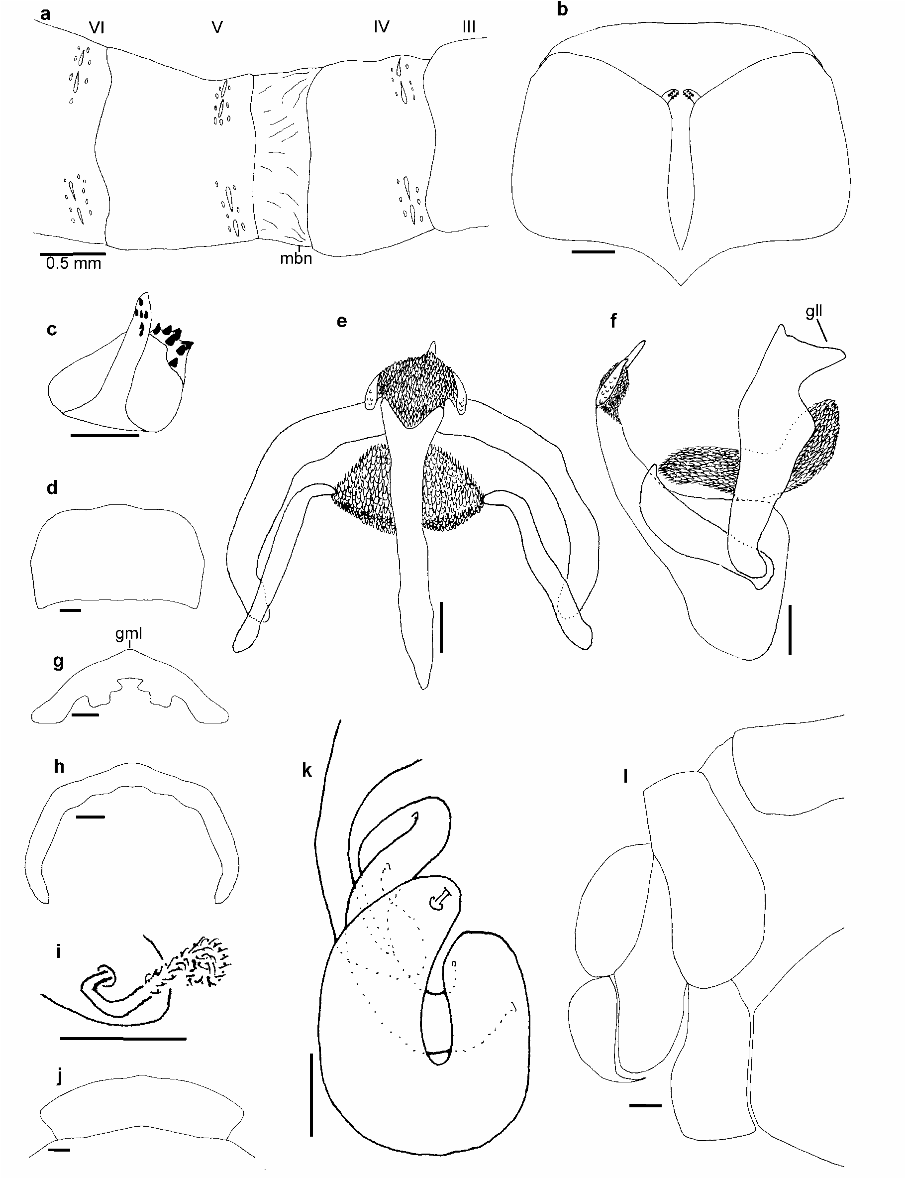
(Figs. 11–12)
Diagnosis. ventromedial lobe completely sclerotized and curved; pseudopenis shorter than pseudopenal membrane; lateral lobe of gonarcus large; hindwing with basal space between C and Sc brown; pronotum white to light yellow, except brown area between anterior border and maculae.
Holotype male. Vertex brown medially, yellow laterally and in a central “Y” shaped spot (Fig. 11d). Head in frontal view almost completely yellow, except for central longitudinal dark brown stripe beginning between antennae and ending at labrum. Stripe narrow at frons. Labrum and clypeus yellow area slightly darker than other head sclerites (Fig. 11a). Mandibles and palpi dark brown at apex, other mouthparts reddish brown. Antenna with scape yellow ventrally and reddish brown dorsally. Pedicel and first three flagellomeres reddish brown; other flagellomeres dark brown (Fig. 11a).
Pronotum: nearly straight in lateral view, with few setae on proximal and distal regions arising directly from it surface. Length-width-ratio at maculae: 6.1. Between anterior border and maculae brown, remainder white to light yellow, except two dark brown small central stripes, one anterior and other posterior (Fig. 11d). Pteronotum: with three longitudinal dark brown stripes, one central and two lateral, with light brown stripes between them (Fig. 11d). Mesoscutellum dark brown, except for yellow lateral border (Fig. 11d). Metascutellum completely dark. Both scutella with 5–6 pores each one. Pteropleura predominantly yellow, mesopreepisternum, mesepisternum, mesanepisternum and mesokatepisternum with dark brown spots at the anterior border (Fig. 11b).
Foreleg: coxa yellow, except reddish brown apex. Trochanter, last four tarsomeres and femur posterior surface reddish brown (Fig. 11f). Femur anterior surface reddish brown except middle region dark reddishbrown (Fig. 11e). Tibia and tarsomere I reddish brown, except for yellow tibia base. Mid and hindlegs yellow, except midcoxa light brown (Fig. 11b). Tarsal claws with six teeth.
Forewing: length: 12.5 mm, 7 costal crossveins and 12 veins extended posteriorly from RP. Hyaline, except 1M, 1Cu, bases of AA and 1AP, radial triangle and space between Sc and RA brown. Cell1AP apex light yellow. Pterostigma reddish-brown. Veins brown, except AP1, AP2 and AA yellow. RA and C light brown basally, yellow medially, and reddish brown apically (Fig. 11g). Hindwing: 6 costal crossveins and 13 veins that extend posteriorly from RP, hyaline except for cell 1M, space between Sc and RA and beginning of space between C and Sc brown. Pterostigma reddish-brown. Veins color similar to forewing, except for AP1 brown, RA, C and Sc yellow, with apex reddish-brown (Fig. 11g).
Abdomen yellow scattered by reddish brown spots ventrally and dorsally. Tergites IV and V separated by a large membrane. Pleura dark-brown. Tergites IV–VI with 2 groups of 6–9 pores in two transverse parallel rows anterolaterally; each group with one or two large pores or a scar between them ( Fig. 12a View FIGURE 12 ).
Terminalia: ectoproct posterior border rounded. Ventromedial lobe completely sclerotized, curved, with wide internal bend ( Fig. 12b, c View FIGURE 12 ) with 8–9 stout setae; and narrow external bend with 5–6 stout setae ( Fig. 12c View FIGURE 12 ). Sternite IX posterior border rounded in ventral view ( Fig. 12d View FIGURE 12 ). Gonarcus with small median lobe, hardly visible in posterior view ( Fig. 12g, h View FIGURE 12 ), and two large lateral lobes, easily seen in posterior and lateral view ( Fig. 12g, f View FIGURE 12 ). Gonocoxite with constant width and with apex bent medially in ventral view ( Fig. 12e View FIGURE 12 ). Basal two-thirds of mediuncus wider in lateral view and apex bifurcate in ventral view ( Fig. 12e, f View FIGURE 12 ). Gonarcal membrane with medial group of spinules, group wider than long, touching gonocoxite apex ( Fig. 12e View FIGURE 12 ). Pseudopenal membrane slightly shorter than pseudopenis and with small scales on dorsal surface ( Fig. 12e View FIGURE 12 ). Hypomere apex rounded with small granules ( Fig. 12e, f View FIGURE 12 ).
Paratype female. Similar to male except length-width-ratio at maculae: 5.6–6.4; pteronotum central stripe slightly wider than others; scutella completely dark brown or with yellow spots laterally; pterothorax pleurites predominantly brown with yellow spots at border (Fig. 11c); forewing length: 9.6–12.9; scutella with 2–6 pores each one.
Terminalia: ectoproct larger than gonocoxite ( Fig. 12l View FIGURE 12 ). Sternite VIII large and easily seen in lateral and ventral view; in ventral view with posterior border larger than anterior ( Fig. 12j View FIGURE 12 ). Spermathecal duct with few bends ( Fig. 12k View FIGURE 12 ), Fertilization canal narrow. Capsule covered by minuscule setae ( Fig. 12i View FIGURE 12 ).
Variation, paratype male. First three flagellomeres dark brown; dark reddish brown spot on femur
Geographical data. Neotropical, with records only for Brazil, except South and Southeast regions.
Bionomy. Specimens collected in April, May, September and October. Nothing is known about its biology.
Discussion. Some characteristics indicate that D. leucophaea is close to D. moulti : space between C and Sc brown in the hindwing; small size of gonarcus median lobe and length of group of spinules in the gonarcal membrane. However, the pronotum color pattern and the length of pseudopenis, gonarcal lateral lobes and forewing, clearly separate these two species.
Etymology. The specific name is a combination of two Greek adjectives leukos (= white) and phaios (= brown, dusky) and refers to the pronotum color pattern.
Type material: Holotype male: Brazil: Mato Grosso: Nova Lacerda, Serra , 14°28’38’’S – 59°33’30’’W, 27.iv.2006, J.A.Rafael, F.F.Xavier F°, arm[adilha] luz (printed on rectangular white paper)–INPA. GoogleMaps
Paratypes: Pará: Serra-Norte, Serraria–Ig [arapé] Azul , isca luminosa, 19.x.1984, T.Pimentel, col., MPEG NEU, 20000154 (1 ♂, 1 ♀ – MPEG) ; Rondônia: Ouro Preto do Oeste, R. INPA/ Ceplac , 10°43’00’’S – 62°14’45’’W, 21.iv.2006, J.A.Rafael, F.F.Xavier F°, arm[adilha] luz (3 ♀ – INPA) GoogleMaps ; Mato Grosso: Nova Lacerda, Serra , 14°28’38’’S – 59°33’30’’W, 27.iv.2006, J.A.Rafael, F.F.Xavier F°, arm[adilha] luz (3 ♀ – INPA) GoogleMaps ; Juina , v.1985, O. Roppa, #37736 (1 ♂ –MZUEFS) ; Maranhão: Caxias: Res. Ecol. Inhamum , 02.ix.2005, luz, 04°53’27’’S – 43°24’54’’W, F. L. Oliveira ( CZMA) (1 ♂ –CZMA) GoogleMaps .
Holotype condition: excellent. Abdomen dissected, cleared and preserved in a microvial with glycerin.
| MPEG |
Museu Paraense Emilio Goeldi |
| INPA |
Instituto Nacional de Pesquisas da Amazonia |
No known copyright restrictions apply. See Agosti, D., Egloff, W., 2009. Taxonomic information exchange and copyright: the Plazi approach. BMC Research Notes 2009, 2:53 for further explanation.
|
Kingdom |
|
|
Phylum |
|
|
Class |
|
|
Order |
|
|
Family |
|
|
Genus |