
Cratera crioula ( Froehlich, 1955 )
|
publication ID |
https://doi.org/ 10.11646/zootaxa.4500.4.3 |
|
publication LSID |
lsid:zoobank.org:pub:70672C0A-EC78-40BA-85EE-6206184CE0F0 |
|
persistent identifier |
https://treatment.plazi.org/id/4F7187CF-C557-FFEB-D5A8-F3CDFC29F8E6 |
|
treatment provided by |
Felipe |
|
scientific name |
Cratera crioula ( Froehlich, 1955 ) |
| status |
|
Cratera crioula ( Froehlich, 1955) View in CoL
Synonymy
Geoplana crioula Froehlich, 1955 View in CoL
Material examined. All specimens collected in Parque Estadual da Cantareira, State of São Paulo, Brazil, (-23.41, -46.60). Holotype ( MZUSP PL 2114 ): E. M. Froehlich & C. G. Froehlich, August 1952, sagittal sections of pharynx and copulatory apparatus on 7 slides (slides S809-S815; illustrated in Fig. 33 and 34 in Froehlich, 1955). F2946 ( MZUSP PL 423 ): F. Carbayo et al. coll., 26 October 2008, sagittal sections of pharynx and copulatory apparatus on 50 slides. F3709 ( MZUSP PL 1078 ): F. Carbayo et al. coll., 19 April 2009, transverse sections of cephalic region on 18 slides; horizontal sections of portion behind cephalic region on 28 slides; transverse sections of pre-pharyngeal region on 8 slides; sagittal sections of pharynx and copulatory apparatus on 49 slides. F3712 ( MZUSP PL 2116 ): F. Carbayo et al. coll., 19 April 2009, sagittal sections of pharynx and copulatory apparatus on 27 slides. F3715 ( MZUSP PL 1079 ): F. Carbayo et al. coll., 19 April 2009, sagittal sections of pharynx and copulatory apparatus on 42 slides. F3744 ( MZUSP PL 2117 ): F. Carbayo et al. coll., 20 April 2009, sagittal sections of copulatory apparatus on 9 slides.
Distribution. Only known from the type locality, Parque Estadual da Cantareira, State of São Paulo, Brazil.
Diagnosis. Species of Cratera some 40 mm long. Dorsum dark brownish, usually with a thin median cream- color stripe and a submarginal stripe on each side of the body of the same color. Dorsal eyes initiating in cephalic region. Proximal portion of prostatic vesicle bifurcated, extrabulbar, detached from penis bulb. Penis bulb extends 0.5 mm anteriorly to penis papilla. Ejaculatory duct may be distally widened. Dorsal insertion of penis papilla slightly anterior to the ventral. Penis papilla projects into female atrium. Female:male atrial length ratio, 0.5-1. Common glandular duct present.
Description. We agree with Froehlich's (1955) description in most aspects. Herein we complement the description and make some observations on the ejaculatory duct.
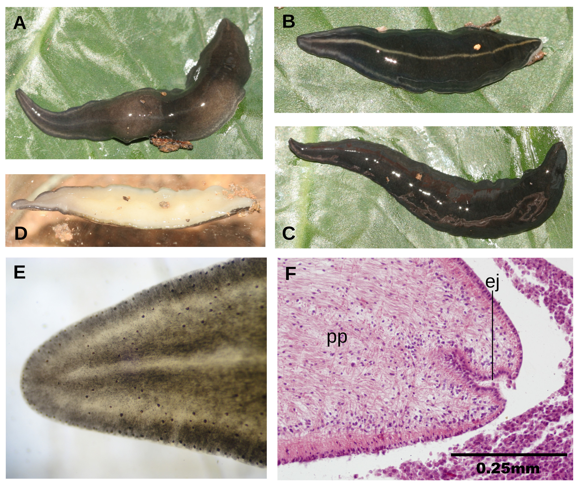
External aspect. Body margins nearly parallel when crawling; anterior end rounded, posterior pointed. Dorsum convex, ventral side nearly flat. At rest and when contracted, the body is lanceolate ( Fig. 2 View FIGURE 2 A-C). The largest preserved specimen was 60 mm in length and 5 mm in width (specimen F2946); specimen F3709 measured 39 mm long, and 6 mm wide. Dorsal surface with dark brownish black pigment spots, excepting a thin median stripe and a submarginal stripe on each side of the body, where the cream ground color of the body appears ( Fig. 2 View FIGURE 2 A-B). These stripes may also be unconspicuous behind anterior extremity ( Fig. 2C View FIGURE 2 ). The ventral side is creamcolored, passing into greyish in the anterior extremity ( Fig. 2D View FIGURE 2 ).
Eyes monolobated, 35 µm in diameter, contouring cephalic region. They spread over dorsum from anterior extremity ( Fig. 2E View FIGURE 2 ), backwards. At the level of the pharynx they extend on a lateral band, on each side of the body, 1/3 of the body width. From that region towards the posterior extremity they spread on a band progressively narrower. Unconspicuous small halos around each eye present. Sensory pits 20 µm deep, contouring cephalic region and extending ventro-marginally backwards on a length equal to at least 20% of body length. Relative mouth:body length and relative gonopore:body length, 58% and 78%, respectively in holotype, and 72% and 85% in specimen F3709.
Internal morphology. Glandular margin present, comprising erythrophil granular glands and xanthophil granular glands. Cutaneous musculature with the usual three layers present in the subfamily Geoplaninae : a thin subepithelial circular layer (2.5 µm thick) followed by a diagonal with decussate bundles (10 µm thick), and then a longitudinal layer (50 µm thick dorsally, 25 µm ventrally) arranged in bundles. Cutaneous musculature as thick as 14% of body height.
The three most common parenchymal muscle layers are also present, i.e., a weak dorsal layer of decussate diagonal fibers (20 µm thick), a very well developed supraintestinal layer of transverse fibers (80 µm thick), and a subintestinal layer (80 µm thick). Parenchymal longitudinal musculature absent. There are no subepidermic nor parenchymal muscle modifications in cephalic region.
Relative position mouth:pharyngeal pouch length, of 62%. Pharynx bell-shaped. Esophagus present, with 19% of pharynx length. Outer epithelium of the pharynx underlain by a thin layer (3 µm thick) of longitudinal muscle, followed by a thin layer of circular fibers (6 µm thick). Inner pharynx musculature consisting of a subepithelial layer (2 µm thick) of longitudinal musculature, followed by a circular layer (150 µm thick) with scattered longitudinal fibers interspersed and an outermost 10 µm thick layer of longitudinal fibers.
Testes arranged in a row of 2-3 testes on each side of the body. They extend from 2 mm behind the level of the ovaries (equal to 5% of the body length) near to the root of the pharynx (equal to 60% of the body length) (specimen F3709). Lateral to the mid-region of the prostatic vesicle, sperm ducts curve medially to communicate with the two very short branches of the prostatic vesicle. Prostatic vesicle large and extrabulbar, detached from penis bulb. Anterior half of the unpaired section of this vesicle C-shaped, posterior half runs sinuously backwards, subsequently penetrates the penis bulb to continue as a straight ejaculatory duct. The ejaculatory duct traverses the central region of the penis papilla to open at its tip. The lumen of this duct is 20 µm in diameter, and widens 1.5 times distally in the holotype ( Fig. 2F View FIGURE 2 ); widening of distal section of this duct not remarkable in specimen F3712; it passes from 15 µm to 20 µm distally. In specimen F3744 there is no widening at all.
Penis bulb well developed; extending anteriorly to penis papilla 0.5 mm. The prostatic vesicle is lined with a ciliated epithelium, and is pierced by glands producing erythrophil granules. This epithelium is surrounded by a circular muscle (15 µm thick). The penis papilla is conical, large and extends into the female atrium. This papilla is lined with a non-ciliated, cuboidal epithelium, and is underlain by a 25 µm thick circular muscle, followed by a 6 µm thick of longitudinal muscle. The ejaculatory duct is lined with a cuboidal ciliated epithelium, and is underlain by a circular muscle 10 µm thick. The male atrium is broad, not folded, and mostly occupied by the penis papilla ( Fig. 3 View FIGURE 3 A-B). The muscularis of the male atrium consists of a thin 10 µm thick circular muscle, followed by a thinner longitudinal muscle. There is no accumulation of cyanophil glands that pierce its roof.
Ovaries roughly rounded, 250 µm in diameter. They are located at a distance from anterior end equal to 23% of body length. Ovovitelline ducts arise from the dorso-lateral aspect of the ovaries. Behind the level of the gonopore, ovovitelline ducts ascend to communicate with each other above dorsal section of female atrium. Distal ascending section of ovovitelline ducts receives shell glands ( Fig. 3B View FIGURE 3 ). Common ovovitelline duct horizontal; female genital duct is a projection of the postero-dorsal region of female atrium and is dorso-anteriorly oriented. The f emale atrium is an ample cavity continued with the male atrium; female:male atrial length, 1:1. Female atrium is lined with a 40 µm high epithelium with stratified aspect and internal gaps ( Fig. 3 View FIGURE 3 A-B), and is underlain by a 5 µm thick circular muscle, followed by a 5 µm thick longitudinal muscle.
Remarks. In E. M. Froehlich’s collection, there is a number of slides labeled only ‘crioula’. However, the histological slides S809-S815 fully coincide with drawings of copulatory apparatus and pharynx supported recognition of the holotype.
Original description makes no mention to the distal widening of the ejaculatory duct present in the holotype. This widening is relatively small and may not exist in some specimens.

| MZUSP |
Museu de Zoologia da Universidade de Sao Paulo |
No known copyright restrictions apply. See Agosti, D., Egloff, W., 2009. Taxonomic information exchange and copyright: the Plazi approach. BMC Research Notes 2009, 2:53 for further explanation.
|
Kingdom |
|
|
Phylum |
|
|
Order |
|
|
Family |
|
|
Genus |
Cratera crioula ( Froehlich, 1955 )
| Lago-Barcia, Domingo & Carbayo, Fernando 2018 |
Geoplana crioula
| Froehlich 1955 |