
Corydoras apiaka, Espíndola & Spencer & Rocha & Britto, 2014
|
publication ID |
https://doi.org/ 10.1590/0031-1049.2014.54.03 |
|
persistent identifier |
https://treatment.plazi.org/id/595787E0-431B-FF84-D620-FC374675D90E |
|
treatment provided by |
Carolina |
|
scientific name |
Corydoras apiaka |
| status |
sp. nov. |
Corydoras apiaka View in CoL , new species
Fig. 1A View FIGURA 1 , Fig. 1B View FIGURA 1 , Table 1
Holotype: MNRJ 40720 View Materials , 28.4 mm SL, Ribeirão Oito de Julho, a tributary of the right margin of rio Arinos, rio Tapajós Basin , at road MT-338 between Tapurah- Nova Paraná, Porto dos Gaúchos, Mato Grosso, 11°41’44”S, 57°06’42”W, 19 January 2002, P. Buckup, A. Aranda, F. Silva & C. Figueiredo. GoogleMaps
Paratypes: BRAZIL, Mato Grosso, rio Tapajós basin: MNRJ 23358 View Materials , 11 View Materials (+1 cs) 28.9-34.9 mm SL, collected with the holotype. MNRJ 23334 View Materials , 1 View Materials , 31.6 mm SL, Tapurah, rio São Venceslau on the Tapurah-Nova Paraná Road (MT-338), rio Arinos basin, 12°19’32”S, 56°40’31”W, 19 January 2002, P. Buckup, A. Aranda, F. Silva & C. Figueiredo. MNRJ 23657 View Materials , 2 View Materials , 33.7-34.6 mm SL, rio Arinos , Porto dos Gaúchos, 11°31’51”S, 57°25’08”W, 19 January 2002, P. Buckup, A. Aranda, F. Silva & C. Figueiredo. MZUSP 45340 View Materials , 8 View Materials , 30.8-39.5 mm SL, riacho Monjolinho, a tributary of rio Preto , Diamantino 14°20’00”S, 56°22’00”W, 24 October 1992, N. Menezes, H. Britski, O. Oyakawa & M. Triques. MZUSP 95885 View Materials , 1 View Materials , 34.05 mm SL, rio Teles Pires , Itaúba, 11°03’44”S, 55°19’08”W, 26 September 2007, J.L.O. Birindelli & P. Holanda Carvalho. MZUSP 99037 View Materials , 1 View Materials , 37.64 mm SL, rio Teles Pires basin unnamed stream at BR-163, Itaúba 11°06’51”S, 55°18’25”W, 18 February 2008, P. Holanda Carvalho & S. Lima GoogleMaps .
Diagnosis
Corydoras apiaka differs from its spotted congeners by having the dorsal-fin spine smaller than the three anterior branched dorsal-fin rays (vs. dorsal-fin spine smaller than just two anterior dorsal-fin rays, or equal in size). Corydoras apiaka is distinguished from its congeners, except C. albolineatus , C. araguaiaensis , C. haraldschultzi , C. julii , C. polystictus , C. multimaculatus and C. xinguensis by the presence of numerous dark-brown spots over the head and body (vs. spots absent). Corydoras apiaka differs from C. albolineatus , C. araguaiaensis and C. haraldschultzi by having several spots scattered all over the head and trunk in a somewhat random pattern (vs. spots more concentrated and with a single pale horizontal stripe in the middle of the body in C. albolineatus ; or spots arranged in three or more stripes along the trunk in C. araguaiaensis and C. haraldschultzi ); pectoral, pelvic and anal fins hyaline (vs. several series of small blotches over fin rays), and adipose fin hyaline or bearing one spot (vs. two or more spots). The new species can be further distinguished from C. multimaculatus and C. xinguensis by blotches on interradial membrane of dorsal fin absent (vs. present); adipose fin hyaline or with a single spot (vs. two or three well defined spots); spots not reaching over the anal fin base (vs. spots extending to anal-fin base). Corydoras apiaka is distinguished from C. xinguensis by spots on anal fin absent (vs. present); body spots with diffuse border (vs. spots with well delimited edge). Corydoras apiaka further differs from all other spotted species, with the exception of C. multimaculatus , by the naked ventral region (vs. belly with small platelets).
Description: Morphometric data presented in Table 1. Head compressed with slight convex dorsal profile ( Fig. 1 View FIGURA 1 ); roughly triangular in dorsal view. Snout rounded. Head profile convex from upper lip to tip of parieto-supraoccipital expansion. Dorsal profile of body slightly convex from tip of parieto-supraoccipital expansion to base of last dorsal-fin ray. Body slightly concave from last ray of dorsal fin to base of adipose- fin spine; straight to slightly concave from that point to caudal-fin base. Ventral profile of body straight from isthmus to pelvic-fin origin, slightly convex from that point to anal-fin origin. Profile slightly concave from first anal-fin ray to caudal-fin base. Body roughly triangular in cross section at pectoral girdle, gradually becoming more compressed toward caudal fin.
Eye round, dorsolateral on head; orbit delimited dorsally by frontal and sphenotic, ventrally by infraorbitals. Anterior and posterior nares close to each other and only separated by flap of skin. Anterior naris tubular. Posterior naris close to anterodorsal margin of orbit, separated from orbit by distance slightly smaller than naris diameter. Mouth small, subterminal, width nearly equal to bony orbit diameter. Maxillary barbel not reaching anteroventral limit of gill opening. Length of maxillary barbel nearly equal to that of outer mental barbel. Inner mental barbel fleshy. Small rounded papillae covering entire surface of all barbels, upper and lower lips, and isthmus. Gill membranes united to isthmus. Four branchiostegal rays covered by thin layer of skin; two distal branchiostegal rays united at their tips by branchiostegal cartilage. Teeth on upper pharyngeal tooth plate 46(1), and on fifth ceratobranchial 45(1).
Posterior area of mesethmoid, frontal, sphenotic, compound pterotic, and parieto-supraoccipital visible externally, all covered by thin layer of skin and bearing minute scattered odontodes. Frontal fontanel elongate, ellipsoid, covered by thin layer of skin; posterior portion extending into parieto-supraoccipital. Nasal slender, curved laterally, mesial border contacting frontal. Frontal rectangular; anterior expansion in contact with nasal bone, posterior portion contacting sphenotic and parieto-supraoccipital. Sphenotic trapezoid in shape, contacting parieto-supraoccipital dorsally, compound pterotic posteriorly, second infraorbital ventrally. Compound pterotic roughly pipe-shaped, with posterior expansion contacting first lateral-line ossicle. Ventral margin of compound pterotic contacting opercle and cleithrum. Parietosupraoccipital quadrangular with posterior expansion notched at its tip, sutured with nuchal plate.
Two infraorbital bones, externally visible, covered by thin layer of skin. First infraorbital with anterior expansion. Second infraorbital bone contacting only sphenotic posteriorly. Opercle exposed, slender in shape, with smooth free border. Preopercle externally visible, slender and covered by thin layer of skin.
Trunk lateral line with three laterosensory canals; two anteriormost canals reduced to small ossicles. Last lateral-line canal encased in second dorsolateral body plate. Lateral-line canal entering neurocrani- um through compound pterotic, splitting into three branches before entering sphenotic: pterotic and preoperculomandibular, each with single pore. Sensory canal continuing through compound pterotic, entering sphenotic as temporal canal, which splits into two branches: one branch giving rise to infraorbital canal, the other branch entering frontal through supraorbital canal. Supraorbital canal not branched, running through nasal bone. Epiphyseal pore opening at supraorbital main canal. Nasal canal with single opening at each end. Infraorbital canal running through entire second infraorbital, extending to infraorbital 1 and opening into three pores. Preoperculomandibular branch giving rise to preoperculomandibular canal, which runs through entire preopercle with three openings, leading to pores 3, 4, and 5, respectively.
Body plates with minute odontodes restricted to posterior margins. Nuchal plate exposed. Posterior tip of cleithrum along vertical through dorsal-fin spinelet. Cleithrum and mesial process of scapulocoracoid exposed. Body plates not touching counterparts ventrally, leaving narrow naked area. Dorsolateral body plates 23(2), 24*(24); ventrolateral body plates 21*(25), 22(1); dorsolateral body plates along dorsalfin base 6(7), 7*(19); dorsolateral body plates from adipose fin to caudal-fin base 8*(23), 9(3); preadipose platelets 3(1), 4(3), 5*(18), 6(4). Precaudal vertebrae 10, and caudal vertebrae 11. Six pairs of ribs, first pair conspicuously larger than others.
Dorsal fin roughly triangular, located just posterior to second dorsolateral body plate. Dorsal-fin spine shorter than first branched ray. Anterior bor- der of dorsal-fin spine smooth; posterior border with 3-18 minute serrations (26). Dorsal-fin rays II,8* (26). Adipose fin roughly triangular, separated from base of last dorsal-fin ray by 7-8* dorsolateral body plates. Anal fin roughly triangular, located just posterior to 13th ventrolateral body plates, and at vertical through anterior margin of adipose-fin spine. Analfin rays ii,5,i (26). Pectoral fin triangular, its origin located just posterior to gill opening. Ossified portion of pectoral-fin spine shorter than first branched ray. Distal tip of spine with minute, segmented, unossified portion. Pectoral-fin spine with 16-25 small serrations along entire posterior border. Pectoral serrations more developed than those of dorsal spine. Pectoral-fin rays I,8*(22), or I,7(4). Pelvic fin ellipsoid, located just below first ventrolateral body plate, and at vertical through first branched dorsal-fin ray. Pelvic-fin rays i,5*(26). Caudal fin forked; upper lobe slightly longer. Principal caudal-fin rays i,6/6,i(26). Upper and lower procurrent caudal-fin rays both 5(26). All fins with minute odontodes scattered over all rays.
Color in alcohol: Ground coloration of head light brown to brown, light brown ventrally. Interorbital to supraoccipital region darker than snout, anterior and posterior margins of the eye to opercle. Several small irregular chromatophores scattered over snout to parieto-supraoccipital. Chromatophores less concentrated over lower anterior and posterior margins on opercle and superior surfaces. Mental barbels dark brown, remaining barbels yellowish light brown.
Ground color of trunk yellowish white to light brown. Brownish white on dorsal surface near dorsal fin. Small white area on cleithrum surface. Long dark brown stripe from compound pterotic to pectoral fin posterior tip line. Small irregular chromatophores scattered over dorsolateral plates to upper portions of ventrolateral plates. Lower portions of ventrolateral plates yellowish brown. In smaller specimens, small irregular chromatophores over lower portions of ventrolateral plates. Scattered irregular chromatophores on dorsal spine, dorsal-fin rays, preadipose platelets, adipose-fin spine and membrane, and caudal-fin rays. Pectoral, pelvic and anal fins hyaline.
Phylogenetic relationships: Insertion of the new species in Britto’s (2003) data matrix, recovered the following clade: ( Corydoras araguaiensis ( Corydoras metae + Corydoras apiaka )). The monophyletic clade including Corydoras apiaka and C. metae is supported by two synapomorphies: anterior portion of mesethmoid long and uncinate process of epibranchial 3 triangular. The monophyletic clade including Corydoras araguaiensis ( C. metae + C. apiaka ) is supported by just one synapomorphy: pointed process on the maxilla for insertion of the retractor tentaculi muscle. Also, the new species shows the following homoplastic features: process on anterolateral margin of frontal bone (also present in Corydoras britskii , C. rabauti , C. aeneus , C. pygmaeus , C. vittatus , C. septentrionalis , C. stenocephalus , C. gracilis , C. ephippifer , C. julii , C. difluviatilis , C. flaveolus , C. arcuatus , C. habrosus , C. axelrodi , C. cochui , C. nattereri , C. aurofrenatus , and C. loretoensis ), complex vertebra slender (also including Corydoras vittatus , C. acutus , C. ellisae , C. stenocephalus , Corydoras difluviatilis , C. undulatus , C. gracilis , C. osteocarus , C. garbei , C. ornatus , C. paleatus ), a fully interdigitated junction between metapterygoid and hyomandibular (including Corydoras zygatus , C. acutus , C. ellisae , C. stenocephalus , C. undulatus , C. gracilis , C. nanus , C. napoensis , C. elegans , C. bicolor , C. trilineatus , C. osteocarus , C. julii , C. araguaiensis , C. flaveolus , C. arcuatus , C. baderi , C. habrosus and C. cochui ) and dorsal lamina on anguloarticular triangular (present in Corydoras reticulatus , C. difluviatilis , C. ornatus , and C. agassizi ).
Sexual dimorphism: No sexually dimorphic characters found. Corydoradine catfishes often have dimorphic genital papillae (see Britto, 2003), but specimens of Corydoras apiaka have no pectoral-fin spine serration modifications.
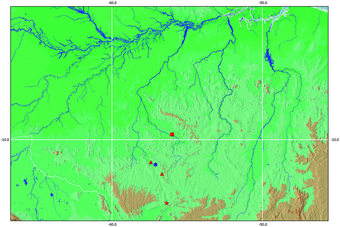
Distribution: Corydoras apiaka is only known from tributaries of the rio Arinos, rio Teles Pires and rio Preto, clearwater tributaries of the upper rio Tapajós in Mato Grosso State, Brazil ( Figs. 3 View FIGURA 3 , 4 View FIGURA 4 ).
Habitat and ecological notes: Corydoras apiaka was mostly found in lotic habitats in the rio Arinos and its tributaries. The rio Arinos has a muddy-brown color, with soft bottom of clay and sand. Most of the specimens were captured in the small forest streams of black or clear water, or in marginal ponds.
Etymology: The specific name apiaka is treated as a noun in apposition and is named for the indigenous tribe Apiaká (means “people” in Tupi language), which originally occupied the middle and lower rio Arinos, lower rio Juruena, but is nowadays restricted to the lower rio Juruena ( Menéndez, 1992). The tribe is known for facial tattoos and bravery in battles, as well as by anthropophagic rites after fights ( Castelnau, 1850).


No known copyright restrictions apply. See Agosti, D., Egloff, W., 2009. Taxonomic information exchange and copyright: the Plazi approach. BMC Research Notes 2009, 2:53 for further explanation.