
Chironephthya cornigera, Imahara & Chavanich & Viyakarn & Kushida & Reimer & Fujita, 2020
|
publication ID |
https://doi.org/ 10.11646/zootaxa.4780.2.6 |
|
publication LSID |
lsid:zoobank.org:pub:ADDCAA3B-FBBD-4F0B-859A-EC62DAF3DAD6 |
|
DOI |
https://doi.org/10.5281/zenodo.3854345 |
|
persistent identifier |
https://treatment.plazi.org/id/03A1CD0A-B562-FFB9-FF44-61ADA2E81175 |
|
treatment provided by |
Plazi |
|
scientific name |
Chironephthya cornigera |
| status |
sp. nov. |
Chironephthya cornigera View in CoL sp. nov.
( Figs. 8–3 View FIGURE 8 )
urn:lsid:zoobank.org:act:3D0575F0-A3AB-45F8-AAA2-E56ACCD1FD5B
Japanese name: Tsuno-kudayagi
Holotype. NSMT-Co 1704, at 11 m depth, Koh Rin , off Pattaya, Chonburi Province, Thailand (12°47.44'N, 100°42.18'E), on 13 October 2013. No. MT 424942 View Materials (mtMutS), MT 414717 View Materials (COI), MT 419963 View Materials (28SrDNA). GoogleMaps
Paratype. NSMT-Co 1705, at 13 m depth, same locality and same date as the holotype. No. MT 424943 View Materials (mt- MutS), MT 414718 View Materials (COI) .
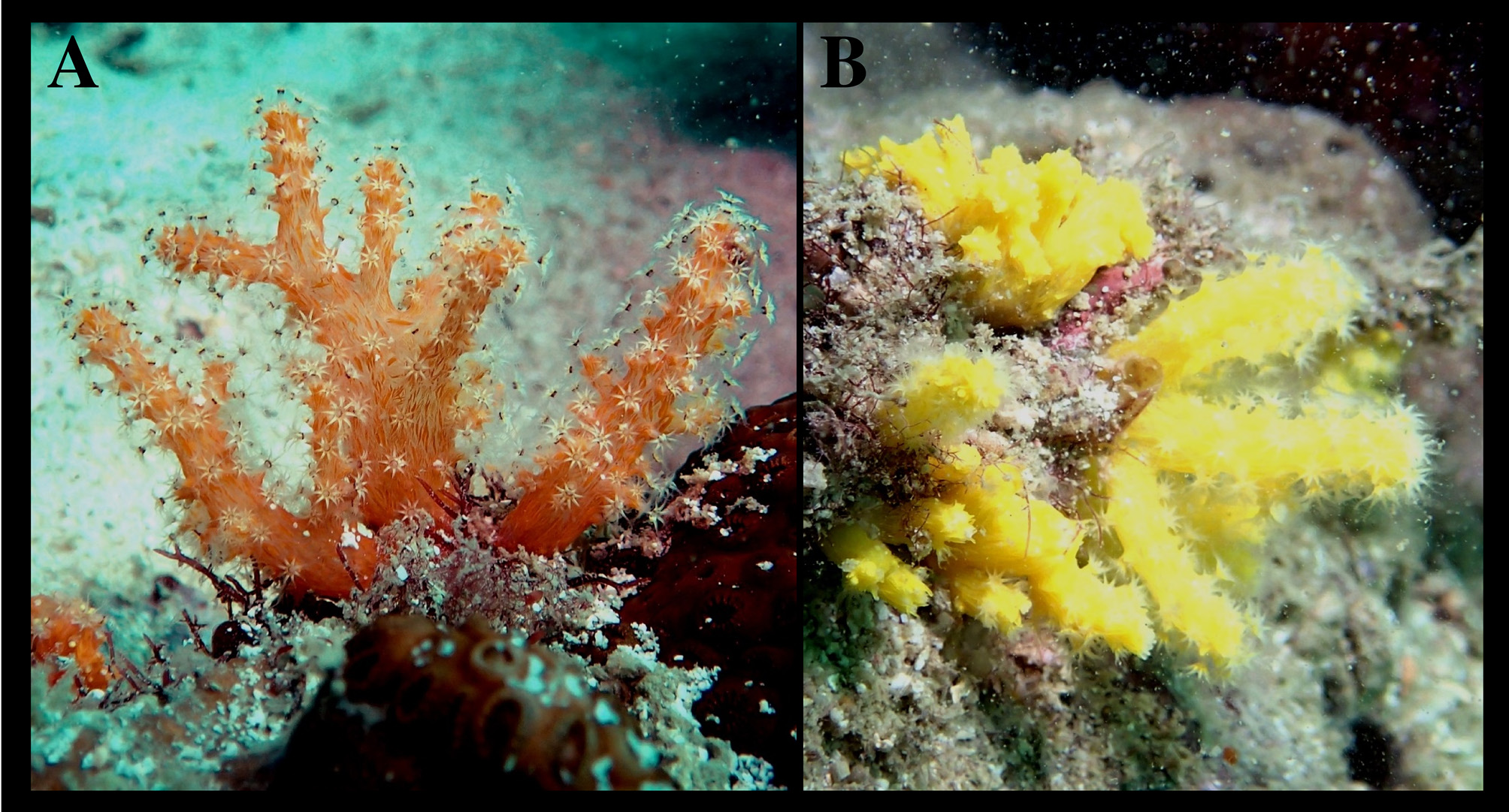
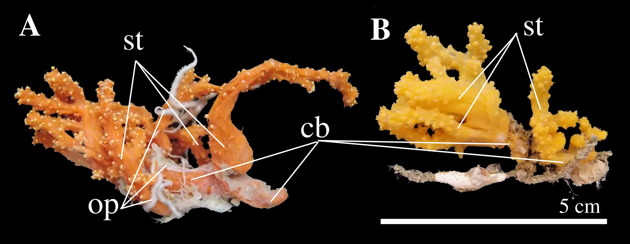
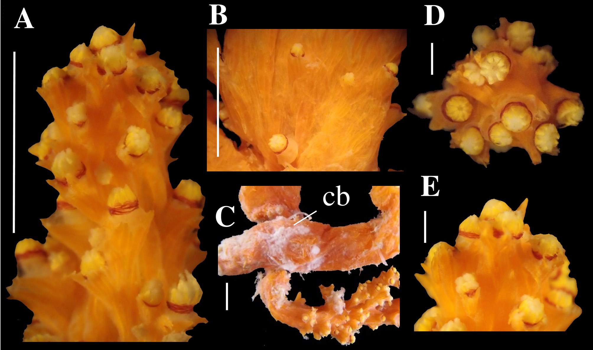
Description of the holotype. Colony form: Colony, 40 mm high, consisting of seven polyparia arising from a thick cylindrical common base, 6 x 7 mm in the maximal diameter and 25 mm long, attached to a dead scleractinian coral, soft in life ( Fig. 8A View FIGURE 8 ) but stiff in ethanol ( Fig. 9A View FIGURE 9 ). Common base covered with many hydrozoan polyps and membranous bryozoans, and in the interior, there are several thick canals, 0.5–1.5 mm in diameter, running through its length. Lower portion of each polyparium are short stems, 3–7 mm long and 3 x 3.5–3 x 4 mm in diameter near the proximal portion. Polyparia 11–47 mm high (including lower stem), sparsely branched in all directions. Some of these branches branch off into a few secondary branches. Terminal branches are cylindrical and long, reaching 19 mm long and 3.5 x 4 mm in diameter, whether primary or secondary branch. In the interior of stem five to seven thick canals leading to apical polyps and an additional three to five thin canals. Canal walls are thin, soft and limp.
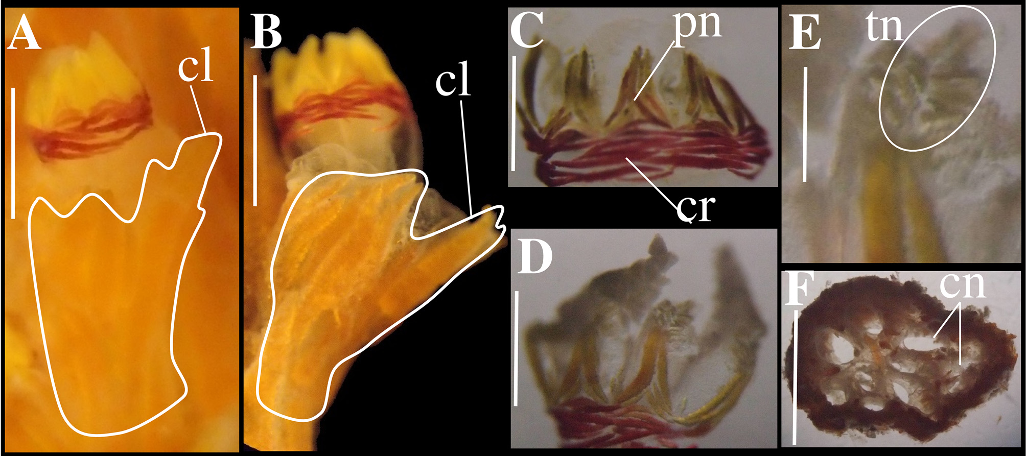
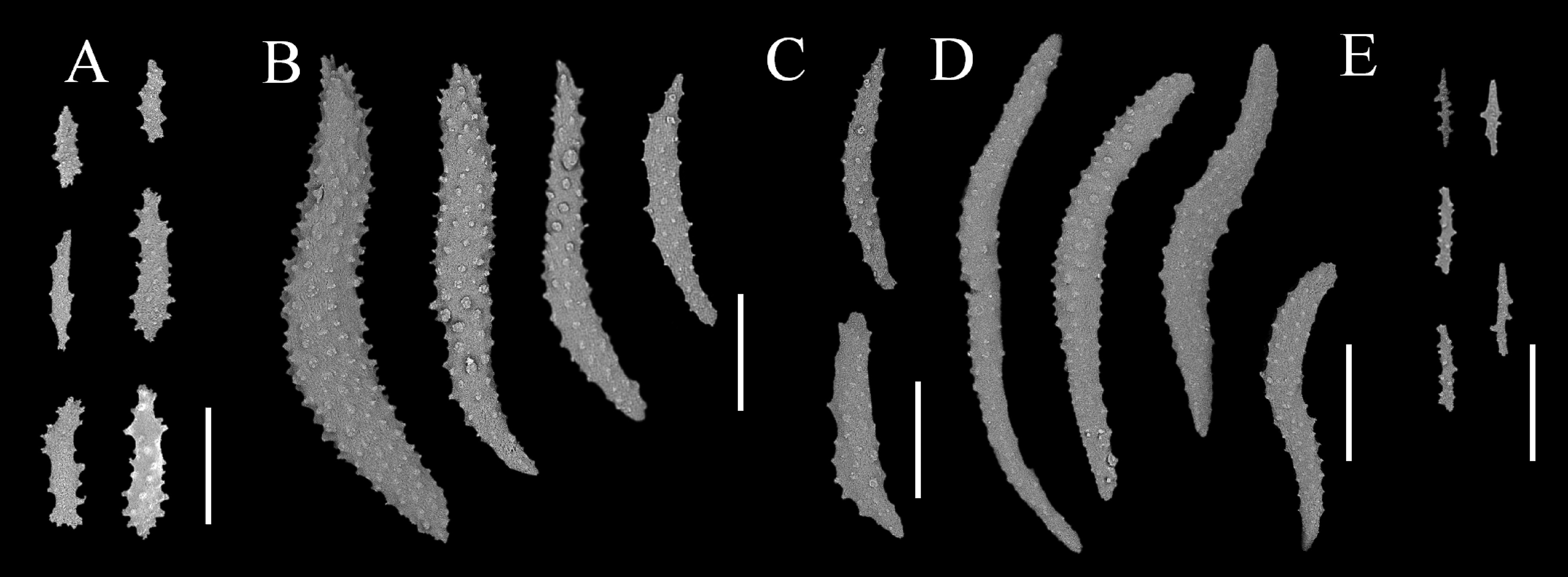
Polyps: Monomorphic polyps with calices, occur independently at wide intervals, 2–3 mm, at the lower portion of the stem ( Fig. 10B View FIGURE 10 ), but at the upper portion, the interval gradually narrows, and in some cases, they occur together. At the distal end portion of the branch, 5–7 polyps gathered, and just below which, two to eight polyps sur- round the branch ( Fig. 10D, E View FIGURE 10 ). Below these, single polyps occur in a generally oblique row around the branch ( Fig. 10A View FIGURE 10 ). In a few cases, a young polyp arises from the proximal portion of the calyx of large polyps. Calyces, in two types, cup-shaped ( Fig. 10D View FIGURE 10 ) at distal portion of terminal branches, and shelf-like ( Fig. 11A, B View FIGURE 11 ) on lateral sides of branches and stem. Both types of calyces supported by two to three teeth formed by seven to nine large, sometimes bifurcated spindles of <1.3 mm long with many complex warts ( Fig. 13A View FIGURE 13 1 View FIGURE 1 ) on the outer surface, and having small slender spindles or needles of <0.43 mm long with some conical warts ( Fig. 13A View FIGURE 13 2 View FIGURE 2 ) embedded in the inner surface. Anthocodiae, <1.0 mm high and <1 mm wide, armed by eight points and a crown ( Fig. 11 View FIGURE 11 C–D). Each point consists of one pair of large spindles of <0.42 mm with many complex warts, and 1–2 pairs of small spindles of <0.22 mm long with some conical warts ( Fig. 12B View FIGURE 12 ). Crown bearing 6–8 rows of horizontally-disposed curved spindles <0.45 mm long with many complex wart ( Fig. 12D View FIGURE 12 ). Between two adjacent points, intermediate sclerites of 1–2 pairs of rods, <0.19 mm long ( Fig. 12C View FIGURE 12 ). Anthocodial formula: 1P+(1–2)p+(6–8)Cr+(1–2)M. Tentacle axis provided with several rods, <0.12 mm log, arranged in chevron towards the distal end ( Fig. 11E View FIGURE 11 ). These rods with a few conical and complex warts. Almost completely smooth slender rods, <0.06 mm long, entering pinnules. Pharynx with a few minute spiny spindles, <0.07 mm long ( Fig. 12E View FIGURE 12 ).
Sclerites of coenenchyme: Surface layer of branches covered tightly with vertically aligned many large spindles of <3.85 mm long with many complex warts ( Fig. 13B View FIGURE 13 1 View FIGURE 1 ), and a few slender blunt ended spindles of <0.77 mm long with many complex warts ( Fig. 13B View FIGURE 13 2 View FIGURE 2 ). Several large spindles branched at one end or in the middle. Interior of branches containing a few slender spindles or needles of <0.47 mm long with a few large conical warts ( Fig. 13C View FIGURE 13 ). Surface layer of stem and common base covered tightly with large spindles of <2.1 mm long and short and thick club-like sclerites of <1.2 mm long both with many complex warts ( Fig. 13D View FIGURE 13 1 View FIGURE 1 ) and slender blunt spindles at one end of <0.47 mm long with some complex warts ( Fig. 13D View FIGURE 13 2 View FIGURE 2 ). Most spindles curved and line up vertically but some crosses across each other. Interior of stem and common base containing a few long spindles of <0.35 mm long with many complex warts ( Fig. 7E View FIGURE 7 1 View FIGURE 1 ), and a few needles of <0.28 mm long with a few conical warts ( Fig. 13E View FIGURE 13 2 View FIGURE 2 ).
Color: Colony orange, reflecting the color of large spindles, with whitish tentacles and red crown in life. These colors hardly change in ethanol.
Etymology. Specific name cornigera is a Latin word corniger = bearing horns, referring to the colony shape in life.
Variability among type material. Paratype similar in colony form, distribution of polyps, sclerites, and anthocodial formula, but different in its yellowish color from the holotype.
Remarks. This new species differs from other species of Chironephthya in that, like C. sirindhornae sp. nov., it has multiple stems arising from a common base and lacks large spindles in the interior of coenenchyme. This new species is distinguished clearly from the former species C. sirindhornae sp. nov. due to its limited branching, the small number of small spindles of the point, the presence of sclerites in its pinnules, and its many branching spindles at the cortex of the branches. The branched horn-like appearance of the living colony of this species is similar to that of Nidalia macrospina Kükenthal, 1906 . However, the calyces of these specimens are shelf-like as in the ge- nus Chironephthya , not truncated and conical as in the genus Nidalia . The appearance of this species in life is also similar to the branched colony Eleutherobia dofleini ( Kükenthal, 1906) . However, E. dofleini is completely different from this species because in Eleutherobia , the calyx is fleshy and the sclerites of the cortex of coenenchyme are barrel-shaped. Further, the appearance of living colony of this species is similar to that of the genus Nephthyigorgia ( Fabricius & Alderslade (2001: p. 133, fig. 4), but the calyces of it are cylindrical or conical, which are different from the shelf-like form of the genus Chironephthya .


| MT |
Mus. Tinro, Vladyvostok |
No known copyright restrictions apply. See Agosti, D., Egloff, W., 2009. Taxonomic information exchange and copyright: the Plazi approach. BMC Research Notes 2009, 2:53 for further explanation.
|
Kingdom |
|
|
Phylum |
|
|
Class |
|
|
Order |
|
|
Family |
|
|
Genus |