
Cetopsis jurubidae ( Fowler, 1944 )
|
publication ID |
https://doi.org/ 10.1590/S1679-62252005000200001 |
|
publication LSID |
lsid:zoobank.org:pub:DEDABC86-3340-4797-9561-5D1E0D07A76D |
|
persistent identifier |
https://treatment.plazi.org/id/E56BC71F-0E1A-FF8E-3E51-FE74FA04E5EC |
|
treatment provided by |
Carolina |
|
scientific name |
Cetopsis jurubidae ( Fowler, 1944 ) |
| status |
|
Cetopsis jurubidae ( Fowler, 1944) View in CoL
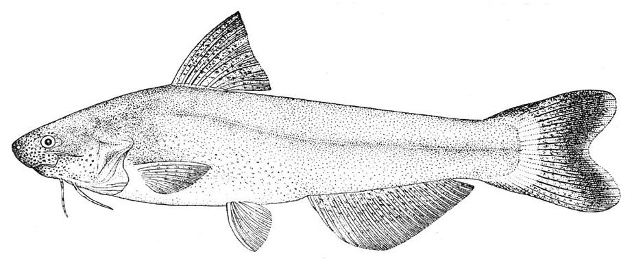
Figs. 23-25 View Fig View Fig View Fig , Tables 9 -15
Pseudocetopsis jurubidae Fowler, 1944: 235 View in CoL , figs. 7-9 [type locality: Colombia, río Jurubidá, Nuquí].– Böhlke, 1984: 40 [holotype despository].–Vari and Ferraris, 2003: 258 [in check list; distribution].
Diagnosis. Cetopsis jurubidae can be distinguished from all of its congeners by the combination of the presence of an eye, the conical teeth on the vomer and dentary, the rounded posterior nares that is distinctly separated from the contralateral nares by a distance greater than the width of the posterior nares, the absence of a dark humeral spot, the lack of dark basal pigmentation on the middle portion of the dorsal fin, the absence of a posteriorly-rounded, variably-developed, bilobed patch of dark pigmentation at the base of the caudal fin, the lack of dark pigmentation on the distal portions of the anal and pectoral fins, and the possession of 9 pectoral-fin rays, 43 total vertebrae, 12 precaudal vertebra, 18 preanal vertebrae, 31 caudal vertebrae, 23 branched anal-fin rays, and 28 total anal-fin rays.
Description. Body relatively elongate, slightly transversely compressed anteriorly and becoming progressively distinctlycompressed posteriorly, but with relative degree of compression impossible to determine because of desiccation of only known specimen. Body depth at dorsal-fin origin approximately 0.25 of SL, and approximately equal to HL. Lateral line on body complete, unbranched, and midlateral; extending from vertical through pectoral-fin base onto hypural plate and terminating prior to posterior margin of hypural plate. Dorsal profile of body cannot be determined because of desiccation that specimen experienced at some point, but holotype illustrated ( Fowler, 1944, fig. 8; reproduced herein as Fig. 24 View Fig ) with dorsal profile slightly convex from point slightly anterior to dorsal-fin origin to posterior of base of dorsal fin and nearly straight from that point to caudal peduncle. Caudal-peduncle depth approximately equal to caudal-peduncle length.
Head in profile acutely triangular overall with somewhat pointed snout, but with profile modified by noted desiccation of holotype and illustrated by Fowler (see Fig. 24 View Fig ) as more rounded. Dorsal profile of head nearly straight from tip of snout to nape. Ventral profile of head cannot be determined because of desiccation of specimen, but shown as nearly straight in drawing by Fowler (see Fig. 24 View Fig ). Margin of snout in dorsal view obtusely triangular overall and rounded anteriorly. Postorbital margins of head posteriorly divergent from dorsal view (see also Fowler, 1944, fig. 9). Condition of specimen makes it impossible to determine presence or absence of enlarged jaw musculature externally evident on dorsal surface of postorbital portion of head in some species of Cetopsinae .
Opercular membrane attaching to isthmus only to region anterior to vertical through pectoral-fin insertion. Opercular opening moderate; extending ventral of pectoral-fin insertion by distance equal to snout length and extending dorsal of pectoral-fin insertion by distance equal to width of orbit.
Eye situated on lateral surface of head; located entirely dorsal to horizontal extending through pectoral-fin insertion; eye visible in dorsal view, but not in ventral view, of head. Middle of orbit at approximately anterior one-fourth of HL. Eye diameter approximately equal to one-half of snout length. Interorbital width approximately equal to distance from tip of snout to posterior margin of orbit. Form of anterior narial opening cannot be evaluated because of desiccation of specimen. Opening of anterior nares located dorsal to horizontal extending through maxillary-barbel origin. Distance between anterior nares approximately equal to snout length. Posterior narial opening located on dorsal surface of head, situated along vertical through anterior margin of orbit; opening nearly round and nearly completely surrounded by flap of skin around anterior two-thirds of aperture and with anterior portion of flap highest.
Mouth inferior, apparently very wide in somewhat desiccated holotype; its width approximately one-half of HL. Margin of lower jaw gently rounded, its posterior limit reaching to vertical through posterior margin of orbit. Premaxillary tooth patch in form of gently-arched band continuous across midline, with anterior margin convex and posterior margin concave and running in parallel to anterior margin. Teeth on premaxilla small, conical, sharply-pointed, and arranged in three irregular rows (see also Fowler, 1944, fig. 7). Teeth of innermost premaxillary tooth row slightly larger than remaining teeth on that bone. Vomerine teeth conical and arranged in single row continuous across midline; tooth row extending transversely on medial portion of vomer and then curving posteriorly laterally. Vomerine teeth similar in form and size to largest teeth on premaxilla. Dentary teeth comparable in size and shape to premaxillary teeth, with two rows medially that taper to one row laterally (see also Fowler, 1944, fig. 7).
Maxillary barbel slender, its length slightly greater than distance from tip of snout to posterior margin of orbit, approximately 0.40 of HL; barbel origin located ventral to anterior margin of pupil. Mental barbels approximately equal in length to maxillary barbel and to each other. Medial mentalbarbel origin located along vertical through rictus. Lateral mental-barbel origin situated posterior of vertical slightly short of middle of adpressed medial-mental barbel. Tips of adpressed lateral mental barbels nearly reaching posterior margin of opercle.
Dorsal fin moderately large overall with length of dorsal-fin base approximately 0.33 of HL. Dorsal fin damaged and relative proportions of longest ray cannot be determined. Dorsal-fin spinelet apparently absent. First dorsal-fin ray missing. Form of distal margin of dorsal fin indeterminate as consequence of condition of specimen but shown as nearly straight by Fowler (see Fig. 24 View Fig ). Dorsal-fin origin located at approximately anterior 0.30 of SL and along vertical extending through distal one-half of adpressed pectoral fin. Position of tip of adpressed dorsal fin cannot be determined because of poor condition of specimen. Posterior most dorsal-fin ray without posterior, membranous attachment to body. No filaments illustrated for first ray of dorsal fin by Fowler (1944; see Fig. 24 View Fig ).
Caudal fin shallowly-forked, apparently symmetrical (see also Fowler, 1944, fig. 8); tips of lobes slightly rounded. Length of longest caudal-fin ray approximately 1.5 times length of middle fin rays.
Base of anal fin comparatively long, but length as proportion of SL cannot be determined because of condition of holotype. Anal-fin origin located well posterior of middle of SL, and slightly anterior to vertical through middle of TL. Analfin margin relatively straight, but exact form indeterminate as consequence of poor condition of holotype, but shown as convex anteriorly and straight along rest of its length by Fowler (1944, fig. 8). Posterior most unbranched anal-fin ray longest in fin, with subsequent rays becoming gradually shorter. Posterior most anal-fin ray without posterior, membranous attachment to body.
Pelvic fin moderately long; distal margin apparently nearly straight, with first ray longest. Pelvic-fin insertion apparently located anterior to middle of SL and posterior of vertical through posterior terminus of base of dorsal fin ( Fig. 24 View Fig ). Tip of adpressed pelvic fin extending beyond middle of SL and to beyond anterior limit of vent but falling short of anal-fin origin. Medial most pelvic-fin ray with membranous attachment to body along basal two-thirds of its length.
Pectoral-fin length approximately two-thirds of HL. Pectoral-fin margin very gently convex, with first ray longest. First pectoral-fin ray not spinous and without distal filament (see also Fig. 24 View Fig ).
Coloration in alcohol. Coloration on head and body cannot be determined as a consequence of desiccation of the holotype, the only known specimen. Although the pigmentation was not discussed in detail in the original description of Cetopsis jurubidae , the illustration of the holotype by Fowler (1944, fig. 8) showed dark pigmentation scattered on head and body with larger spots of pigmentation on snout, upper jaw, region ventral to orbit, and on lower portion of opercle.

No known copyright restrictions apply. See Agosti, D., Egloff, W., 2009. Taxonomic information exchange and copyright: the Plazi approach. BMC Research Notes 2009, 2:53 for further explanation.
|
Kingdom |
|
|
Phylum |
|
|
Class |
|
|
Order |
|
|
Family |
|
|
Genus |
Cetopsis jurubidae ( Fowler, 1944 )
| Vari, Richard P., Ferraris Jr, Carl J. & de Pinna, Mário C. C. 2005 |
Pseudocetopsis jurubidae
| Bohlke, E 1984: 40 |
| Fowler, H 1944: 235 |