
Campsurus latipennis (Walker), 1853
|
publication ID |
https://doi.org/ 10.11646/zootaxa.4300.3.1 |
|
publication LSID |
lsid:zoobank.org:pub:554D8B46-D396-42FA-9604-6DA9DFA3EFE7 |
|
DOI |
https://doi.org/10.5281/zenodo.6033461 |
|
persistent identifier |
https://treatment.plazi.org/id/0386A43A-EF75-A230-8B85-FA62FAB3FA75 |
|
treatment provided by |
Plazi |
|
scientific name |
Campsurus latipennis (Walker) |
| status |
|
Campsurus latipennis (Walker) View in CoL
( Figs. 1–24 View FIGURES 1 – 12 View FIGURES 13 – 24 , 163 View FIGURES 163 – 166 )
Palingenia latipennis Walker, 1853: 554 ; Hagen, 1861: 304.
Campsurus latipennis View in CoL ; Kimmins, 1960: 313 (male); Domínguez et al. 2006: 572.
Type material. Photographs of the lectotype male subimago and lectoallotype alate female (both pinned), in the British Museum , from " Para, 49–I " ( Kimmins 1960: 313).
Additional material. BRAZIL: 200 ♂ i from Espírito Santo, Linhares, Lagoa Nova, S 19° 25' 7.2" – W 40° 9' 26.4", 17 m, 12.ix.2009, light-trap, Salles F. leg. ( CZNC); 3 ♂ i, 1♀ msi, alate 7 ♀ and 3 last instar ♂ nymphal cuticles from Espírito Santo, Linhares, Palminhas, Rio Doce, 9–10.ix.2014, Salles F. leg. (IBN); and 6 ♂ i (wings on slide IBN527CM) from Tocantins (TO), Ilha Bananal, rio Araguaia , (approx. S 5° 24' / W 48° 37'), 10.xi.1953, AA Pesle col. (IBN). GoogleMaps
Male imago. Length (mm): body, 9.8–17.3; fore wing, 8.4–13.5; hind wing, 4.3–6.5; cerci, 22.0–35.0; fore leg, 4.2–5.5. General coloration whitish with well-defined gray markings. Head yellowish white, almost completely shaded with black dorsally ( Fig. 163 View FIGURES 163 – 166 ). Antennae whitish translucent, shaded with gray. Thorax ( Fig. 163 View FIGURES 163 – 166 ). Pronotum translucent, anterior portion almost completely shaded with gray, darker at medial line, pronotal hump (ph in Fig. 163 View FIGURES 163 – 166 ) translucent; posterior pronotal portion translucent, shaded with black on margins; prosternum with area between coxae gray. Mesonotum yellowish, almost completely shaded with gray except for unpigmented anteronotal projection (ap), and blackish medioparapsidal suture (mps in Fig. 163 View FIGURES 163 – 166 ) and area between posterolateral protuberances ( PSP in Fig. 163 View FIGURES 163 – 166 ); pleura yellowish except PLsS (superior pleural suture) blackish and sternum shaded black on area between anterior furcasternal protuberances; metanotum, shaded medially with gray, metasternum with submedian black subcircular mark. Legs: fore legs whitish, almost completely shaded with gray; fore coxa with two dark-grayish marks; middle and hind legs yellowish translucent. Wings. Membrane hyaline except base and fore margin purplish gray; fore wings with veins C, Sc and R1 purplish gray, lighter toward apex; other longitudinal and cross veins shaded slightly with gray except from MP2 toward base; hind wing with base and subcostal veins purplish grayish. Abdomen ( Fig. 163 View FIGURES 163 – 166 ) translucent whitish shaded with gray on terga as in Fig. 163 View FIGURES 163 – 166 , slightly paler medial line present on some terga; abdominal sterna pale without shading, except remnants of gill insertions and apical third of sterna VII–VIII shaded light grayish. Genitalia ( Figs. 1–6, 9–12 View FIGURES 1 – 12 ): sternum IX with grayish medial triangle and blackish hind margin, with blunt medial projection ( Figs. 1 View FIGURES 1 – 12 mp) and microsculptured with fine, long and simple microtrichiae ( Fig. 10 View FIGURES 1 – 12 ); pedestal bases well separated from each other ( Figs. 1, 3, 9 View FIGURES 1 – 12 ), pedestals yellowish white, with outer apical corner projected forming relatively short and blunt parastylus (ps in Fig. 2 View FIGURES 1 – 12 ); forceps whitish; penes whitish except dorsal sclerotized margin yellowish, base of penes (bp in Figs. 1, 9 View FIGURES 1 – 12 ) large and subquadrangular, main lobe of penes (ml in Figs. 1, 9 View FIGURES 1 – 12 ) subconical with subapically indented dorsal margin (in lateral view, arrows in Figs. 4–5 View FIGURES 1 – 12 ), apical portion twisted, secondary lobe of penes (sl in Figs. 1, 9 View FIGURES 1 – 12 ) small, membranous and subcylindrical. Caudal filament translucent whitish.
Alate female. Length (mm): body, 11.0–16.5; fore wing, 12.5–16.0; hind wing, 5.3–6.5; cerci, 3.8–4.5; fore leg, 1.4–1.5. General aspect and color pattern similar to male imago. Abdominal sternum VIII with anteromedian paired sockets, sockets fused with each other medially, shallow ( Figs. 7–8 View FIGURES 1 – 12 ).
Egg. Length, 320–335 µm; maximum width, 235–250 µm. Ovoid outline, bowl-shaped, as usual for Campsurinae; no polar cap present.
Nymphal cuticle (last instar, male). Length (mm): body, 13.5–14.5; cerci, 9.0–10.0; terminal filament, 7.0. Head with frons almost completely covered with short and long setae (setal alveoli marked as dots in Fig. 13 View FIGURES 13 – 24 ), straight anterior margin with row of long setae, two anterolateral blunt tubercles at base on antennae (larger apical one and smaller one, t in Fig. 13 View FIGURES 13 – 24 ), anterior half of gena protruded and covered with long, strong setae, posterior half of gena bare, except for small area before eye also covered with long, strong setae (not shown in Fig. 13 View FIGURES 13 – 24 ); inner margin of compound eye without row of setae. Pedicel 1.4× length of scape, dorsally covered with two groups of long strong setae (one group near base and another group on distal half), in-between group of slightly shorter and much thinner setae present; scape dorsally with short and strong setae; flagellum (apical portion broken off and lost) with submedian short seta on each segment. Mandibular tusk relatively long and slender, apex strongly curved inward ( Fig. 16 View FIGURES 13 – 24 ); exposed area 0.5× length of head capsule; basal U-row of filtering setae present (fs in Figs. 14– 15 View FIGURES 13 – 24 ); dorsal surface with numerous very long, strong setae; outer margin covered with long setae and 25–30 strong blade-like spines (increasing in size distally, bls in Fig. 16 View FIGURES 13 – 24 ); inner margin with 10 small tubercles (subequal in size, except basal one slightly larger, t in Fig. 16 View FIGURES 13 – 24 ), some of them alternating with short strong setae (ss in Fig. 16 View FIGURES 13 – 24 ), tubercles and setae ordered in slightly curved row (in occlusal view, Fig. 14 View FIGURES 13 – 24 ); ventral surface almost without setae. Maxilla (apparently hard to see in described exuviae) with small ventral gill. Thorax. Posterolateral corner of pronotum and propleura without strong setae; metasternum with few long setae. Legs. Fore legs with coxa and trochanter bare; dorsal surface of femur with outer submarginal row of long setae on distal third (arrows on Fig. 17 View FIGURES 13 – 24 ), near base of outer margin with rounded projection covered by long, weak setae (rp in Fig. 17 View FIGURES 13 – 24 ); base of femur and part of inner margin with row of marginal long, strong setae (st in Fig. 17 View FIGURES 13 – 24 ); ventrally long U-row of filtering setae (fs in Fig. 18 View FIGURES 13 – 24 ); tibia-tarsus wide, with margins subparallel, dorsal surface almost completely covered with short spines and long setae ( Fig. 17 View FIGURES 13 – 24 ), ventral surface with 2 rows of filtering setae: basal W-shaped transversal row extending along ventral surface and hind margin (wr in Fig. 18 View FIGURES 13 – 24 ), and double longitudinal row along anterior margin (ar in Fig. 18 View FIGURES 13 – 24 ); apex of tibia-tarsus rounded, slightly projected (projection ca. 1/4 length of claw); tarsal claw slightly curved and with row of about 18 triangular denticles ( Fig. 19 View FIGURES 13 – 24 ). Middle leg with coxa and trochanter with strong setae; dorsal surface of femur covered with many long setae forming mediolongitudinal group, anterior group along margin, and subdistal transversal group, hind margin bordered by much longer setae ( Fig. 20 View FIGURES 13 – 24 ), ventral surface bare; tibia with anterior margin distally projected and with crown of strong spines (cs in Fig. 20 View FIGURES 13 – 24 ), posterior margin completely covered with very long setae, anterior margin basally bare, distal half densely covered with thick yellowish setae; tarsus with long setae on hind margin and relatively shorter setae on apical third of fore margin; tarsal claw long and slender, slightly curved, with row of 16 denticles increasing in size distally ( Fig. 21 View FIGURES 13 – 24 ). Hind leg ( Fig. 22 View FIGURES 13 – 24 ) similar to middle leg, except as follows: femur with dense row of short setae on anterior margin, anterior margin of tibia with similar dense row of setae; crown of spines on tibia absent, and tarsal claw with row of 27–30 small denticles ( Fig. 23 View FIGURES 13 – 24 ). Abdomen. Lateral margins of all segments with row of long setae. Terga IV–VII with mediolongitudinal row of setae. Sterna without setae. Gills. Abdominal gill I bilobed, dorsal lamella almost 3× width of ventral lamella, ventral lamella 2/3× length of anterior portion. Cercus 0.7× length of body, covered with setae dorsally and ventrally, except distal half bare. Terminal filament 0.5× length of body, thinner than cercus, covered with long scattered setae completely. Paraproct with distal sublateral spine (arrow in Fig. 24 View FIGURES 13 – 24 ).
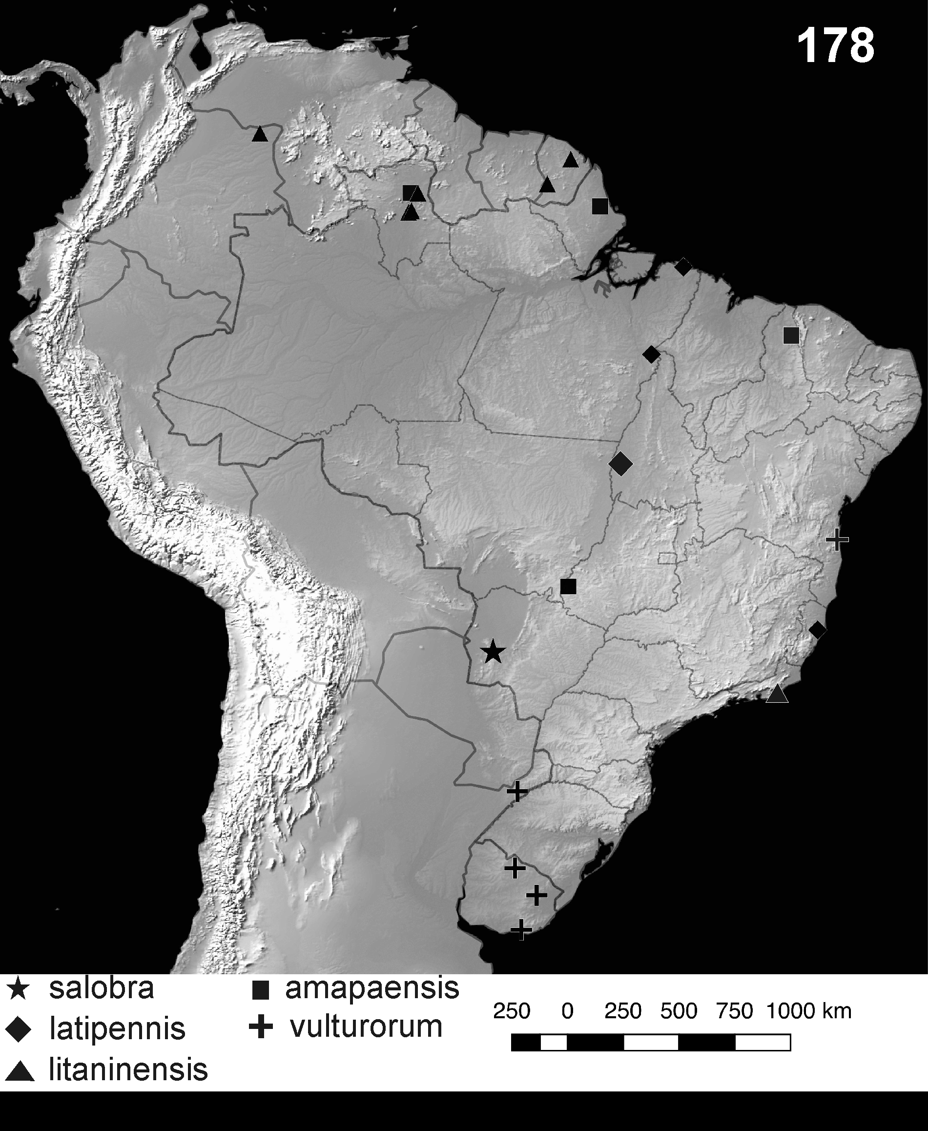
Distribution ( Fig. 178 View FIGURES 178 ). Brazil (Pará orig. New records: Espírito Santo and Tocantins).
Diagnosis. Campsurus latipennis can be distinguished from all other species of the genus by the following combination of characters. In the adult: 1) posterior margin of male abdominal sternum IX convex and subtriangular, medially rounded ( Figs. 1, 3, 9 View FIGURES 1 – 12 ), surface covered with microtrichiae ( Fig. 10 View FIGURES 1 – 12 ); 2) pedestal bases well separated from each other, pedestals subquadrate with outer-posterior margin projected forming a short parastylus ( Figs. 2, 11 View FIGURES 1 – 12 ); 3) penes separated and diverging slightly distally, each arm formed by a large and twisted sclerotized lobe, and a small ventral membranous lobe ( Figs. 1, 3, 4–6, 9, 12 View FIGURES 1 – 12 ), in lateral view the main lobe presents a subapical indentation (arrows in Figs. 4–5 View FIGURES 1 – 12 ); 4) medium to large size (length of male fore wings 8.4–13.5 mm); 5) female sternum VIII with anteromedian paired sockets, sockets fused with each other medially, shallow ( Figs. 7– 8 View FIGURES 1 – 12 ); 6) egg without polar cap. In the nymph: 1) with small paired tubercles at base of each antennae (t in Fig. 13 View FIGURES 13 – 24 ), scape and pedicel with setae; 2) pre-ocular group of setae not mounted on a tubercle; 3) mandibular tusks with apex strongly curved inwards ( Fig. 16 View FIGURES 13 – 24 ), inner margin with ten tubercles of subequal size except the basal one slightly larger (t in Fig. 16 View FIGURES 13 – 24 ), outer margin with 25–30 strong marginal blade-like spines and long setae ( Fig. 16 View FIGURES 13 – 24 ), 10 tubercles on inner margin; 4) fore femur wide (max. width 1/2 of max. length), fore tibiae with parallel margins (rectangular in form) ( Figs. 17–18 View FIGURES 13 – 24 ); 5) tarsal claws with one row of denticles (18 on fore claw, 16 on middle claw, 27–30 on hind claw) ( Figs. 19, 21, 23 View FIGURES 13 – 24 ).
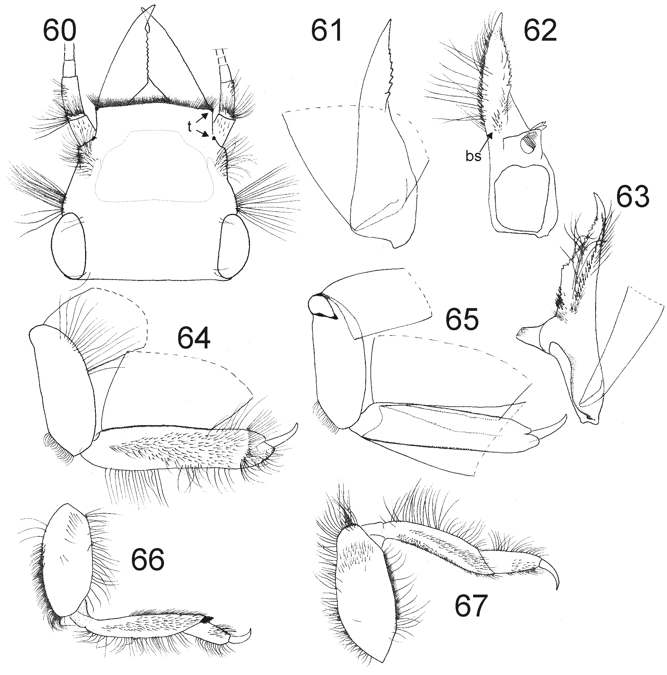
Discussion. Some mistakes appear in the use of the name C. latipennis . Walker (1853) described it based on male subimagos from Pará; the genitalia were later illustrated by Kimmins (1960: 313, Fig. 62 View FIGURES 60 – 67 ). Eaton (1883: Plate V, Fig. 8 View FIGURES 1 – 12 c) worked with imagos, obviously from a different species, collected in the same expedition by Wallace and Bates. The genitalia of the lectotype are damaged and could not be studied, but Kimmins’ (1960) figure 62 can be used to identify the species. Eaton’s (1883) imagos were not studied by us, but from his figures we identify them as C. essequibo Traver (1947) , treated below. Other authors also treated C. essequibo under the misidentification of C. latipennis ; please see the former species’ section, below, for further discussion.
Campsurus latipennis View in CoL is very similar to Campsurus salobra sp. nov. and C. evanidus Needham & Murphy 1924 View in CoL , but it can be separated from them because the hind margin of the styliger is medially projected but rounded (acute and triangular in C. evanidus View in CoL ), the pedestal presents a slightly thinner and longer parastylus (thin but short in C. salobra , short and basally wide in C. evanidus View in CoL ) and the penes present an apically twisted primary lobe and a well-developed secondary lobe (apex not twisted in the others, secondary lobe absent or reduced in C. evanidus View in CoL ). Also, the penes of C. latipennis View in CoL and C. evanidus View in CoL , in lateral view, show an indented dorsal margin, with marked subapical concave area (arrow in Figs. 4–5 View FIGURES 1 – 12 ) (smoothly convex in C. salobra sp. nov.). Campsurus cuspidatus Eaton (1871) View in CoL and C. vulturorum View in CoL share the distomedial projection of the hind margin of sternum IX, thus probably forming a monophyletic group. The absence of a polar cap on the egg is an autapomorphy of C. latipennis View in CoL . Among the species of Campsurus View in CoL , only C. evanidus View in CoL presents a very small polar cap (Emmerich & Molineri 2011).


| PSP |
Parasitic Seed Plants |
No known copyright restrictions apply. See Agosti, D., Egloff, W., 2009. Taxonomic information exchange and copyright: the Plazi approach. BMC Research Notes 2009, 2:53 for further explanation.
|
Kingdom |
|
|
Phylum |
|
|
Class |
|
|
Order |
|
|
Family |
|
|
Genus |
Campsurus latipennis (Walker)
| Molineri, Carlos & Salles, Frederico F. 2017 |
Campsurus latipennis
| Dominguez 2006: 572 |
| Kimmins 1960: 313 |
Palingenia latipennis
| Hagen 1861: 304 |
| Walker 1853: 554 |