
Callyspongia (Euplacella) abnormis Pulitzer-Finali, 1993
|
publication ID |
https://doi.org/ 10.5852/ejt.2020.698 |
|
publication LSID |
lsid:zoobank.org:pub:623BBCE3-12A5-45A9-802A-2ED2E15164A3 |
|
DOI |
https://doi.org/10.5281/zenodo.4335454 |
|
persistent identifier |
https://treatment.plazi.org/id/F52B791A-FFED-E92B-82FF-FA05C55CF36E |
|
treatment provided by |
Valdenar |
|
scientific name |
Callyspongia (Euplacella) abnormis Pulitzer-Finali, 1993 |
| status |
|
Callyspongia (Euplacella) abnormis Pulitzer-Finali, 1993 View in CoL
Fig. 7 View Fig
Callyspongia abnormis Pulitzer-Finali, 1993: 340 View in CoL , fig. 78.
Material examined
PONTA DO OURO • 5 fragments preserved dry, the larger is about 10 × 3 cm and 4 mm thick; 26°49′17.512″ S, 32°53′42.518″ E; Kev’s; 23.4 m deep; 3 Feb. 2017; Cerrano leg.; PO5 GoogleMaps • 1 fragment alcohol preserved, 1 dried fragment about 13 × 6 cm × 4 mm; 26°47′34.8″ S, 32°53′57.665″ E; Close; 18.6 m deep; 9 Feb. 2017; Cerrano leg.; PO30 GoogleMaps • Slide preparations of the holotype, labelled MBA 167, belonging to the author’s personal collection.
Description
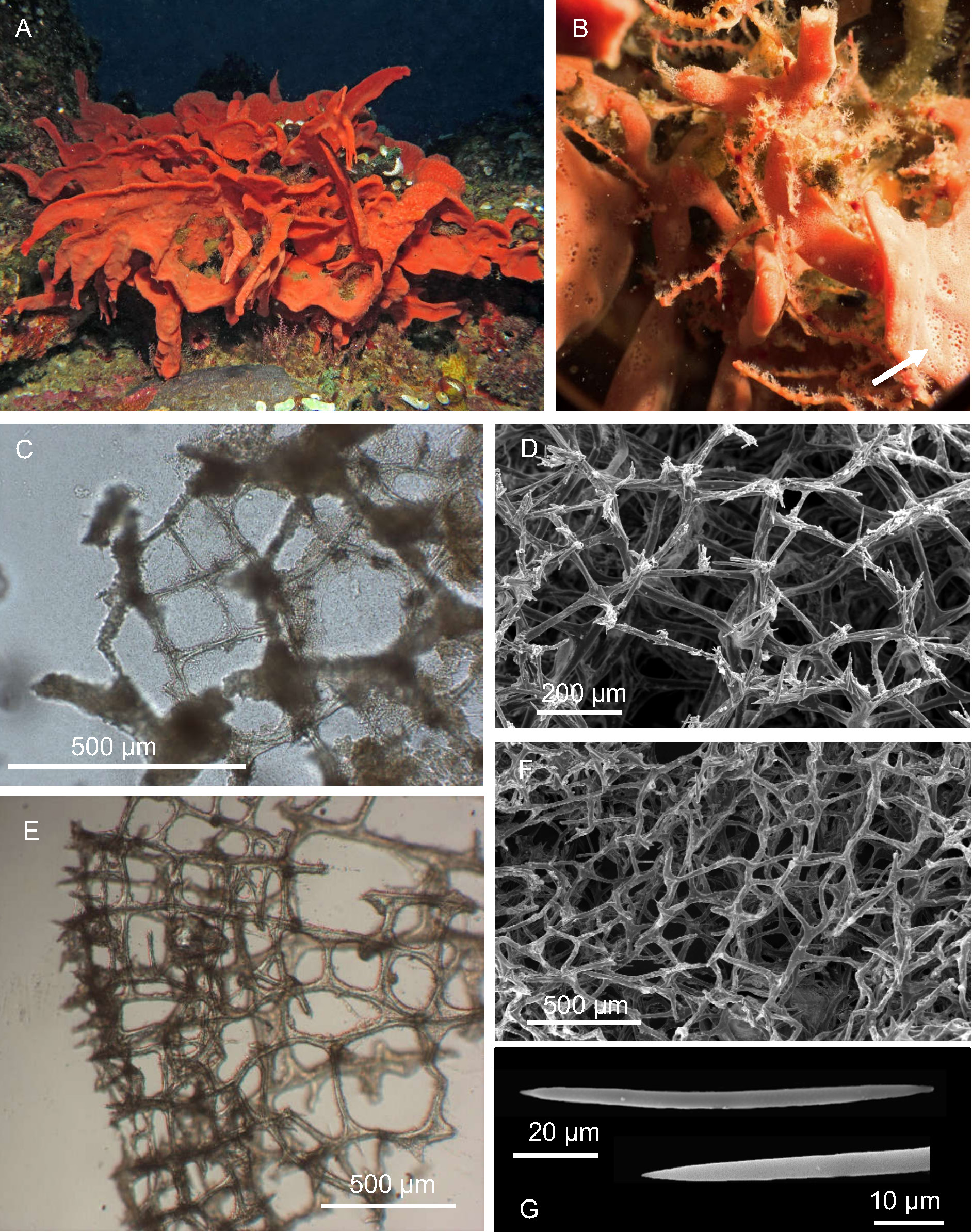
Live sponges erect, foliaceous, lamellar, about 4–5 mm thick, and with scattered, digitate processes ( Fig. 7 View Fig A–B). On one side of the sponge, roundish vents, about 300–400 μm in diameter, are grouped ( Fig. 7B View Fig ), while on the opposite side, numerous pores are scattered and not visible to the naked eye. The
color in life is brick red; ochre in preserved specimens. Surface optically smooth, but microhispid at microscope magnification. The consistence is soft and compressible, rough at touch.
SKELETON. Ectosome ( Fig. 7 View Fig C–D) formed by a reticulation of paucispicular (2–5 oxeas) fibers, 18–35 µm in section, with no marked difference between primary and secondary fibers; the meshes are irregular and 150–350 µm wide, and are echinated by free spicules. Choanosome ( Fig. 7 View Fig E–F) formed by reticulation of thicker, paucispicular (4–8 oxeas) fibers, 30–45 µm, that form irregular, rounded or polygonal meshes, 230–360 µm wide.
SPICULES. Slender and slightly curved oxeas with acerate tips, 90–(102.5, 11.9)– 115 µm × 2–(5, 1.4)– 6 µm ( Fig. 7G View Fig ).
Remarks
These samples fit with C. abnormis Pulitzer-Finali, 1993 from Mombasa, Kenya, in external morphology, skeleton structure, and spicule shape and size. The examination of the slides of the holotype confirms the identification. This new finding allowed us to assign the species to the subgenus Euplacella , considering that the ectosomal skeleton is made by one size of small rounded mesh, echinated by spicules. This is the first record of the species since its original description.
No known copyright restrictions apply. See Agosti, D., Egloff, W., 2009. Taxonomic information exchange and copyright: the Plazi approach. BMC Research Notes 2009, 2:53 for further explanation.
|
Kingdom |
|
|
Phylum |
|
|
Class |
|
|
Order |
|
|
Family |
|
|
Genus |
Callyspongia (Euplacella) abnormis Pulitzer-Finali, 1993
| Calcinai, Barbara, Belfiore, Giuseppe, Pica, Daniela, Torsani, Fabrizio, Palma, Marco & Cerrano, Carlo 2020 |
Callyspongia abnormis
| Pulitzer-Finali G. 1993: 340 |