
Callibaetis (Callibaetis) radiatus Navás 1920
|
publication ID |
https://doi.org/ 10.11646/zootaxa.4231.4.2 |
|
publication LSID |
lsid:zoobank.org:pub:99D539A1-D4BF-48C4-AEE1-0CA8F198C631 |
|
DOI |
https://doi.org/10.5281/zenodo.5315580 |
|
persistent identifier |
https://treatment.plazi.org/id/039E3278-FFAC-FFF9-DDDA-23670DDE90E7 |
|
treatment provided by |
Plazi |
|
scientific name |
Callibaetis (Callibaetis) radiatus Navás 1920 |
| status |
|
Callibaetis (Callibaetis) radiatus Navás 1920 View in CoL
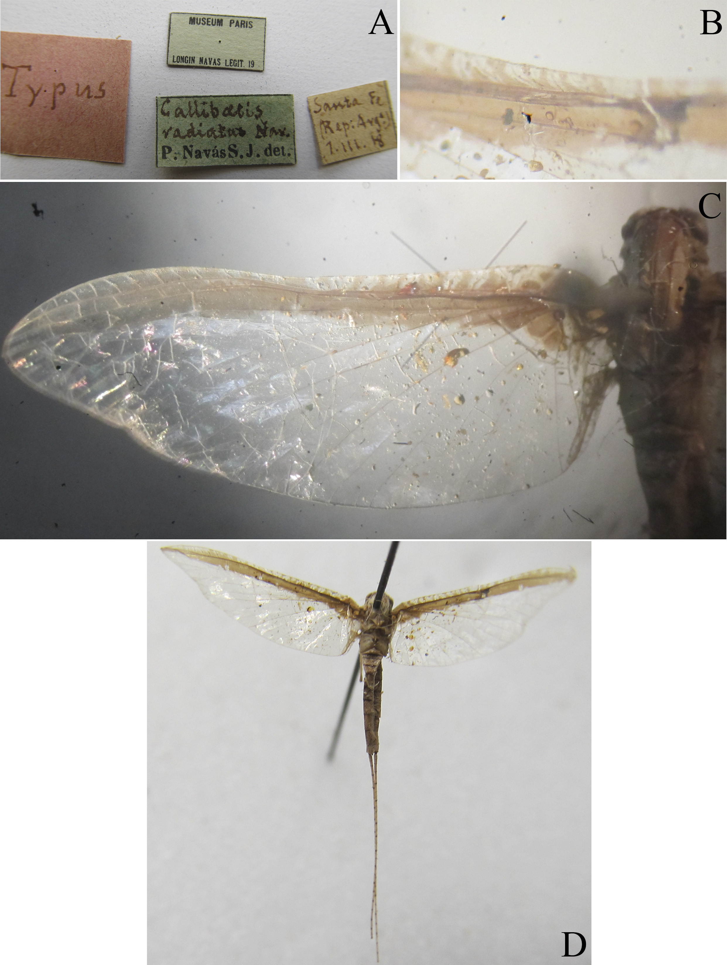
( Figs. 11 View FIGURE 11 A–14D)
Callibaetis radiatus Navás 1920a: 132 View in CoL ; Gillies 1990: 26; Salles et al. 2003: 13; Domínguez et al. 2006: 116; Cruz et al. 2014: 58; García 2014: 41; Lima et al. 2016: 214; Vinasco-Mondragón & Zúñiga 2016: 92.
Callibaetis venulosus Navás 1933: 114 . (syn. by Gillies 1990)
Known stages. I ♀♂, N.
Diagnosis. Male imago: 1) dorsal portion of turbinate eye oval ( Fig. 21 View FIGURE 21 in Salles et al. 2003); 2) dorsal portion of turbinate eyes without constriction; 3) height of dorsal portion of turbinate eye stalk 0.85 × height of dorsal portion; 4) turbinate portion of compound eyes (in lateral view) with divergent anterior and posterior margins; 5) forewings hyaline (Fig. 167 in Cruz et al. 2014); 6) thick cross veins in C and Sc areas (Fig. 167 in Cruz et al.
2014); 7) marginal intercalary vein single (Fig. 167 in Cruz et al. 2014); 8) hind wing hyaline (Fig. 168 in Cruz et al. 2014); 9) hind wing with quadrangular costal process (Fig. 168 in Cruz et al. 2014); 10) hind wing with thick cross veins (Fig. 168 in Cruz et al. 2014); 11) marginal intercalary veins present on hind wing (Fig. 168 in Cruz et al. 2014); 12) abdominal terga with black mark anterolaterally (Fig. 169 in Cruz et al. 2014); 13) abdominal sterna with a weakly pigmented medioanterior and medioposterior sigilla (Fig. 172 in Cruz et al. 2014); 14) segment I of forceps wide at base (Fig. 166 in Cruz et al. 2014); 15) segment III of forceps truncated apex (Fig. 166 in Cruz et al. 2014).
Female imago: 1) forewing with brown pigmented C, Sc and R1 areas, extending beyond R1 but not forming bands ( Fig. 11 View FIGURE 11 C); 2) forewing with thick and black cross veins in C and Sc areas ( Fig. 11 View FIGURE 11 B); 3) marginal intercalary veins single ( Fig. 11 View FIGURE 11 C); 4) hind wing hyaline (Fig. 171 in Cruz et al. 2014); 5) hind wing with quadrangular costal process (Fig. 171 in Cruz et al. 2014); 6) marginal intercalary veins on hind wing present (Fig. 171 in Cruz et al. 2014); 7) hind wing with thick cross veins (Fig. 171 in Cruz et al. 2014); 8) abdomen with brown spots; 9) anterolateral spot on abdominal terga and sterna present (Figs. 169 and 172 in Cruz et al. 2014); 10) abdominal sterna with a weakly pigmented medioanterior and medioposterior sigilla (Fig. 172 in Cruz et al. 2014).
Mature nymph: 1) maxillary palp 1.2 × length of galea-lacinia ( Fig. 12 View FIGURE 12 F); 2) tuft of robust spine-like setae below insertion of maxillary palp absent ( Fig. 12 View FIGURE 12 F); 3) ventral surface of paraglossa with row of spine-like setae ( Fig. 12 View FIGURE 12 G); 4) segment III of labial palp rounded apically ( Fig. 12 View FIGURE 12 G); 5) metanotum without spines; 6) anterior surface of foretarsus without spine-like setae ( Fig. 13 View FIGURE 13 A).
Redescription. Mature nymph: Length: body, 7.2–8.0 mm; broken cerci and terminal filament (n=1). Head. Coloration: faded in alcohol. Turbinate portion of male compound eyes yellowish brown. Antenna with small spines and fine, simple setae ( Fig. 12 View FIGURE 12 A). Labrum ( Fig. 12 View FIGURE 12 B) maximum length about 1.4 × maximum width; anterolateral margins with long spine-like setae; distal margin with spine-like setae medially; dorsal surface with many, long, fine and simple setae; distal margin with one row of fine spine-like setae ventrally; ventral surface with short, spine-like setae near lateral margin. Right mandible ( Fig. 12 View FIGURE 12 C) with 4 + 3 denticles; margin between prostheca and mola convex; basal half with short, fine, simple setae and pores scattered over dorsal surface. Left mandible ( Fig. 12 View FIGURE 12 D) with 4 + 2 denticles; margin between prostheca and mola straight; basal half with short, fine and simple setae and pores scattered over dorsal surface. Hypopharynx ( Fig. 12 View FIGURE 12 E). Lingua with lobe covered with small simple setae; short, fine and simple setae scattered over distal margin of superlingua. Maxilla ( Fig. 12 View FIGURE 12 F). Medial protuberance of galea with 1+ 3 spine-like setae. Maxillary palp short, 1.2 × length of galea-lacinia; palp segment II 1.2 × length of segment I; outer margin of segment I scattered with long, fine, simple setae; inner margin of segment II with few spine-like setae. Labium ( Fig. 12 View FIGURE 12 G). Glossa subequal in length to paraglossa; inner margin with 11 spine-like setae; apex with 3 – 5 long spine-like setae; outer margin with row of long spine-like setae; dorsal surface with one row of long spine-like setae near outer margin and one tuft of setae at apex. Paraglossa. Ventral surface with one row of fine and simple setae; dorsal surface with three rows of long spine-like setae. Labial palp with segment I 1.1 × length of segments II and III combined; segment I covered with micropores; inner and outer margin of segment II with six spine-like setae; dorsal surface with row of six short spine-like setae; segment III with rounded apex. Thorax. Foreleg ( Figs. 13 View FIGURE 13 A). Ratio of foreleg segments 0.9:1:0.6:0.3. Forefemur. Length about 4.0 × maximum width; dorsal margin with row of short, spine-like setae; apex with two robust spinelike setae; length of setae about 0.1 × maximum width of femur; anterior surface near dorsal margin with one row of spine-like setae. Tibia. One row of short spine-like setae ventrally; anterior surface with few fine and simple setae. Tarsus. Anterior and posterior surface without spine-like setae. Tarsal claw 0.5 × length of tarsus ( Figs. 13 View FIGURE 13 B). Femur of hind leg without trifid setae; tarsal claw with small denticles ( Fig. 13 View FIGURE 13 C). Abdomen. Terga. Posterior margin with regular spines ( Fig. 14 View FIGURE 14 A). Sterna. Surface with fine, simple setae. Paraproct ( Fig. 14 View FIGURE 14 B) with 24 marginal spines; surface with micropores and short, fine, simple setae. Cercus and terminal filament at base as in Figures 14 View FIGURE 14 C and 14D respectively.
Comments. The female imagoes of C. (C.) radiatus , C. viviparus and C. camposi are similar and could not be distinguished based on re-evaluation of the morphological evidence. However, as stated in the comments of C. camposi , the type locality of C. viviparus can be accessed and new specimens can be collected in the future.
García (2014) recorded C. (Callibaetis) radiatus in Venezuela based on reared specimens (female, male and its nymphal exuviae). Taking into account the similarity between C. (C.) radiatus , C. viviparus and C. camposi and, in order to enable the comparisons to confirm this record, we re-describe and present new diagnosis and illustrations of the nymph described by Salles et al. (2003), as well figures of the specimen originally described by Navás (1920), here designated as lectotype ( Figs. 11 View FIGURE 11 A–11D).
Recently, Lima et al. (2016) and Vinasco-Mondragón & Zúñiga (2016) recorded C. (C.) radiatus respectively from Bahia state ( Brazil) and Colombia, all based on female imagoes. Taking into account the morphological evidence here presented, both 2016 records of C. radiatus could be applicable to C. viviparus or C. camposi . Associations between stages should be made in order to confirm or refute these records.
Material examined. Callibaetis radiatus , photographs of one female imago (lectotype by present designation), Argentina, Santa Fé, 1.iii.1918, MNHN ; one female imago, Argentina, Formosa, Reserva EL, Bagual , xi-2004, Serochi coll.; one female imago, Paraguay, Asunción, 15.x.1919, MZB ; male imago, female imago (reared) and two nymphs, BRAZIL, Minas Gerais, Viçosa, Ranário—Universidade Federal de Viçosa , 27.i.1997, E.R. Silva coll.
Distribution. Argentina: Santa Fé. Paraguay: Asunción. Brazil: Bahia; Minas Gerais.


No known copyright restrictions apply. See Agosti, D., Egloff, W., 2009. Taxonomic information exchange and copyright: the Plazi approach. BMC Research Notes 2009, 2:53 for further explanation.
|
Kingdom |
|
|
Phylum |
|
|
Class |
|
|
Order |
|
|
Family |
|
|
Genus |
Callibaetis (Callibaetis) radiatus Navás 1920
| Cruz, Paulo Vilela, Salles, Frederico Falcão & Hamada, Neusa 2017 |
Callibaetis venulosus Navás 1933 : 114
| Navas 1933: 114 |
Callibaetis radiatus Navás 1920a : 132
| Lima 2016: 214 |
| Vinasco-Mondragon 2016: 92 |
| Garcia 2014: 41 |
| Dominguez 2006: 116 |
| Salles 2003: 13 |
| Gillies 1990: 26 |
| Navas 1920: 132 |