
Boana gracilis ( Melin, 1941 )
|
publication ID |
https://doi.org/ 10.11646/zootaxa.4750.1.1 |
|
publication LSID |
lsid:zoobank.org:pub:FC39AEF5-7190-4C94-8C14-6BACBA41E311 |
|
DOI |
https://doi.org/10.5281/zenodo.3706257 |
|
persistent identifier |
https://treatment.plazi.org/id/03D85941-E361-BB0A-FF4F-6F1CF81AFCC4 |
|
treatment provided by |
Plazi |
|
scientific name |
Boana gracilis ( Melin, 1941 ) |
| status |
|
Boana gracilis ( Melin, 1941) , revalidated, new combination
( Figs. 2 View FIGURE 2 , 4 View FIGURE 4 , 9 View FIGURE 9 )
Hyla granosa gracilis Melin 1941: 21 View in CoL ; Lutz 1951 a [1949]: [map].
Hyla granosa Rivero 1961: 100 View in CoL ; Rivero 1972: 182; Duellman 1974: 8; Hoogmoed 1979: 5 (partly, material from Canaripó); Galatti et al., 2007 (only Fig. 5 View FIGURE 5 which shows a specimen from PNJ).
Hyla punctata Cochran & Goin 1970: 224 View in CoL .
Hypsiboas granosa Faivovich et al., 2005: 85 .
Hypsiboas cinerascens Frost et al., 2006: 65 View in CoL ; Pyron & Wiens 2011: 570; Faivovich et al., 2013: 50; Duellman et al., 2016 (partly); Pinheiro et al., 2019
Boana cinerascens Frost, 2019 : [website].
Diagnosis. Based on the molecular phylogenetic analysis, Boana gracilis should be considered a member of the B. punctata group. Boana gracilis is diagnosed morphologically by the following combination of characters: (1) medium sized Boana (SVL males 33.7–37.2, mean 35.9 mm, N = 26); (2) snout truncate in both dorsal and lateral views; (3) skin on dorsum granular; (4) forearm slightly hypertrophied; (5) prepollex small prepollical spine not protruding out of skin; (6) mental gland present; (7) number of vomerine teeth up to 10; (8) lacking small tubercles on postaxial side forearm and supernumerary tubercles on hand; (9) round subarticular tubercles on fingers I and IV, by not having supernumerary tubercles on hands; (10) in life, dorsum predominantly yellowish-green with large yellow blotches, scattered reddish-brown dots (melanophores) and reddish-brown irregular blotches within the white blotches (most of the examined specimens), a reddish-brown transversal interorbital bar (expanded on eyelid); (11) in preservative, dorsum cream with large white blotches and scattered brown small melanophores, and brown irregular blotches within the white blotches (most of the examined specimens); a brown transversal interorbital bar (expanded on eyelid); (12) dorsolateral stripe absent; (13) subgular, single vocal sac.
Comparisons with congeners in the Boana punctata group. Because of the phylogenetic position of this taxon, we limit the comparisons to species in the B. punctata group. We made the comparisons by direct examination of preserved specimens, photographs (especially for color in life) and/or the literature. Character states for the species under comparison are given in parentheses. Boana gracilis differs from B. alemani in dorsal color pattern (in preservative: cream with dark spots; color in life unknown) and its larger size (SVL 30.5 mm). Boana gracilis differs from B. atlantica in dorsal color pattern (in preservative: white with yellow dorsolateral stripes and sparse spots; in life: green with reddish brown dorsolateral stripes and sparse spots) and its smaller size (male SVL> 37 mm) (examined specimens and Caramaschi & Velosa 1996; Haddad et al. 2013). Boana gracilis differs from B. cinerascens by its higher number of vomerine teeth (up to 10 vs. up to six in B. cinerascens ), by lacking small tubercles on postaxial side forearm (present), and supernumerary tubercles on hand (present), dorsal color pattern (in preservative: cream with scattered, very small, white spots; in life: green, with sparse small white spots). Boana gracilis differs from B. hobbsi in dorsal color pattern (cream with few and sparse dark brown spots and two dorsolateral dark brown stripes) and its small size (holotype B. hobbsi SVL = 42.5 mm) (examined specimen, and Cochran & Goin 1970). It differs from B. picturata ( Boulenger, 1899) in dorsal color pattern (in preservative: cream with reticulate violet blotches margined by purplish red) and much smaller size (SVL> 47mm in all examined specimens). Boana gracilis differs from B. punctata in dorsal color pattern (in preservative: cream with sparse white spots and, in most individuals, dorsal lateral white and red stripes) (examined specimens and photo of the holotype provided in Milto & Barabanov 2011). It differs from B. sibleszi in dorsal color pattern (sexual dimorphism [ Hoogmoed, 1979], in life: green speckled by white or brown, with or without yellow dorsolateral stripes; in preservative: cream speckled by brown and a transversal brown bar between eyes), mental gland (absent) ( Cole et al. 2013; Hoogmoed, 1979; Kok & Kalamandeen 2008; Rivero 1972). Boana gracilis and B. sibleszi are not sister species in our phylogeny ( Fig. 5 View FIGURE 5 ). Boana gracilis differs from B. jimenezi by the shape of the subarticular tubercles on fingers I and IV (bifid), by not having supernumerary tubercles on hands (present), by the presence of a mental gland (absent) and dorsal color pattern (in life: pale green with numerous small tan or reddish-brown chromatophores, with or without dorsolateral white stripes; in preservative: cream with numerous small chromatophores, with or without immaculate white dorsolateral stripes) ( Señaris & Ayarzagüena 2006).
Description. SVL adult males 32.7–37.2 mm (35.4 ± 1.4; N = 22) and SVL adult females 28.1–33.2 mm (30.4 ± 2.1; N = 4). Head as wide as long (HW/HL = 0.88–1.13, 1.04 ± 0.05; N = 26), widest at corner of the mouth; snout truncate in both dorsal and lateral views; interorbital distance more than two and a half times the distance between the nostrils (IOD/IND = 2.44–3.72, 2.84 ± 0.33; N = 26); eye diameter slightly smaller than eye–nostril distance (ED/END 0.80–1.17, 0.99 ± 0.09; N = 26); canthus rostralis indistinct; nostrils protuberant, nearly elliptical, directed dorsolaterally; internarial area slightly concave, interorbital area flat, loreal area concave. Eyes large and protuberant, directed laterally, larger than tympanum diameter (ED/TD 1.52–2.31, 1.76 ± 0.18; N = 26); pupil horizontally elliptical; nictitating membrane transparent without any trace of reticulation, its border with brown spots.
Supratympanic fold barely evident; tympanum small (TD/ED 0.43–0.66, 0.58 ± 0.05; N = 26), round, completely covered by skin with tympanic annulus barely evident. Vocal sac subgular, single, extending slightly over the forearms. Choanae small, elliptical, not concealed by palatal shelf, larger than vomerine odontophores; a pair of vomerine odontophores present with 4–10 (7.5 ± 1.5; N = 17) vomerine teeth on right side; tongue cordiform, nearly one fourth of posterior end free; vocal slits present, extending diagonally from lateral base of tongue (anterior third) almost to angle of jaw.
Arms slender, only slightly hypertrophied; axillary membrane absent. Lateral margins of upper arm and fore- arm without tubercles or fringes; finger tips round; finger disks present on all fingers, disk on FI smallest; relative lengths of fingers: I <II <IV <III; subarticular tubercles round, narrower than finger width; subarticular tubercles on FIV larger than those of FI–III; supernumerary tubercles absent; inner metacarpal tubercle flat, elliptical, outer metacarpal tubercle absent; prepollex small prepollical spine not protruding out of skin; outer metacarpal tubercle small, almost round, barely distinguishable. Webbing between FI and FII absent, present but not extensive between FII–FIII and FIII–FIV. Variation in finger webbing: II (2 - –2)–(2½–3 +) III (2 + –2½)–(2–2 +) IV.
Legs long and slender, lacking appendages (e.g., fringes, folds, or tubercles). Ankle without appendices or tubercles. All toes well developed, disks present on all toes, disk on TI and TII smallest; relative lengths of toes: I <II <V <III <IV. Subarticular tubercles round, single; inner metatarsal tubercle flat, slightly elliptical; outer metatarsal tubercle hardly visible. Variation in foot webbing formula: I 2 - –(2–2 +) II (1–1 +)–(2–2 +)III(1–1 +)–(2–2 +) IV (2–2 +)–(1– 1 +) V. Skin on dorsum, head, dorsal surfaces of limbs, flanks and groin granular; skin on chest, belly and undersurfaces of thigh areolate; oval mental gland; skin on vocal sac granular; skin on ventral parts of fore limbs and tibiae smooth. Cloacal opening directed posteriorly; cloacal region with tubercles.
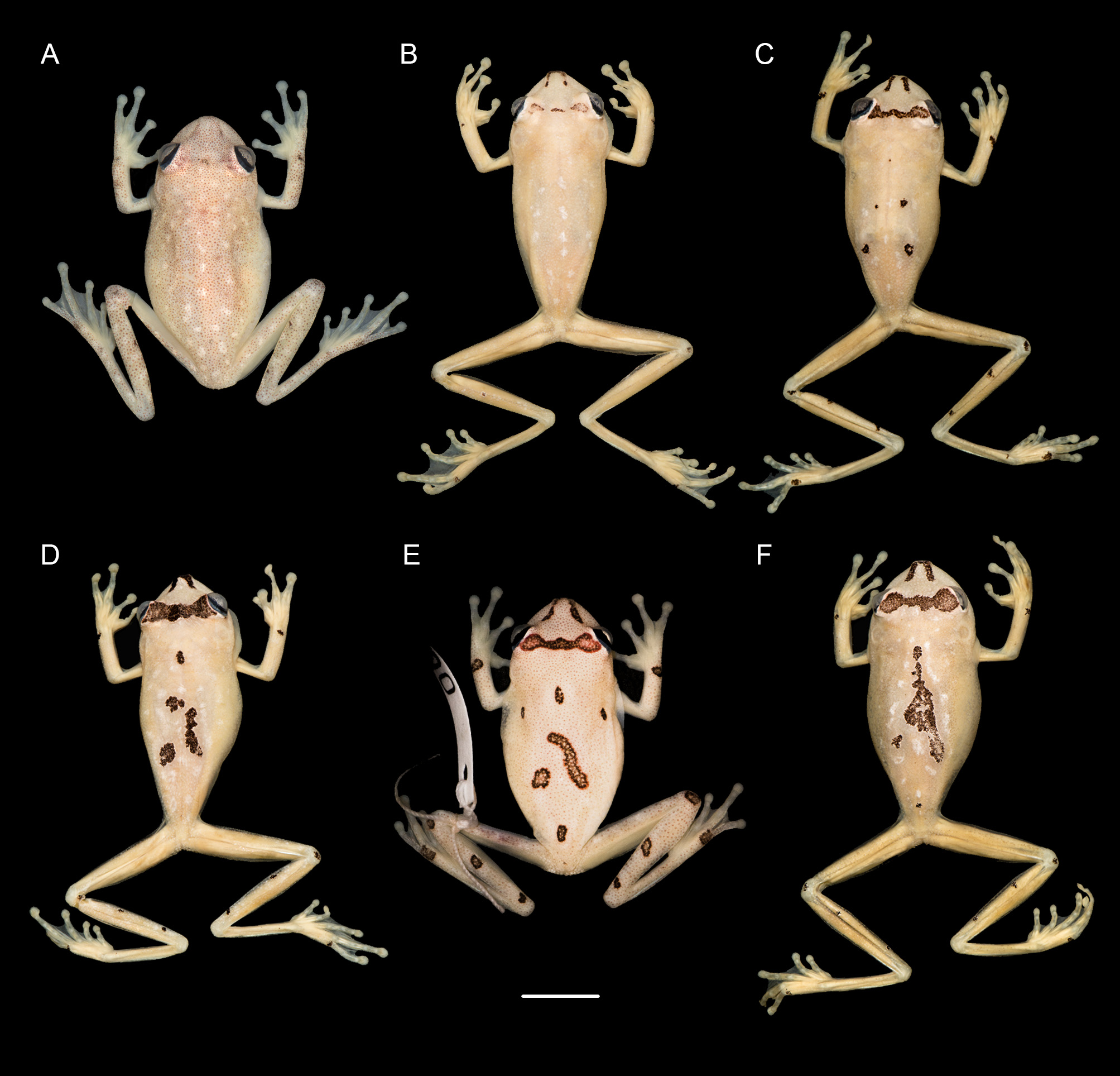
Color in preservative ( Fig. 9 View FIGURE 9 ). Dorsum cream with large white blotches, with brown and reddish-brown small melanophores. Many individuals with a brown transverse interorbital bar (expanded on eyelid). Dorsum may have large brown spots. Flanks predominantly cream with brown and reddish-brown melanophores on upper region. Inguinal region, anterior and posterior region of the thighs cream. Thigh dorsally cream with white blotches and brown and reddish-brown small melanophores; anteriorly and posteriorly cream. Upper arms, forearms, and tibiae dorsally cream with white blotches and brown and reddish-brown small melanophores—commonly showing a high- er concentration of melanophores forming spots and stains. Gular region cream with light-brown melanophores. Chest and belly translucent cream, with light-brown melanophores, and white visceral peritonea covering the organs visible. Ventral surface of forearms and hindlimbs cream with light brown spots. Ventral surface of hands and feet cream.
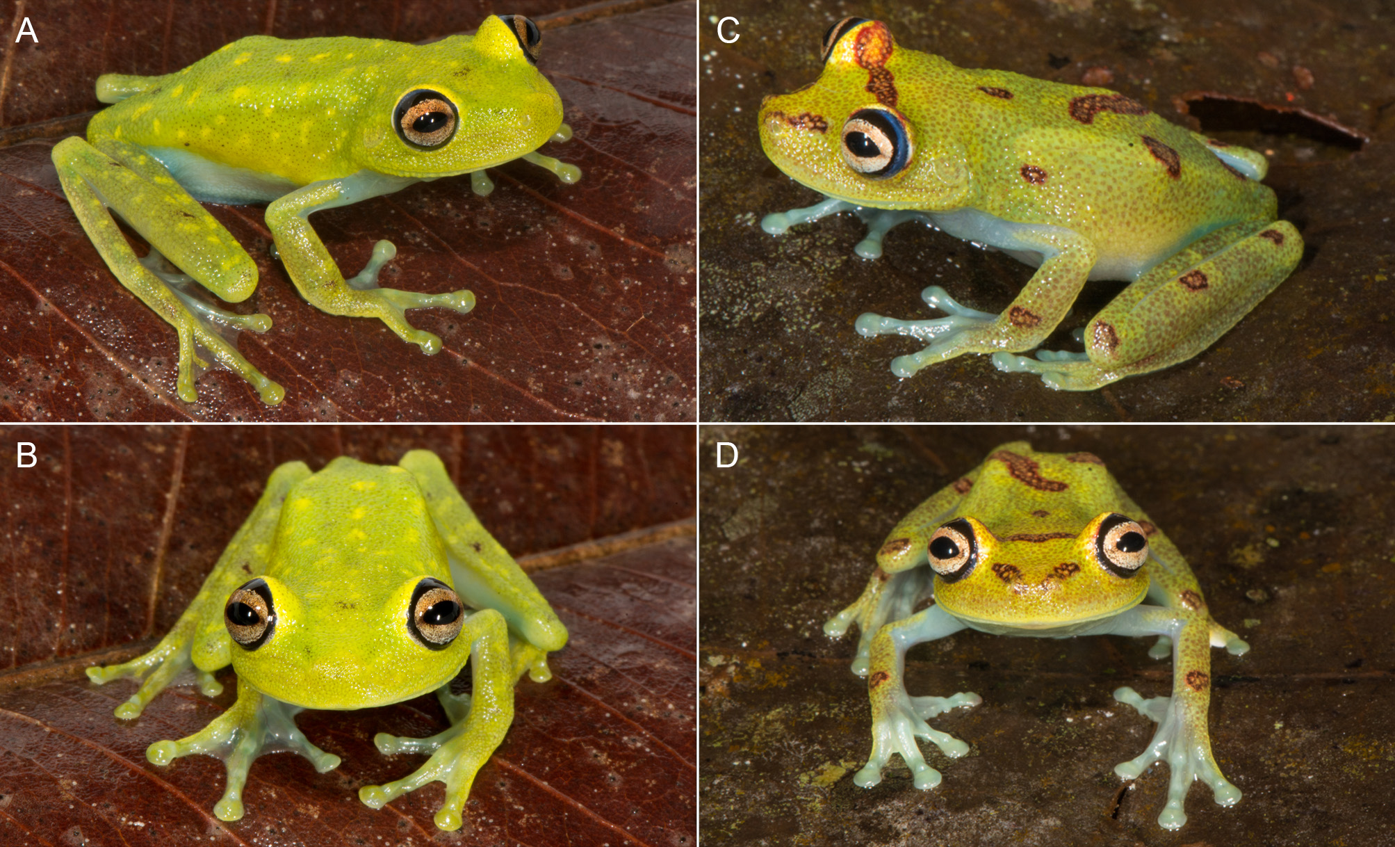
Color in life ( Fig. 4 View FIGURE 4 ). Dorsum yellowish green with light yellow blotches, with or without concentrations of brown and reddish-brown small melanophores ( Fig. 4 View FIGURE 4 ); region between eyes with brown blotches. Flanks predominantly yellow; light yellow blotches and reddish-brown melanophores on upper region. Inguinal region, anterior and posterior region of the thighs light lemon green. Dorsal surface of thighs, forearms and tibiae as same as dorsum ( Fig. 4 View FIGURE 4 ). Gular region, chest and belly translucent light lemon green.
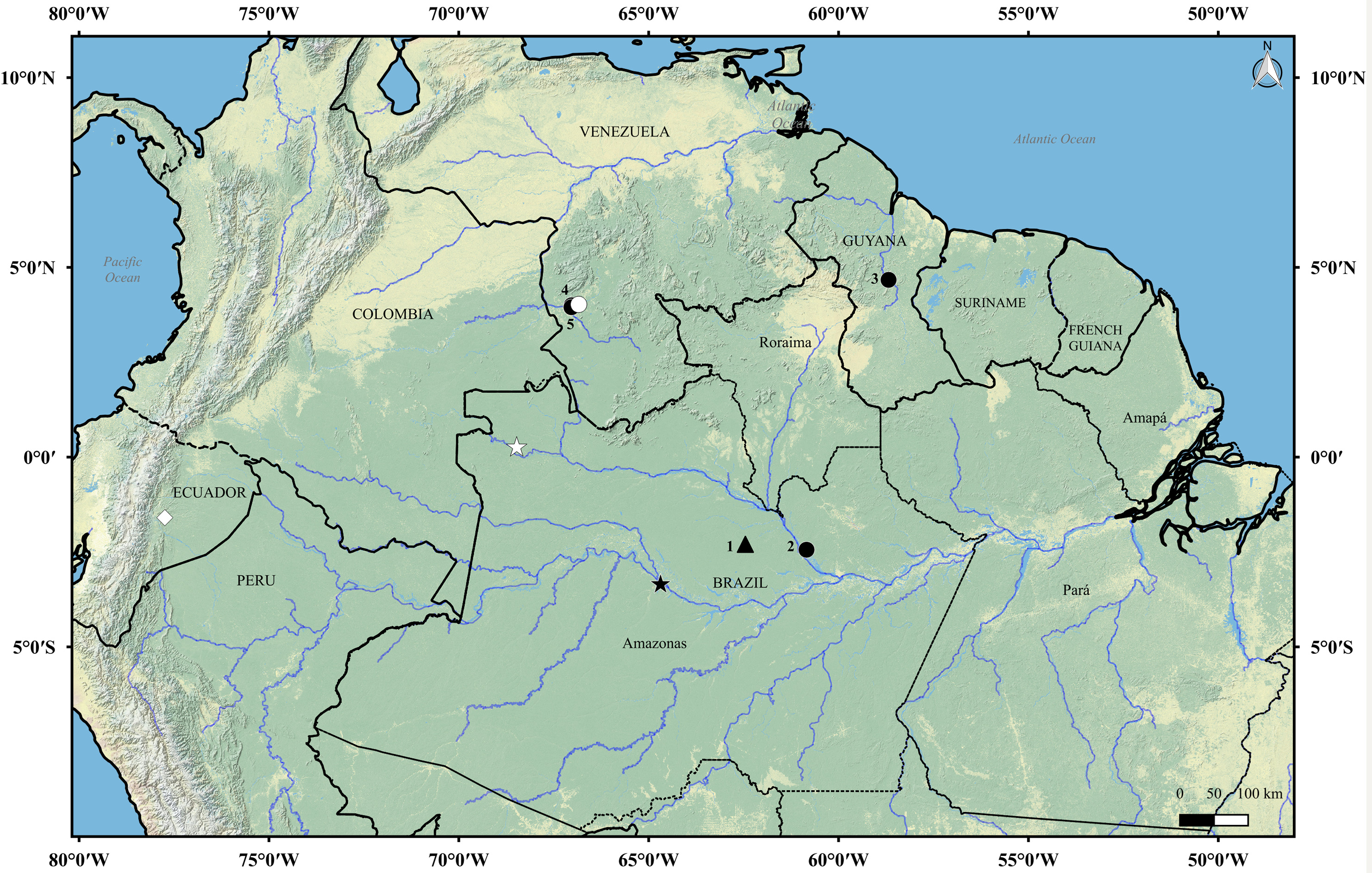
Distribution. Boana gracilis is known with certainty from the type locality “Rio Uaupes (north of Ipanoré), Brazil; the Municipality of Novo Airão (Parque Nacional do Jaú, and Parque Nacional de Anavilhanas), State of Amazonas, Brazil; Canaripó, Venezuela ( Hoogmoed 1979), San José de Chipiro, boca del río Ventuari, Venezuela, Macuruco, confluencia Orinoco-Ventuari, Venezuela; and from Iwokrama, Guyana ( Fig. 10 View FIGURE 10 ). This species occurs syntopically with B. cinerascens and may occur more widely.


No known copyright restrictions apply. See Agosti, D., Egloff, W., 2009. Taxonomic information exchange and copyright: the Plazi approach. BMC Research Notes 2009, 2:53 for further explanation.
|
Kingdom |
|
|
Phylum |
|
|
Class |
|
|
Order |
|
|
Family |
|
|
Genus |
Boana gracilis ( Melin, 1941 )
| Sturaro, Marcelo José, Costa, João Carlos Lopes, Maciel, Adriano O., Lima-Filho, Geraldo R., Rojas-Runjaic, Fernando J. M., Mejia, Daniela Pareja, Ron, Santiago R. & Peloso, Pedro L. V. 2020 |
Hypsiboas cinerascens
| Faivovich, J. & Mcdiarmid, R. W. & Myers, C. W. 2013: 50 |
| Pyron, R. A. & Wiens, J. J. 2011: 570 |
| Frost, D. R. & Grant, T. & Faivovich, J. & Bain, R. H. & Haas, A. & Haddad, C. F. B. & De Sa, R. & Channing, A. & Wilkinson, M. & Donnellan, S. C. & Raxworthy, C. J. & Campbell, J. A. & Blotto, B. L. & Moler, P. & Drewes, R. C. & Nussbaum, R. A. & Lynch, J. D. & Green, D. M. & Wheeler, W. C. 2006: 65 |
Hypsiboas granosa
| Faivovich, J. & Haddad, C. F. B. & Garcia, P. C. A. & Frost, D. R. & Campbell, J. A. & Wheeler, W. C. 2005: 85 |
Hyla punctata
| Cochran, D. M. & Goin, C. J. 1970: 224 |
Hyla granosa
| Hoogmoed, M. S. 1979: 5 |
| Duellman, W. E. 1974: 8 |
| Rivero, J. A. 1972: 182 |
| Rivero, J. A. 1961: 100 |
Hyla granosa gracilis
| Melin, D. 1941: 21 |