
Austroterobia iceryae Bouček
|
publication ID |
https://doi.org/ 10.11646/zootaxa.4301.1.1 |
|
publication LSID |
lsid:zoobank.org:pub:67A78566-A4FD-4E37-96E9-DCC4CCF321E5 |
|
DOI |
https://doi.org/10.5281/zenodo.6038598 |
|
persistent identifier |
https://treatment.plazi.org/id/4D3D879A-FFA9-FFBC-FF16-C06AFD66FE61 |
|
treatment provided by |
Plazi |
|
scientific name |
Austroterobia iceryae Bouček |
| status |
|
Austroterobia iceryae Bouček View in CoL
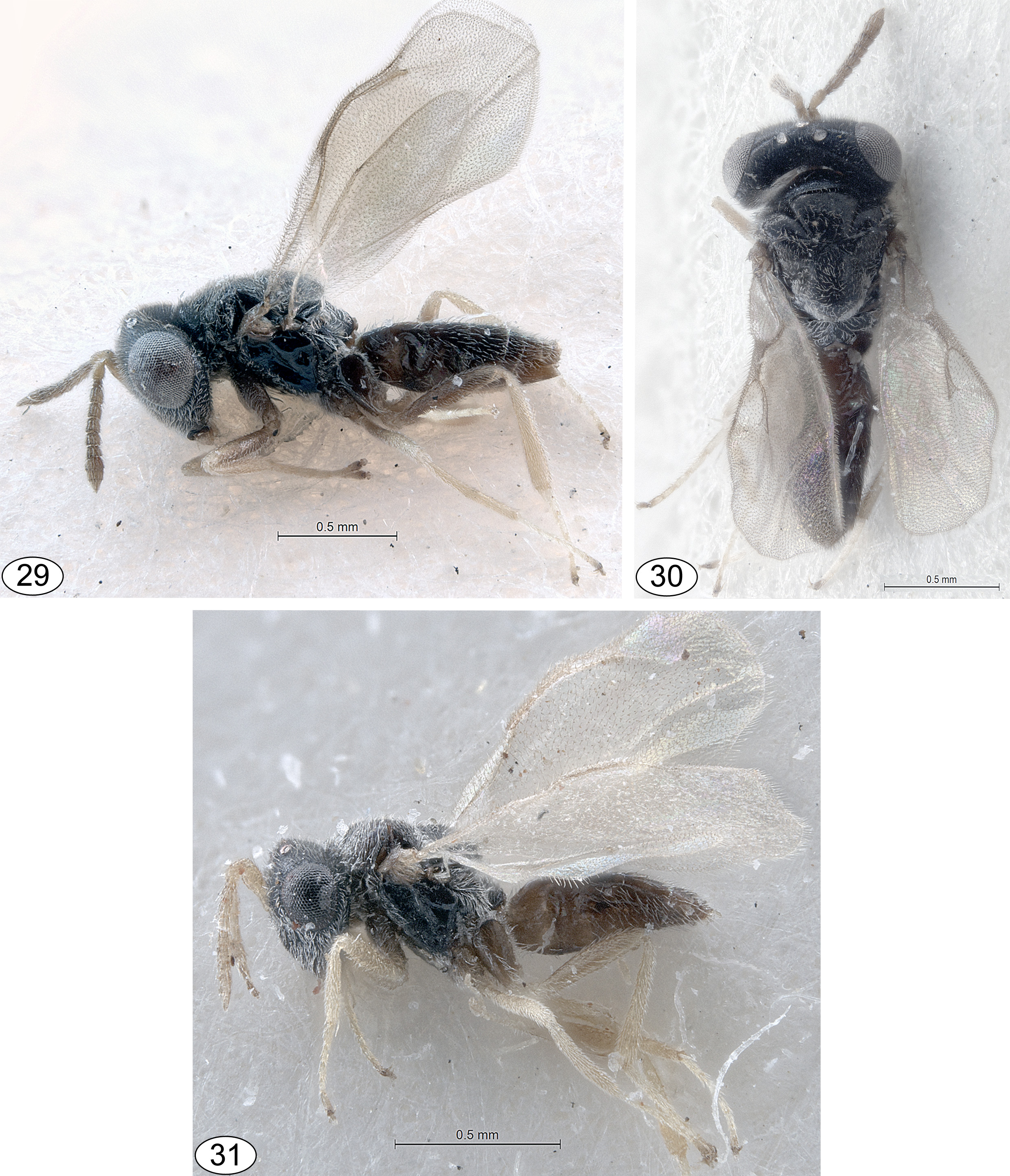
( Figs 29–39 View FIGURES 29 – 31 View FIGURES 32 – 39 )
A. iceryae Bouček, 2004 View in CoL ; in Bouček & Noyes 2004: 138 –140; holotype in BMNH, examined.
Diagnosis. Both sexes: head with at most very faint dark metallic reflections ( Figs 32, 33 View FIGURES 32 – 39 ); mandibular formula 3:3; gena not depressed at mouth corner ( Fig. 34 View FIGURES 32 – 39 ); pronotum visible in dorsal view ( Fig. 36 View FIGURES 32 – 39 ); notauli visible throughout length of mesoscutum ( Fig. 36 View FIGURES 32 – 39 ); axillae virtually touching medially ( Fig. 36 View FIGURES 32 – 39 ); prepectus and dorsal side of propodeum entirely setose ( Figs 37, 38 View FIGURES 32 – 39 ); posterior margin of mesoscutellum densely setose, with small spine ( Fig. 36 View FIGURES 32 – 39 ); anterior margin of propodeum distinctly raised, concealing dorsellum ( Fig. 38 View FIGURES 32 – 39 ); propodeal plicae absent ( Fig. 38 View FIGURES 32 – 39 ).
The spiny mesoscutellum ( Fig. 36 View FIGURES 32 – 39 ) and the flanged anterior margin of the propodeum ( Fig. 38 View FIGURES 32 – 39 ), structures that are found in all species of Teasienna , but no other Austroterobia , together with the entirely setose prepectus and propodeum ( Figs 37, 38 View FIGURES 32 – 39 ), make A. iceryae easily recognizable.
Material examined. Type material. Holotype ♀ (deposited in BMNH) [entire; on rectangular card]. KENYA: ‘ Kenya: Kiambu District, Mchana Estate , ex Icerya pattersoni on coffee 19.vi.1984 (S. T. Murphy)’.
Paratypes. GAMBIA: 1♂ ‘W. Afr . Gambia: Lamin, Abulco, 27./I., 1978. L. Huggert’, ‘ Paratype’ , ‘ Paratype ♂ Austrot. iceryae sp. n., Boucek det. 1989’ ( BMNH) ; KENYA: 2♀ and 2♂ ‘ Kenya, Oaklands Estate, Kiambu Dist., 29.x.85 ’, ‘assoc. with Icerya pattersoni on coffee, CIE A17679 View Materials ’, ‘ Paratype’ , ‘ Paratype ♀ Austroterobia iceryae sp. n., Boucek det. 1989’ ( BMNH) ; 1♂ ‘ Kenya, Mchana Estate, Kiambu Dist., 28.x.85 ’, ‘assoc. with Icerya pattersoni on coffee, CIE A17679 View Materials ’, ‘ Paratype’ , ‘ Paratype ♂, Austroterobia iceryae sp. n., Boucek det. 1989’ ( BMNH) ; 1♂, same information, ‘det. Z. Boucek, 1986’ (BMNH); NIGERIA: 1♀ and 2♂ ‘ Nigeria: Oyo St., Ibadan, IITA compound’, ‘ xi. 1987 Icerya patt., J.S. Noyes’, ‘ Paratype’ , ‘ ♀ Austroterobia iceryae sp. n., Boucek det. 2003’ ( BMNH) ; 1♀ ‘ Nigeria: Ibadan, IITA, ex Icerya pattersoni / coffee’, 23.X. / em. XI.1987, J.S. Noyes’, ‘ Paratype’ , ‘ Paratype ♀ Austroterobia iceryae sp. n., Boucek det. 1989’ ( BMNH) .
Redescription. Female. Body length: 1.4–1.9 mm.
Colour. Head and mesosoma black, with weak, mainly blue-green reflections ( Figs 32, 33 View FIGURES 32 – 39 ). Gaster dark brown ( Figs 29, 30 View FIGURES 29 – 31 ). Eyes pale reddish-grey; ocelli whitish ( Fig. 33 View FIGURES 32 – 39 ). Antenna ( Fig. 34 View FIGURES 32 – 39 ) with scape and pedicel yellowishbrown; flagellum brown, paler ventrally. Mandibles yellowish-brown, teeth reddish-brown. Legs ( Fig. 29 View FIGURES 29 – 31 ) with coxae dark brown to black; trochanters and femora brown; tibiae and tarsi yellowish except for brown pretarsi. Fore wing subhyaline to subinfumate, hind wing hyaline ( Figs 29 View FIGURES 29 – 31 , 39 View FIGURES 32 – 39 ). Tegula and venation brown ( Fig. 39 View FIGURES 32 – 39 ). Body setation whitish to pale brown, wing setation brownish ( Figs 34, 36, 38, 39 View FIGURES 32 – 39 ).
Sculpture. Head coriaceous except for virtually smooth clypeus and smooth, small area at mandibular base ( Figs 32–34 View FIGURES 32 – 39 ); dorsal side of mesosoma, except for propodeum, mostly finely coriaceous, appearing shiny ( Fig. 36 View FIGURES 32 – 39 ); propodeum with median area almost smooth; mesopleuron smooth, with femoral depression conspicuous, but without any indication of a central pit ( Fig. 37 View FIGURES 32 – 39 ); mesepimeron with central lunate depression ( Fig. 37 View FIGURES 32 – 39 ); metapleuron smooth; gaster smooth.
Structure. Head. Toruli closer to median ocellus than to ventral margin of clypeus ( Fig. 32 View FIGURES 32 – 39 ). Scape not reaching median ocellus ( Fig. 34 View FIGURES 32 – 39 ). Gena flat at mouth corner ( Fig. 34 View FIGURES 32 – 39 ), posterior margin not carinate. Malar sulcus absent ( Fig. 34 View FIGURES 32 – 39 ). Eyes broad oval, inner margins diverging in lower part ( Fig. 32 View FIGURES 32 – 39 ). Funicular segments not pedicellate ( Fig. 34 View FIGURES 32 – 39 ). Head width 2.3–2.6× length in dorsal view (53:23) and 1.4–1.6× height in frontal view (53:38). POL about equal to OOL (8:8). Eye height about 1.1× length (22.0:19.5), 1.4–1.6× malar space (22:16), and about 1.8× scape length (22.0:12.5). Head width about 1.4× length of pedicel plus flagellum (53:37). F1 quadrate; F5 width 1.2–1.3× length; clava length 2.0–2.4× width.
Mesosoma. Dorsally setose, including anterior part of metanotum and median area of propodeum ( Fig. 36 View FIGURES 32 – 39 ); prepectus and posterior margin of metapleuron setose ( Fig. 37 View FIGURES 32 – 39 ); mesopleuron bare except along anteroventral margin ( Fig. 37 View FIGURES 32 – 39 ). Pronotum distinctly visible in dorsal view ( Fig. 36 View FIGURES 32 – 39 ). Notauli complete ( Fig. 36 View FIGURES 32 – 39 ). Axillae virtually touching medially ( Fig. 36 View FIGURES 32 – 39 ). Posterior margin of mesoscutellum with small spine ( Fig. 36 View FIGURES 32 – 39 ). Anterior margin of propodeum distinctly raised, concealing dorsellum ( Fig. 38 View FIGURES 32 – 39 ). Propodeum without median carina (although slightly raised along median line) and plicae ( Fig. 38 View FIGURES 32 – 39 ). Fore wing ( Fig. 39 View FIGURES 32 – 39 ) completely setose, without speculum; parastigma without hyaline break; marginal vein thickened. Mesosoma length 1.1–1.2× width (49:42), width about 1.2× height. Mesoscutum width about 2.6× length. Mesoscutellum length about equal to width. Propodeum length about 0.5× mesoscutellum length. Fore wing length 1.8–1.9× width (100:55). MV length about 6× width (18:3); MV 1.1–1.2× as long as SV (18.0:15.5); PV about 1.1× as long as MV (20:18).
Gaster. Ovate, shorter than head plus mesosoma ( Figs 29, 30 View FIGURES 29 – 31 ); length 2.0–2.2× width.
Male. Differs from the female mainly as follows. Body length: 1.2–1.6 mm. Antennae and legs, except for coxae, uniformly yellowish-brown ( Fig. 31 View FIGURES 29 – 31 ). Eyes smaller and rounder ( Fig. 31 View FIGURES 29 – 31 ). Scape with ventral protuberance towards apex ( Fig. 35 View FIGURES 32 – 39 ); flagellum subfiliform, anelli and distal funicular segments longer ( Fig. 35 View FIGURES 32 – 39 ). Head width about 1.3× height in frontal view, width 1.1–1.2× length of pedicel plus flagellum. Eye height very slightly larger than eye length and about 1.2× malar space. POL about 0.9× as long as OOL. Clava length about 2.8× width. Fore wing length about 2.1× width. MV about 1.4× as long as SV. Gaster widening towards apex, length about 1.9× width.
Distribution. Gambia, Kenya, Nigeria ( Bouček & Noyes 2004).
Biology. Egg predator of Icerya pattersoni Newstead and I. nigroareolata Newstead ( Hemiptera : Monophlebidae ) on coffee ( Bouček & Noyes 2004).
Remarks. In the original description the scutellum was stated as posteriorly rounded ( Bouček & Noyes 2004: 138), but in fact a very small spine can be observed among the dense whitish setation ( Fig. 36 View FIGURES 32 – 39 ). This feature, as well as the raised anterior margin of the propodeum, which conceals the dorsellum, and the absence of plicae ( Fig. 38 View FIGURES 32 – 39 ), are characteristic for Teasienna and not Austroterobia . However, the position and structure of the antennae in both sexes ( Figs 32, 34, 35 View FIGURES 32 – 39 ), as well as the structure of the fore wing ( Fig. 39 View FIGURES 32 – 39 ) and the mandibular formula and structure indicate that the species is better placed in Austroterobia than in Teasienna .

| IITA |
International Institute of Tropical Agriculture |
No known copyright restrictions apply. See Agosti, D., Egloff, W., 2009. Taxonomic information exchange and copyright: the Plazi approach. BMC Research Notes 2009, 2:53 for further explanation.
|
Kingdom |
|
|
Phylum |
|
|
Class |
|
|
Order |
|
|
Family |
|
|
Genus |
Austroterobia iceryae Bouček
| Mitroiu, Mircea-Dan 2017 |
A. iceryae Bouček, 2004
| Boucek 2004: 138 |