
Aptilotella erinacea Luk & Marshall, 2014
|
publication ID |
https://doi.org/ 10.11646/zootaxa.3761.1.1 |
|
publication LSID |
lsid:zoobank.org:pub:82E0F1DC-BC98-4E8A-A3D5-21ECB392CC0B |
|
DOI |
https://doi.org/10.5281/zenodo.4909097 |
|
persistent identifier |
https://treatment.plazi.org/id/038487F1-FFB2-FFAB-FDC7-F8EDFE6C0B6D |
|
treatment provided by |
Felipe |
|
scientific name |
Aptilotella erinacea Luk & Marshall |
| status |
sp. nov. |
Aptilotella erinacea Luk & Marshall , sp. n.
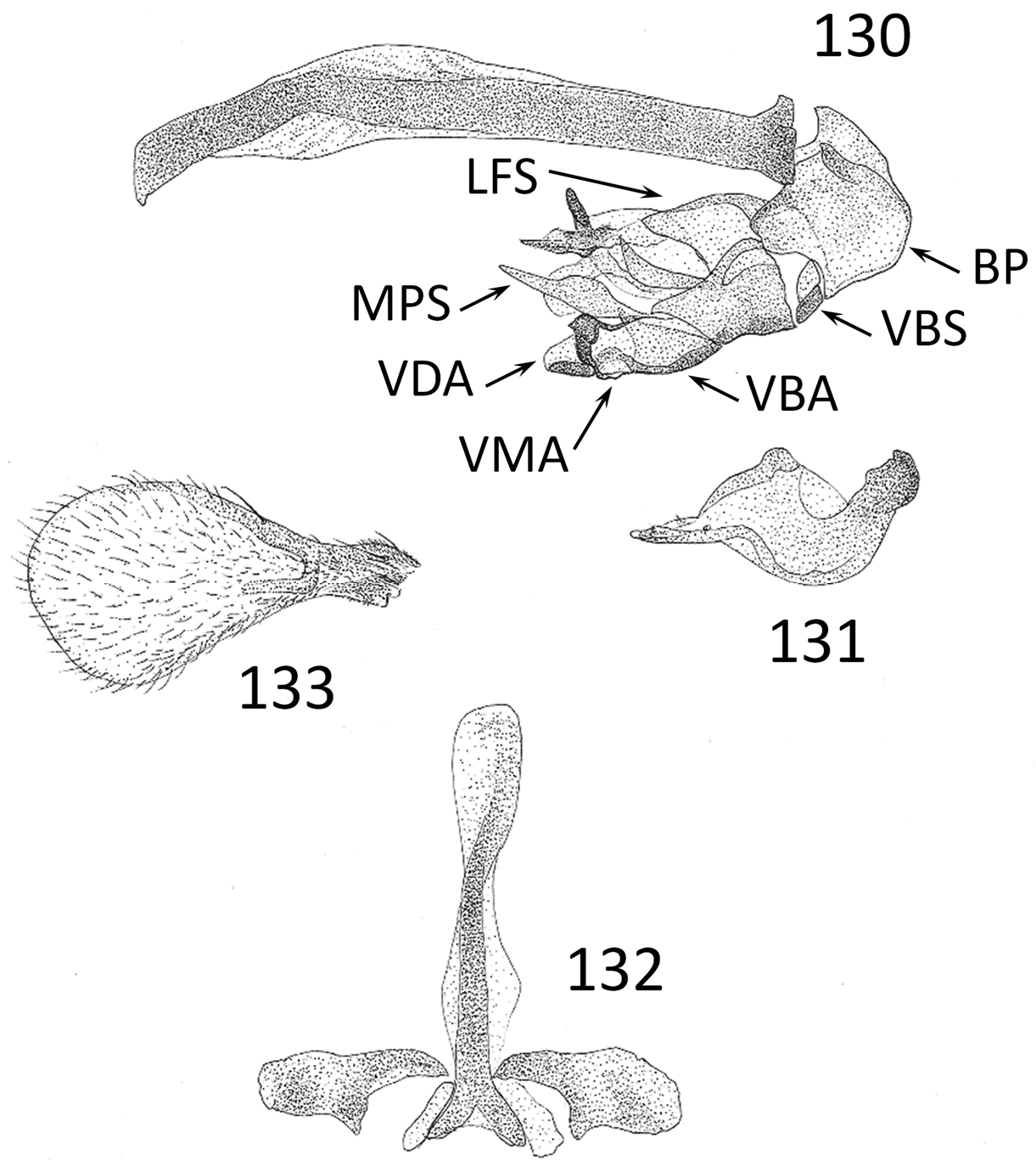
Figures 126–137 View FIGURES 126–129 View FIGURES 130–133 View FIGURES 134–137
Description. Body length 0.8–0.9 mm. Head ground color orange. Frons finely rugose; pale areas silvery, not attaining front margin of frons; brown medial stripes each about one-third the width of frons, tapering anteriorly; brown orbital stripes narrow, not entering ocular emargination. Ocellar tubercle slightly raised; ocelli minute; ocellar bristle two-thirds the length of frons. Orbital bristle present; orbital setulae minute, in three pairs. Interfrontal setae minute, in two pairs. Face shining; facial excavation with a silvery band continuing onto anterior half of gena; gena weakly shining, finely rugose. Antenna brown. Scutum and scutellum dark reddish-brown, shining, finely rugose, finely microtrichose. Scutum uniformly setose; shallowly creased along posterolateral margin. Scutellum flat, twice wider than long, 0.6 times the width of scutum. Apical scutellar bristles twice as long as basal. Pleuron dull. Legs orange; coxae black; basal half of hind femur brown; fore femur, tibia and first tarsomere dark brown; fore tarsus off-white; mid tibia with two anterodorsal bristles. Wing pad ( Fig. 133 View FIGURES 130–133 ) brown in male; reduced in female to a small, brown stub. Abdomen black, shining, finely rugose; tergites uniformly setose and microtrichose; sternites finely microtrichose. Epandrium and synsternite 6+7 dark reddish-brown.
Male terminalia. Sternite 5 ( Fig. 129 View FIGURES 126–129 ) rectangular; posteromedial margin emarginate, with several setae, flanked by three long setae; sternal plate broadly rectangular, densely spinulose, with two dense patches of very stout setae, posteriorly with two pairs of premarginal setae. Synsternite 6+7 ( Fig. 128 View FIGURES 126–129 ) with medial bridge flanked by a strong lobe on both sides. Epandrium ( Fig. 126 View FIGURES 126–129 ) with ventral margins pointed and curled outward. Cercus ( Figs. 126, 127 View FIGURES 126–129 ) swollen, triangular; outer margin with three evenly separated premarginal setae, medially with a long seta; apex with several sensory setae; base and outer half setulose. Surstylus ( Figs. 126, 127 View FIGURES 126–129 ) saddle-shaped, apically rounded; ventral face flat, with setae on outer margin; outer face strongly concave. Postgonite ( Fig. 131 View FIGURES 130–133 ) twice as long as wide; posterior margin rounded; descending portion one-third the length of postgonite, with three sensory setulae; articulatory process for pregonite rounded; articulatory process for basiphallus short-stalked, knobbed. Hypandrium ( Fig. 132 View FIGURES 130–133 ) with slender medial rod slightly skewed to the right; posteromedial fork thick, rounded and divergent; hypandrial arms paddle-shaped, with a large tooth on posteromedial margin; pregonite inconspicuous. Aedeagus as in Figure 130 View FIGURES 130–133 . Basiphallus compressed, squared, with truncate epiphallus; anterior margin weakly arched; articulatory process for postgonite short and divergent. Ejaculatory apodeme discoid, inconspicuous. Ventrobasal sclerite divided. Lateral flanking sclerite narrowly fused ventrobasally; dorsal margin divergent and tapering. Ventral flanking sclerites clustered; the slender basal article originating ventrodistally to lateral flanking sclerite and rising inward, tapering to a point; the convex medial article originating along the basal article and meeting the distal margin of lateral flanking sclerite, margins sclerotized and broadening distally, converging outward to a thickened point; the paddle-shaped distal article originating ventrodistally to medial article, converging inward. Medial paired sclerites originating from apices of lateral flanking sclerites; apices dilated, converging and nearly touching.
Female terminalia. Epiproct ( Fig. 134 View FIGURES 134–137 ) light brown, rectangular, medially setulose. Each half of tergite 8 ( Figs. 134–136 View FIGURES 134–137 ) weakly convex, triangular; setulose. Cercus 2.5 times as long as wide; with one long apical seta and several preapical setae. Sternite 8 ( Figs. 135, 136 View FIGURES 134–137 ) brown, rectangular. Spermathecae ( Fig. 137 View FIGURES 134–137 ) simple; length of sclerotized ducts approximately twice the diameter of a spermatheca.
Variation. The gena is dark yellow in some specimens. Less frequently, the legs have a dark orange ground colour.
Etymology. The species epithet is from the Latin erinaceus, “hedgehog,” referring to the densely spinulose sternal plate.
Type material. Holotype ♂, DEBU. HONDURAS: Cortés, Parque Nacional Cusuco, 18.7 km N Cofradía, 5.4 km W Buenos Aires, Cerro Jilinco , 1960 m, 26.viii.1994, pine/cloud forest berlese, R.S. Anderson.
Paratypes. HONDURAS: same label as holotype (3♂, ♀, DEBU; 3♂, EAPZ) ; same label as holotype but at 2000 m, cloud forest berlese (2♂, DEBU) ; same label as holotype but at 2080 m, elfin cloud forest litter berlese (♂, DEBU) .
Comments. In Aptilotella erinacea , dense spinules are present on the sternal plate of the male and the hypoproct of the female.


| DEBU |
Ontario Insect Collection, University of Guelph |
| EAPZ |
Escuela Agricola Panamericana |
No known copyright restrictions apply. See Agosti, D., Egloff, W., 2009. Taxonomic information exchange and copyright: the Plazi approach. BMC Research Notes 2009, 2:53 for further explanation.