
Aphodius (Chilothorax) variicolor Koshantschikov, 1894
|
publication ID |
https://doi.org/ 10.11646/zootaxa.2169.1.4 |
|
DOI |
https://doi.org/10.5281/zenodo.5333499 |
|
persistent identifier |
https://treatment.plazi.org/id/5B122807-FFAF-FFA0-FF46-B643DF0022BD |
|
treatment provided by |
Felipe |
|
scientific name |
Aphodius (Chilothorax) variicolor Koshantschikov, 1894 |
| status |
|
Aphodius (Chilothorax) variicolor Koshantschikov, 1894
Figs. 16–19, 22–30, 32, 34, 36, 38, 40, 41
This rarely collected species was known from a few localities in western Kazakhstan ( Frolov 2002) and was recently recorded from Russia ( Akhmetova & Frolov 2008).
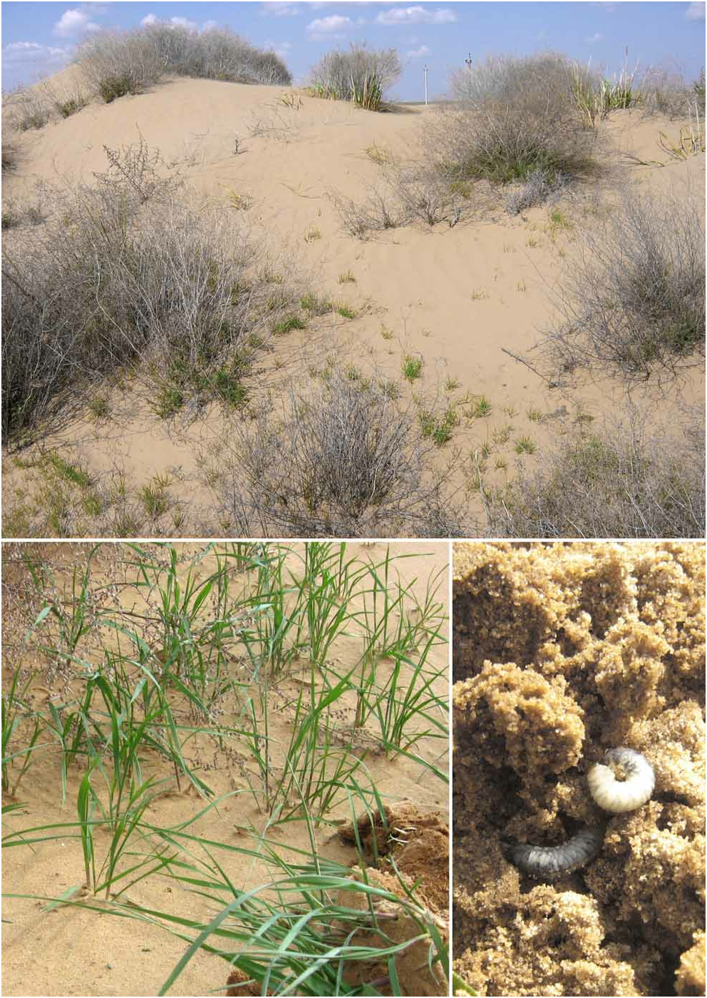
Material examined. More than 50 third-instar larvae were collected from fixed sands in the Dosang environs. Larvae were collected in roots of cheat grass ( Anisantha tectorum (= Bromus tectorum )) ( Fig. 41 View FIGURE 41 ) on 14.IV.2007 (10 specimens) and 4.IV.2008 (majority of specimens). All larvae were about the same size and similar externally. Twenty living larvae collected in 2008 were taken to the ZIN laboratory and in five weeks six adults were obtained .
Third-instar larval description. Larva of typical C-shape form ( Fig. 24 View FIGURES 24–30 ). Head width: 1.39±0.04 mm, length (without labrum): 1.21±0.04 mm. Head surface shiny, brown with unclear pattern of small brown spots on pleural sclerites ( Fig. 26 View FIGURES 24–30 ). Medial part of pleural sclerites and base of frons a bit darker than remaining part of the head capsule. Frontal sutures are visible as very fine, darker lines.
Epicranial suture approximately 2.5 times shorter than frons height. Each pleural sclerite with 4 long setae: 3 near palpifer, 1 near epicranial suture. Two pairs of shorter setae are in the middle of each pleural sclerite. A number of shorter setae located on pleural sclerite without apparent definite pattern. Frons with 5 pairs of setae: 2 short setae in the center of frons, 1 short seta medially and 2 long setae laterally.
Clypeus trapezoidal, yellowish-brown, with 2 pairs of long setae and 1 pair of short setae laterally. Basal part of clypeus (4/5 length of clypeus) is darker than apical fifth. Surface of clypeus without prominent tubercles.
Labrum trilobed, with 2 pairs of long setae on dorsal surface, and 24 short to relatively long setae on the distal margin. Ventral side of labrum with a pair of short setae basally (Fig. 34).
Mandibles triangular, asymmetrical. Left mandible slightly longer than right mandible, its scissorial part wider than in right mandible ( Figs. 27–30 View FIGURES 24–30 ). Base of mandible light brown, scissorial and molar part almost black.
Maxillae symmetrical (Fig. 32). Cardo with 4 short setae: 2 on ventral side and 2 on lateral margin near base of stipes. Ventral side of stipes with long proximal and short distal setae, dorsal side with a row of 10–12 stridulatory teeth and 2 short setae near base of palpifer. Palpifer without stridulatory teeth, with one short setae ventrally. Maxillary palpus with 4 palpomeres; 1st and 4th palpomeres with 1 seta each, 3rd with 2 setae. Ventral side of galea with longitudinal row of 6 short setae. Dorsal side and apex of galea with 6 longer setae. Dorsal side of lacinia with 5 long and thick setae, ventral side with 1 long and thick seta apically and 1 short seta basally. Apex of lacinia tridentate.
Legs are about the same size, anterior legs slightly shorter than others (Fig. 38).
Anal sternite with 48–58 relatively long, strongly sclerotized spinules flattened and abruptly rounded apically (Fig. 22). The spinules are not arranged in rows, in some specimens they are indistinctly divided into 2 groups by a narrow smooth area (Figs. 16–19). Lower anal lobe sinuate in the middle (Fig. 23).
Lateral bulges of abdominal segments with 2 to 3 setae (normally there are probably 3 setae of which one can be abraded). Folds of abdominal tergites with short, thick setae not arranged in transverse row or arranged in a few irregular rows.
Diagnosis. The larvae of A. variicolor are most similar to those of A. distinctus (Müller) , which is also a member of the subgenus Chilothorax Motschulsky ( Frolov 1996) . They can be separated from the later by larger number of anal sternite spinules (about 40 in A. distinctus ) and tridentate apex of lacinia (as opposed to the bidentate lacinia in A. distinctus ).

| ZIN |
Russian Academy of Sciences, Zoological Institute, Zoological Museum |
No known copyright restrictions apply. See Agosti, D., Egloff, W., 2009. Taxonomic information exchange and copyright: the Plazi approach. BMC Research Notes 2009, 2:53 for further explanation.