
Adelosgryllus lucifugus, Merlo & Castro-Souza & Junta & Ferreira, 2021
|
publication ID |
https://doi.org/ 10.11646/zootaxa.4933.1.6 |
|
publication LSID |
lsid:zoobank.org:pub:78BE572F-54FA-426C-A716-399EE6BE3706 |
|
DOI |
https://doi.org/10.5281/zenodo.4558065 |
|
persistent identifier |
https://treatment.plazi.org/id/03F687D2-3126-FFBF-FF37-D32CFD0BFE40 |
|
treatment provided by |
Plazi |
|
scientific name |
Adelosgryllus lucifugus |
| status |
sp. nov. |
Adelosgryllus lucifugus n. sp.
( Figures 2–7 View FIGURES 2–7 , 8–15 View FIGURES 8–15 , 16–18 View FIGURES 16–18 , Table 1 View TABLE 1 )
Material examined. Holotype ³, code ISLA 66149, Brazil, Piauí state, Piripiri municipality, Morcego cave (04° 25’ 44.056’’ S; 41° 40’ 14.044” W), 21.i.2019, Ferreira R. L. leg GoogleMaps . Holotype condition: right tegmen and legs de-tached, kept in the holotype tube. Paratypes, 6 ³³ (ISLA 66144, 66146, 66147, 66150, 66151, 66152) and 2 ♀♀ ( ISLA 66145* and 66148), (* = nymph stage), same data and locality of holotype; 2 ³³ (ISLA 66162 and 66164) and 1 ♀♀ ( ISLA 66163), Onça Morta cave (04° 05’ 12.314’’ S; 41° 41’ 25.034” W), Piracuruca municipality, Piauí state, Brazil GoogleMaps .
Distribution. Known for two caves, the Morcego cave (04° 25’ 44.056’’ S; 41° 40’ 14.044” W—Piripiri mu-nicipality), and the Onça Morta cave (04° 05’ 12.314’’ S; 41° 41’ 25.034” W—Piracuruca municipality), associated with the speleological unit of Canindé ( Fig. 1 View FIGURE 1 ).
Etymology. The specific epithet “lucifugus” refers to the evasive behavior of the specimens in the presence of light. From the Latin luci = light, fugus = escape.
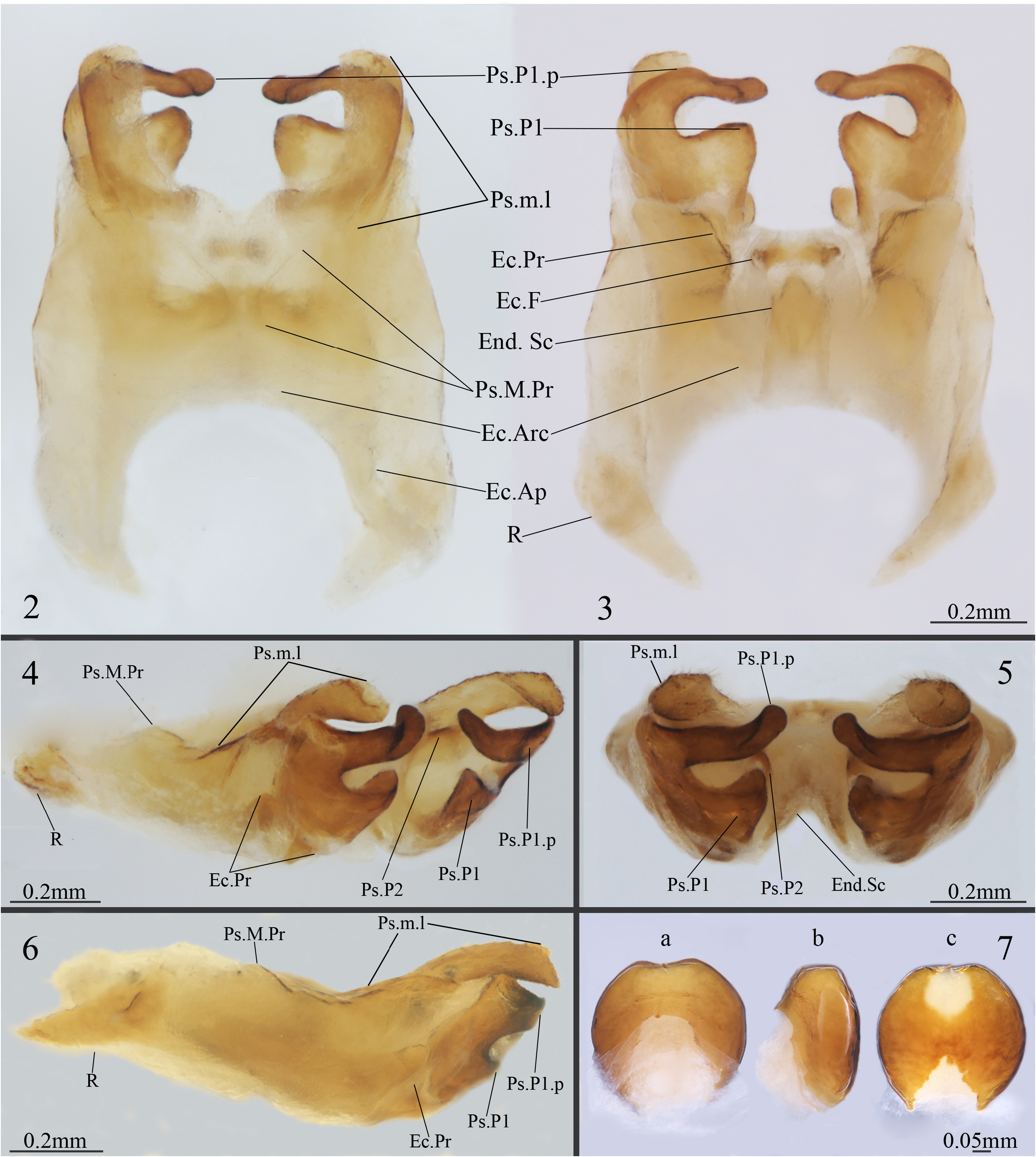
Diagnosis. Combination of the following characters: pseudepiphallic paramere 1 well developed, C-shaped, very similar to A. spurius , but in this new specie the Ps.P1 parameres are far from each other and flattened horizontally ( Figs 2 and 3 View FIGURES 2–7 ), apex dilated and curved inward (Ps.P1.p, Figs 2–5 View FIGURES 2–7 ); ectophallic fold sclerotized, linear-shaped, with the lateral border more sclerotized than that interior base, central part slightly convex at the top and bottom borders (Ec.F, Fig. 3 View FIGURES 2–7 ); endophallus circular-shaped and vertically elongated, forming a short ventral crest (End.Sc), connected to the ectophallic fold by an inverted U-shaped membrane (End.Sc, Fig. 3 View FIGURES 2–7 ).
Description, male holotype. General Coloration. Body dark brown with orange head (in vivo) (head pale yellow after fixation in ethanol 70%) ( Figs 8–15 View FIGURES 8–15 ); Head. slightly pubescent and with long bristles between scapes ( Fig. 8 View FIGURES 8–15 ), few of them are present around the eyes and at the posterior margin and occiput of the head (apparently many were lost after fixation), occiput region is slightly darkened behind the eyes ( Fig. 9 View FIGURES 8–15 ); Eyes. compound eyes with black ommatidia rounded by a margin of depigmented ommatidia, and a superior region more depigmented near the scape insertion ( Fig. 9 View FIGURES 8–15 ); vestigial ocelli ( Fig. 8 View FIGURES 8–15 ); Mouthparts. clypeus and labrum whitish, mandibles dark outlined ( Figs 8 and 9 View FIGURES 8–15 ); maxillary and labial palps lightly darkened between articulations, with distal region outlined in white (maxillary palp) and whitish (labial palp) ( Figs 8 and 9 View FIGURES 8–15 ); maxillary palp slightly pubescent, elongated, with five articulations; the first and second palpomeres of same size and shorter than the others; the third and fourth of same sized and bigger than the first two; fifth palpomere is longer than the third and fourth, claviform, dilated in distal portion ( Figs 8 and 9 View FIGURES 8–15 ); labial palps with three articulations of increasing size, third palpomere claviform ( Figs 8 and 9 View FIGURES 8–15 ); Antennae. scape pubescent, whitish brown coloration, oval shaped and dilated, with long bristles on interior distal portion; pedicel whitish black, narrow, cylindrical and slightly compressed on median portion; antennomeres lightly pubescent, twice shorter than the pedicel; antennomeres with darkened base, distal region slightly whitish, darkened flagel with a median white band. Thorax. pronotum pubescent, darkened brown, marked with a vertical median white stripe; dorsal disc wider than long, lateral lobe rounded, with long bristles at the posterior margin ( Fig. 10 View FIGURES 8–15 ). Legs. Leg I: femur whitish at the proximal part becoming darkened distally; tibia darkened and with two subequal apical spurs, oval tympanum present at the internal proximal face; first tarsomere twice bigger than the second and third together, second tarsomere with one quarter of the third tarsomere length, all tarsomeres darkened between the articulations ( Fig. 18 View FIGURES 16–18 ). Leg II: similar to leg I, with tibial apical spurs longer than in leg I, tympanum absent. Leg III: similar to leg I and II, however, the femur is developed, proximal and median region whitish, with reddish-brown coloration at the articulation between femur and tibia, with black spots at basilateral inner and outer regions, distal portion darkened; tibia darkened, with three inner (S.S. Int., Fig. 17 View FIGURES 16–18 ) and three outer subapical spurs (S.S. Ext., Fig. 16 View FIGURES 16–18 ), and four inner (d, e, f and g, Fig. 17 View FIGURES 16–18 ) and three outer apical spurs (a, b and c, Fig. 16 View FIGURES 16–18 ), first tarsomere developed with two apical spurs, the inner slightly bigger than the outer ( Figs 16 and 17 View FIGURES 16–18 ), tarsomeres II and III broken.
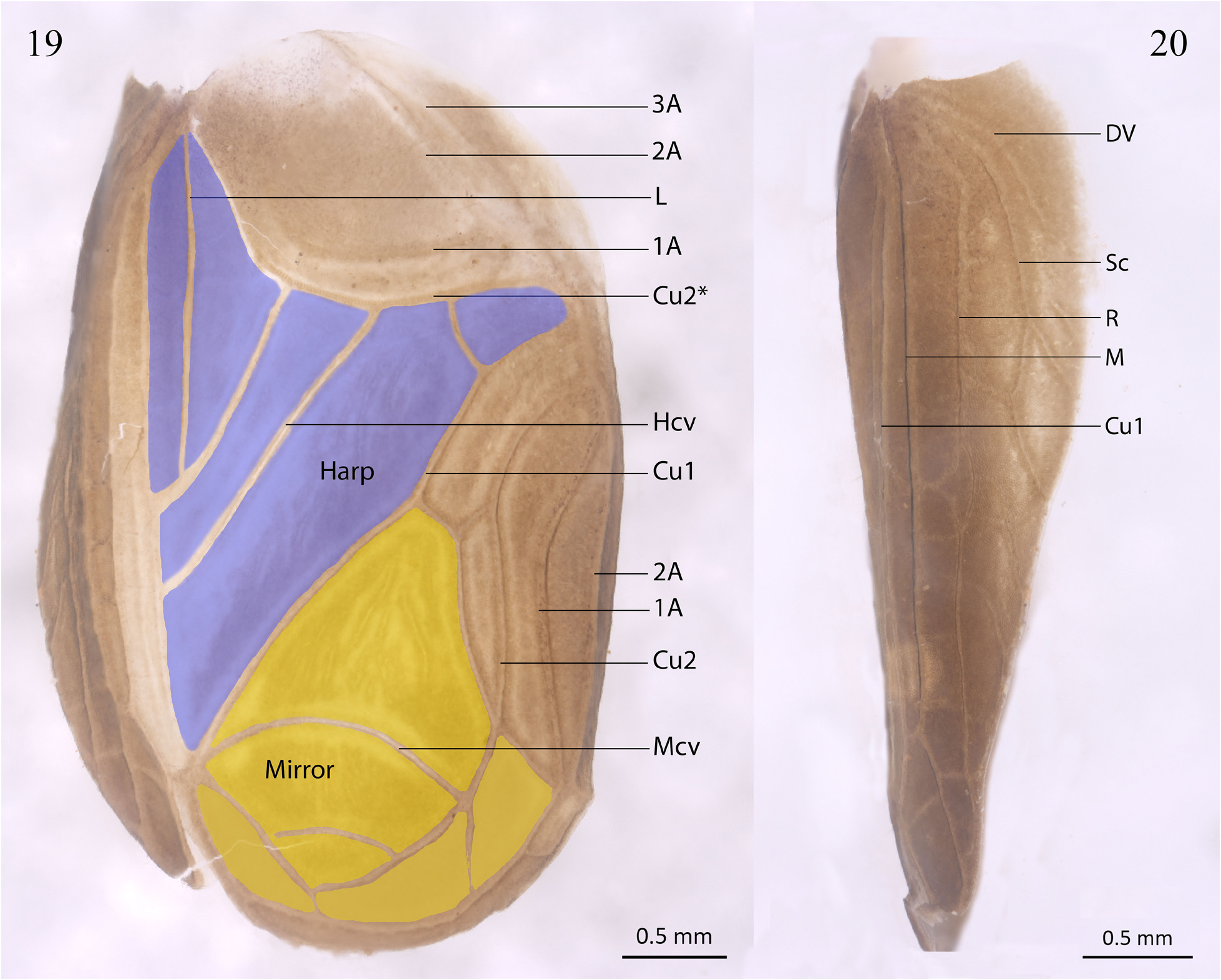
Right tegmen. Darkened brown, covering the first four abdominal tergites ( Fig. 11 View FIGURES 8–15 ). Lateral field (in lateral view) with a diagonal vein (DV) poorly marked in its distal portion and with two little ramifications at the lateral margin of the wing extending parallelly to the subcostal vein (Sc) reaching one-third of the length of the lateral field; subcostal (Sc), radial (R) and medial (M) veins parallelly distributed in the lateral field; Sc with a ramification well marked at the lateral margin with a cross-vein well marked connecting with R at the middle of the wing; R with a small cell undeveloped right after the cross-vein with Sc, two little reticulated veins poorly marked can be observed at subapical region; between the parallel veins M and R can be seen some cross-vein poorly marked (four or more); Field (in ventral view, Fig. 19 View FIGURES 19–20 ): anal area, chordal area, harp area and the mirror area well developed; anal region with veins anal 1 (1A), anal 2 (2A) and anal 3 (3A) poorly demarked; chordal area with veins 1A, 2A poorly marked and cubital 2 (Cu2) well marked; Cu2* modified in stridulatory file; harp with a median-longitudinal vein (L), and three crossed veins (Hcv), two connecting Cu2 to Cu1 towards the lateral field, and one connecting Cu2 to Cu1 at the dorsal proximal portion, forming five cells well marked, the cell above the mirror presents five reticular veins; mirror triangular oval, with a crossed vein (Mcv) well marked at the center and one poorly marked vein at distal region, forming three cells, proximal cell with four reticular veins; stridulatory file with 100 teeth. Abdomen. tergites pubescent, darkened brown ( Figs 14 and 11 View FIGURES 8–15 ); sternites pubescent, slightly whiter than the tergites ( Fig. 11 View FIGURES 8–15 ); subgenital plate darkened, pubescent, quadrangular shape, distal and lateral margins with long bristles, distal central region with a slightly indentation ( Fig. 13 View FIGURES 8–15 ); supra-anal plate slightly whitish comparing to the subgenital plate, pubescent, trapezoidal shaped, with small lateral projections, rounded by two white spots at latero-median portion from structure, distal portion rounded and with long bristles ( Fig. 15 View FIGURES 8–15 ); cerci reddish-brown and whitish at the base, subapical region slightly darkened ( Fig. 14 View FIGURES 8–15 ).
Observations in Paratypes. Male phallic sclerites (paratype ISLA 66144, Figs 2–6 View FIGURES 2–7 ) Pseudepiphallus: median projection curved inward, lobular shape, slightly acuminated at the base, slightly sclerotized (Ps.M.Pr, Figs 2, 4 and View FIGURES 2–7
6); pseudepiphallic median lophy claviform and thin (compared with A. similis and A. cruscastaneus ), acuminated apex with bristles and flattened, (Ps.m.l, Figs 3, 4 and 6 View FIGURES 2–7 ); Paramere 1 well developed, C-shaped (very similar A. spurius ) apex dilated and curved inward (Ps.P1.p, Figs 2–5 View FIGURES 2–7 ); Paramere 2 connected to Paramere 1 by membranous tissue, little protruding and escrerotized in this specie (Ps.P2, Figs 4 and 5 View FIGURES 2–7 ); Rami less elongated and sclerotized (compared to A. rubricephalus , A. similis , A. cruscastaneus , A. parasimilis ), dilated, curved inside and triangular shaped at the tip (R, Figs 3, 4 and 6 View FIGURES 2–7 ). Ectophallic invagination: ectophallic sclerite H-shaped shortened (similar to A. spurius ) ( Figs 2 and 3 View FIGURES 2–7 ), with a apodeme in the distal region (Ec.Ap) weakly sclerotized and slightly dilated to the outer edge of the sclerite ( Figs 2 and 3 View FIGURES 2–7 ) apex of posterior projections quadrangular-shaped in ventral view, dilated and weakly sclerotized, connected to the pseudepiphallic paramere 1 by membranous tissue (Ec.Pr, Figs 2–4 and 6 View FIGURES 2–7 ); ectophallic arc slightly longer as wider (Ec.Arc, Figs 2 and 3 View FIGURES 2–7 ); ectophallic fold sclerotized, linear-shaped, with the lateral border more sclerotized than that interior base, central part slightly convex at the top and bottom borders (Ec. F, Fig. 3 View FIGURES 2–7 ). Endophallus: circular-shaped and vertically elongated, with a short ventral crest (End.Sc), connected to the ectophallic fold by an inverted U-shaped membrane (End.Sc, Fig. 3 View FIGURES 2–7 ).
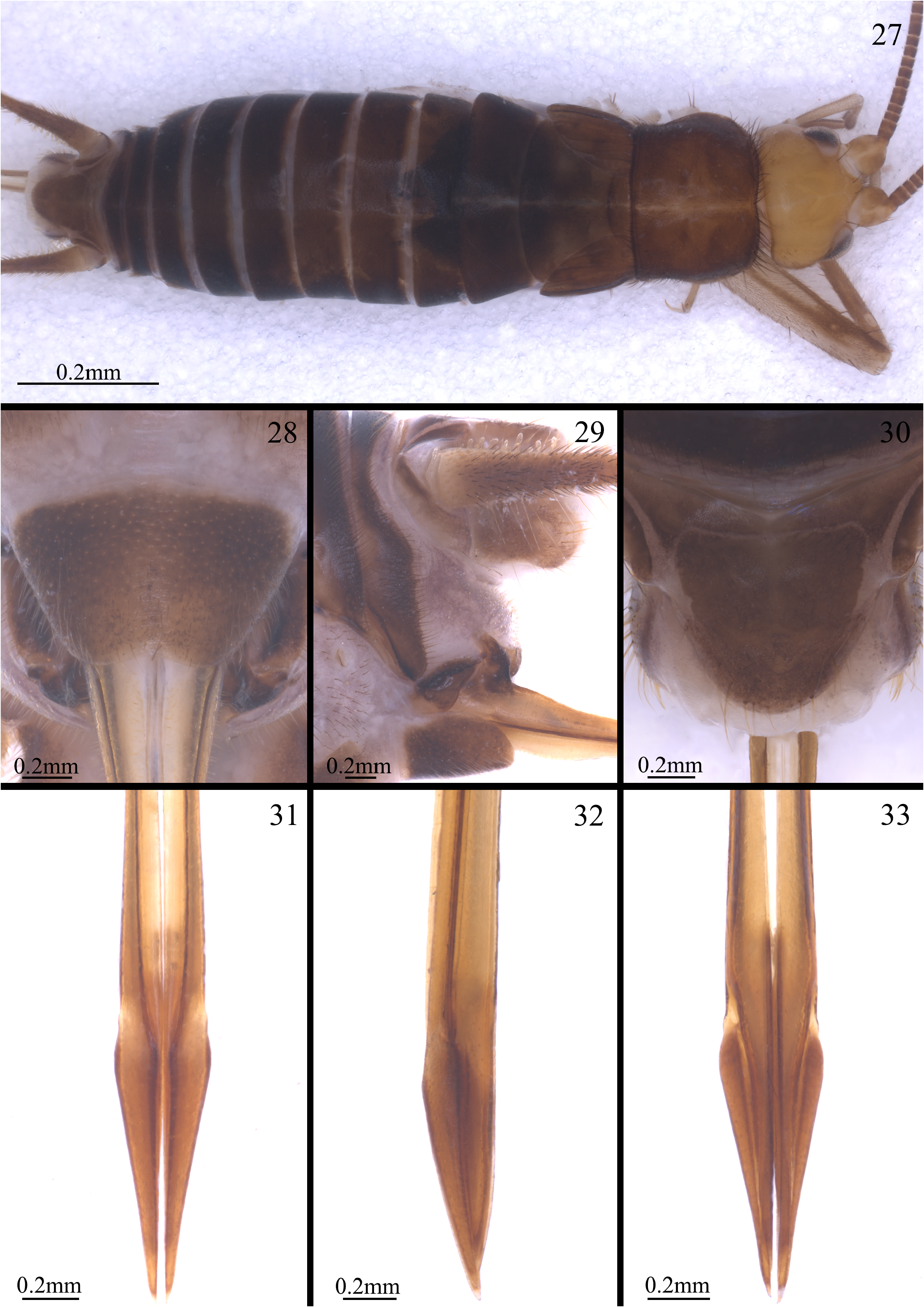
Female ( Figs 27–33 View FIGURES 27–33 , ISLA 66148) Body color similar to holotype ( Fig. 27 View FIGURES 27–33 ), body size bigger than the holotype (13.797 mm); small wings triangular-shaped ( Fig. 27 View FIGURES 27–33 ); supranal plate more whitish than tergites, trapezoidal-shaped elongated, with small lateral projections, surrounded by two white spots on the latero-median region, apex rounded and with long bristles ( Figs 28 and 29 View FIGURES 27–33 ); subgenital plate dark, short, V-shaped, apex with a slight concavity ( Fig. 28 View FIGURES 27–33 ); ovipositor thin and elongated (5.917 mm), proportional to tibia III size, sword format at apex ( Figs 31–33 View FIGURES 27–33 ).
Copulatory Papilla: well sclerotized, circular-shaped, with a large membranous opening area, reaching 2/3 of the structure in dorsal view ( Fig. 7a View FIGURES 2–7 ); lateral face sclerotized in all its extension ( Fig. 7a, 7b and 7c View FIGURES 2–7 ), apex slightly concave, with a ventral indentation followed by a less sclerotized region (in white), base with a membranous opening of triangular shape in ventral view ( Fig. 7c View FIGURES 2–7 ).


| R |
Departamento de Geologia, Universidad de Chile |
No known copyright restrictions apply. See Agosti, D., Egloff, W., 2009. Taxonomic information exchange and copyright: the Plazi approach. BMC Research Notes 2009, 2:53 for further explanation.
|
Kingdom |
|
|
Phylum |
|
|
Class |
|
|
Order |
|
|
Family |
|
|
Genus |