
Acanthocyclops eduardoi Mercado-Salas and Álvarez-Silva, 2013
|
publication ID |
https://doi.org/ 10.1080/00222933.2012.742589 |
|
persistent identifier |
https://treatment.plazi.org/id/03915D7A-FFE1-FFD1-FE05-9243838589D9 |
|
treatment provided by |
Felipe |
|
scientific name |
Acanthocyclops eduardoi Mercado-Salas and Álvarez-Silva |
| status |
|
Genus Acanthocyclops Kiefer, 1927
Acanthocyclops eduardoi Mercado-Salas and Álvarez-Silva n. sp.
( Figures 2–5 View Figure 2 View Figure 3 View Figure 4 View Figure 5 )
Material examined
Holotype. Adult female, dissected, mounted in glycerine sealed with Entellan (ECO- CH-Z-07510), Station 3 December 2010 Eastern Quarry, Ecological Reserve of San Angel Pedregal (19 ◦ 19 ′ 4.42 ′′ N, 99 ◦ 10 ′ 21.4 ′′ W) coll. 3 December 2010 by Carlos Álvarez-Silva. GoogleMaps
Allotype. Adult male, dissected, semi-permanent slide (ECO-CH-Z-07511), same site, date and collector.
Paratypes. (ECO- CH-Z-07512), 20 adult females, three undissected copepodites, same locality and date of collection; ethanol-preserved. Original samples containing several adult males and females and also copepodites are deposited in C. Álvarez-Silva’s laboratory at the Universidad Autónoma Metropolitana Campus Iztapalapa , Mexico City .
Type locality
Station 3 December 2010, Eastern Quarry , Ecological Reserve of San Angel Pedregal, Mexico City, Mexico (19 ◦ 19 ′ 4.42 ′′ N, 99 ◦ 10 ′ 21.4 ′′ W); temperature of water at time of collection 15.1 ◦ C, pH 9.1. Average density of individuals in the sample was 99.7 adult copepods per litre (females 60.2 per litre; males 39.5 per litre) GoogleMaps .
Etymology
The species is warmly dedicated to Dr Eduardo Suárez-Morales for his many contributions to the knowledge of the copepod fauna in Mexico and Latin America and his commitment to training Latin American taxonomists.
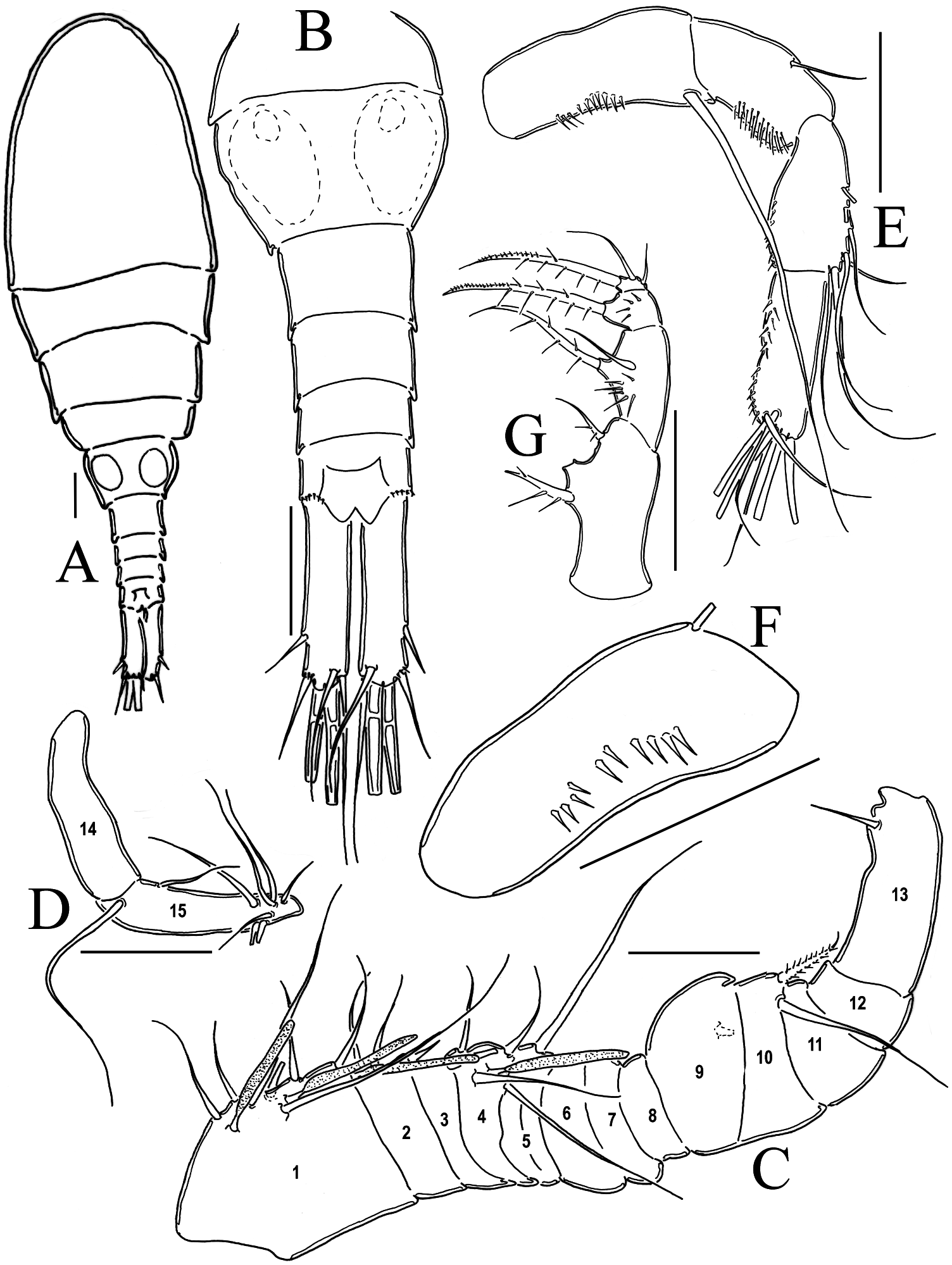
Description – adult female holotype
Total body length 1.62 mm from anterior end of cephalothorax to posterior margin of caudal rami. Body robust, cephalothorax relatively long, slightly expanded laterally at midlength of cephalosome in dorsal view; lateral margins of pedigers 3 and 4 straight, produced posteriorly. Cephalothorax length 1.084 mm, representing 66.66% of total body length. Dorsal surface smooth. Urosome relatively slender, formed by five somites: fifth pediger plus four succeeding somites. Genital double-somite moderately expanded at proximal half. Anal and preanal somites equally sized.
Antennule ( Figure 2D View Figure 2 ). Seventeen-segmented in all specimens examined; armature per segment as follows (s = seta, ae = aesthetasc, sp = spine): 1(8s), 2(3s), 3(1s), 4(6s), 5(3s), 6(1s + 1sp), 7(1s), 8(1s), 9(1s), 10(0s), 11(2s), 12(1s +1ae), 13(0s),14(1s), 15(2s), 16(2s), 17(7s). Antennules reaching middle of second thoracic somite. Dorsal surface of first segment without row of spinules.
Antenna ( Figure 2E,F View Figure 2 ). Four-segmented, basis with three groups of spines on caudal surface and three groups on frontal surface. Eight largest spines arranged longitudinally close to inner margin, additional group adjacent to first one, with four spines of the same size, third row of spines present on outer margin of basis with five small spines. Basis with long exopodal seta biserially pinnate plus two equally long basipodal setae. First segment of endopodite with single outer seta and inner row of small spinules. Second endopodal segment with five lateral and four terminal outer setae; inner margin with row of short spinules. Third endopodal segment with seven terminal setae, inner margin with row of spines.
Labrum ( Figure 2G View Figure 2 ). Distal margin with 11 blunt teeth between rounded lateral protuberances; two rows of long, slender spinules on ventral surface.
Maxillule ( Figure 2H,I View Figure 2 ). Precoxal arthrite with naked surface, with three strong chitinized distal claws and one spiniform seta on frontal side. Basis of palp furnished apically with one biserially pinnate seta and two simple setae. Endopod subquadrate, with three unequally long apical setae.
Maxilla ( Figure 2J View Figure 2 ). Precoxa and coxa not fused; precoxal endite armed with two strong biserially setulated setae. Coxa with single long seta on distal inner margin; coxal caudal surface naked. Proximal basipodal endite well developed, with two apical setae, one furnished with spinules and noticeably thicker than the other. Claw-like basal endite relatively short, endite with two setae, one large, one small, slender, both inserted near base of claw. Endopod with one segment, bearing two strong, long spiniform setae plus two short, slender setae.
Maxilliped ( Figure 2K View Figure 2 ). Four-segmented. Syncoxa with three spiniform setae along inner margin. Basis with strong setulated setae, one longitudinal row of long spines on inner margin, and two transverse rows of spines on outer margin. Endopod two-segmented, first segment with wide-based, stout basal spine sparsely spinulated; longitudinal row of four spines, close to the insertion of seta. Second endopodal segment armed with three elements: one proximal strong, spiniform sparsely spinulated seta plus two shorter, naked setae.
Leg 1 ( Figure 3A View Figure 3 ). Intercoxal sclerite with row of four spinules on each side of anterior surface, distal margin with two rounded chitinized projections. Coxa with strong biserially setulated inner coxal seta. Basis with long basipodal seta on outer margin, inner margin moderately expanded, with strong spiniform seta reaching midlength of third endopodal segment; small spines along insertion of endopod. Endopod and exopod three-segmented. Armature as in Table 1.
Leg 2 ( Figure 3B View Figure 3 ). Intercoxal sclerite naked, with two rounded chitinized projections. Coxa with row of spinules along outer margin. Coxal spiniform seta biserially setulated. Basis with slender basipodal seta on outer margin; inner corner and middle distal margin of basis forming spiniform expansions. Endopod and exopod three-segmented. Armature as in Table 1.
Leg3 ( Figure 3C View Figure 3 ). Intercoxal sclerite naked, with slightly rounded chitinized projections. Coxa with inner coxal seta biserially setulated and row of setules along outer margin. Basis with slender basipodal seta on outer margin, with spinules on insertion of seta; inner corner and middle distal margin of basis forming spiniform expansions. Endopod and exopod three-segmented. Small spines along insertion of endopod. Endopod as long as exopod. Armature as in Table 1.
Leg 4 ( Figure 3D View Figure 3 , Table 2). Intercoxal sclerite with single row of 11 spinules on anterior surface; distal margin smooth, not expanded. Coxal surface with one row of five spines close to proximal margin, one row of 10 spines along proximal margin and one row of spinules along outer margin; inner coxal seta biserially setulated, reaching distal margin of first endopodal segment. Basis with seta on outer margin and row of
of Acanthocyclops eduardoi sp. nov. Sequence follows external to internal positions. spinules on the insertion of seta; row of small spinules on the insertion of endopod. Endopod and exopod three-segmented. Endopod slightly longer than exopod. Inner spine slightly longer than outer spine; length ratio of outer / inner terminal spines of third endopodal segment (Enp 3) 0.97. Length / width ratio Enp 3 = 3.1. Insertion point of seta on outer margin of Enp 3 = 0.56 of the length of segment. Length ratio of inner endopodal spine / Enp 3 = 0.75. Armature as in Table 1.
Leg 5 ( Figure 3E View Figure 3 ). Leg consisting of two free segments, first (proximal) subrectangular, with moderate lateral expansion bearing long regular seta; second (distal) segment about 1.63 times longer than broad, bearing long seta inserted distally. Short spiniform element inserted subdistally; spiniform element slightly longer than distal margin of segment.
Urosome ( Figure 2C View Figure 2 ). Posterior margins of genital double somite, urosomites and anal somites smooth both dorsally and ventrally; relative ratio of each urosomite as: 52: 14: 14: 20 = 100. Genital double-somite representing 17.5% of body length (excluding caudal rami). Genital double-somite smooth on ventral and dorsal surfaces. Anterior half of genital double-somite expanded laterally. Ventral surface of anal somite smooth; distal margin with rows of 18–20 stout spines along insertion of caudal rami of ventral surface and ascending along the flanks and reaching dorsal margin.
Caudal ramus ( Figure 2B View Figure 2 , Table 2). Ramus 0.33 times as long as urosome. Length / width ratio = 4.92. Inner margin, ventral and dorsal surfaces with small spinules not showing an established pattern. Lateral seta short, 0.29 times as long as caudal ramus, inserted at about three-quarters of the outer margin of ramus. Dorsal seta 0.56 times as long as caudal ramus. Innermost terminal seta 0.97 times as long as caudal ramus and longer than dorsal seta. Outermost terminal seta about 0.58 times as long as caudal ramus.
Description – adult / male allotype
Total body length 1.25 mm from anterior end of cephalothorax to posterior margin of caudal rami.
Antennule ( Figure 4C,D View Figure 4 ). Geniculate, 15-segmented.
Antenna ( Figure 4E,F View Figure 4 ). As in female except for the lack of row of tiny spinules on outer margin of basis.
Mouth parts. As in female.
Leg 1 ( Figure 5A View Figure 5 ). As in female except for the number of spinules on intercoxal sclerite being seven instead of four on each side of anterior surface.
Leg 2 ( Figure 5B View Figure 5 ). As in female except for the ornamentation of intercoxal sclerite, the male presents a row of seven spinules on medial margin.
Leg 3 ( Fig 5C View Figure 5 ). As in female except for the ornamentation of intercoxal sclerite, the male presents a row of six spinules on each side of anterior surface.
Leg 4 ( Fig 5D,E View Figure 5 ). As in female except for two characters: (1) the ornamentation of intercoxal sclerite stronger than in female, the male presents an additional row of spinules on anterior surface and a group of strong spinules above the normal row of spinules present on medial margin; and (2) lateral outer seta of male is modified into a tubular spine.
Leg 5. As in female.
Leg 6. Small low plate at distal corner of genital somite with one strong inner spine, one medial seta as long as inner spine and one outer seta about two times longer than inner spine.
Urosome ( Figure 4B View Figure 4 ). Genital somite subrectangular, length / width ratio = 0.625. Genital somite and succeeding three urosomites without ornamentations. Distal dorsal margin of anal somite with rows of five stout spines.
Caudal ramus ( Figure 4B View Figure 4 ). Length / width ratio = 3.93, and not ornamented with spinules as female. Lateral spiniform seta short, 0.31 times as long as caudal ramus, inserted at about three-quarters of the outer margin of ramus. Dorsal seta 0.74 times as long as caudal ramus. Innermost terminal seta slightly longer than caudal ramus (1.04 times) and longer than dorsal seta. Outermost terminal seta about 0.50 times as long as caudal ramus.
Remarks
The characters that allowed us to include A. eduardoi sp. nov. in the genus Acanthocyclops are: (1) the general body shape with the fifth somite broader than the genital somite and lateral margins triangular; (2) fifth leg represented by two segments, the distal one armed with a small subapical spine reaching the distal end of basal segment; (3) female antennule with 17 segments; (4) endopodal and exopodal rami of legs 1–4 all with three segments ( Reid 1985; Einsle 1996; Dussart and Defaye 2001).
The new species was assigned as a member of the A. vernalis – robustus complex because of the number of antennular segments, the length of the basipodal spine, the spine formula, the length of the inner caudal seta and its habitat and geographical distribution. The main character to separate all the species of this complex from the strict A. robustus is through differences in the ornamentation of the antennal basipodite, which bears spinules near the insertion of the exopodal seta, a character lacking in the new species and its congeners of the complex. As in A. marceloi Mercado-Salas and Suárez-Morales, 2009 , A. caesariatus Mercado-Salas and Suárez-Morales, 2009 , A. robustus , A. trajani , A. einslei and A. brevispinosus , A. eduardoi sp. nov. has 17 segmented antennules. In all of these species with exception of A. marceloi the distal end of the long aesthetasc on segment 12 reaches the distal margin of segment 14 (Mercado- Salas et al. 2009). One of the main differences between A. trajani and A. eduardoi sp. nov. is the absence of spinules on inner side of the claw-like seta of the basipodite of maxilla. The length / width ratio of the caudal ramus in A. eduardoi sp. nov. is 4.3–4.95, this proportion is within the range of variation of A. robustus (4–5.8), A. trajani (4.15–5.8) and A. einslei (4.15–6.40), but differs from A. brevispinosus (5.20–6.85), A. marceloi (2.8) and A. caesariatus (3.79). A distinctive character of A. eduardoi sp. nov. is the ornamentation of the ventral and dorsal surface of caudal ramus of the female, it has an extended field of tiny spinules, a similar pattern was previously reported in A. einslei and A. marceloi , but it differs among these species. In A. marceloi the spinules are arranged in symmetrical patches on the ventral surface and irregular ones on the dorsal surface. In the new species spinules are irregularly distributed both dorsally and ventrally, the pattern in A. einslei has not been described.
A useful character to separate A. eduardoi sp. nov. from its congeners is the length ratio of the dorsal caudal seta / caudal ramus (0.47–0.56), which is similar to A. brevispinosus (0.53). A longer seta is present in A. marceloi (0.63), A. trajani (0.64), A. einslei (0.71) and in A. caesariatus (0.75). In A. eduardoi sp. nov. the innermost terminal caudal seta is slightly shorter than the caudal ramus (0.80–0.97) as in A. caesariatus (0.9), A. robustus (0.50–0.88) and A. einslei (0.90), whereas in A. brevispinosus it is much shorter (0.4–0.5) and slightly longer in A. marceloi (1.15) and A. trajani (0.84–1.04). Most of the species belonging to the A. robustus – vernalis complex, including the new species, have a 3444 spine formula, with the exception of some populations of A. robustus (2334 also) ( Mirabdullayev and Defaye 2002) and A. marceloi (3443) (Mercado-Salas et al. 2009). Another important character to separate A. eduardoi sp. nov. from its congeners is the ornamentation of the first leg coupler; in this species it has two groups of spinules, four on each side of the coupler ( Figure 2A View Figure 2 ) as in A. caesariatus (Mercado-Salas et al. 2009) . In A. trajani this structure is armed with two groups of five spinules, in A. marceloi with two rows of 8–10 spinules, and in A. robustus , A. brevispinosus and A. einslei this ornamentation is absent. Couplers of second and third legs in A. eduardoi sp. nov. are naked as in A. marceloi , A. caesariatus , A. einslei and A. brevispinosus ; A. trajani is the only species with an ornamentation represented by a single row of six spinules at each side of coupler of the second leg. It also has one row with six spinules on each side, close to the anterior margin, and one transversal row of approximately 20 tiny spinules on medial margin of the coupler of the third leg, so diverging from the pattern observed in the new species. In A. eduardoi sp. nov., the ornamentation of the fourth leg coupler consists of one transverse row of 11 strong spinules on medial margin as in A. marceloi , A. brevispinosus and A. caesariatus but differs from A. trajani with a row of 15 strong spinules, from A. robustus with a row of 18 tiny spinules and from A. einslei , bearing 11 small spinules. All endopodal and exopodal setae of swimming legs 1–4 in females of A. eduardoi sp. nov. are normal as in A. marceloi and A. caesariatus . In A. robustus and A. trajani modified setae are present on legs 2–4; also, A. einslei and A. brevispinosus have modified setae on legs 3 and 4. An additional distinguishing character of A. eduardoi sp. nov. is the length / width ratio of third segment of endopodite of fourth leg (3.1–3.8), which is shorter in its congeners; in A. trajani it ranges from 2.25 to 3.10 and in the rest of the species it varies between 2.2 and 2.5. The length ratio of inner spine / length of End 3 of fourth leg of A. eduardoi sp. nov. (0.68–0.76) is similar to A. einslei (0.72–0.77) and differs from A. trajani , A. marceloi and A. caesariatus (0.82–0.88), A. robustus and A. brevispinosus present a wider range than the rest of species (0.71–0.95). In A. eduardoi sp. nov. the insertion point of the outer seta on End 3 of fourth leg is 0.57–0.60, whereas it is 0.56–0.66 in A. trajani , 0.60–0.71 in A. robustus , in 0.61 in A. marceloi , 0.67 in A. caesariatus and in A. einslei and A. brevispinosus the value ranges between 0.75 and 0.82.
The male of A. eduardoi sp. nov. shows a unique ornamentation pattern on the coxal plate of the fourth leg, with one transversal row of 13 strong spinules equal in size on medial margin, one group of strong spinules not equal in size at each side above the transversal row and a third row of slender spines of different size close to the anterior margin of coupler ( Figure 4E View Figure 4 ). Finally, the lateral seta of outer margin of End 3 of fourth leg is modified in A. eduardoi sp. nov. as in A. einslei and A. brevispinosus whereas in A. robustus and A. trajani this seta is normal; males of A. marceloi and A. caesariatus have not been described.


No known copyright restrictions apply. See Agosti, D., Egloff, W., 2009. Taxonomic information exchange and copyright: the Plazi approach. BMC Research Notes 2009, 2:53 for further explanation.
|
Kingdom |
|
|
Phylum |
|
|
Class |
|
|
Order |
|
|
Family |
|
|
Genus |