
Xeropicta krynickii (Krynicki, 1833)
|
publication ID |
https://doi.org/ 10.37828/em.2014.1.27 |
|
DOI |
https://doi.org/10.5281/zenodo.8029093 |
|
persistent identifier |
https://treatment.plazi.org/id/03D7AE6E-CE0B-FF9F-FF44-FE58A17CF9C4 |
|
treatment provided by |
Felipe |
|
scientific name |
Xeropicta krynickii (Krynicki, 1833) |
| status |
|
Xeropicta krynickii (Krynicki, 1833) View in CoL View at ENA
Montenegrin populations
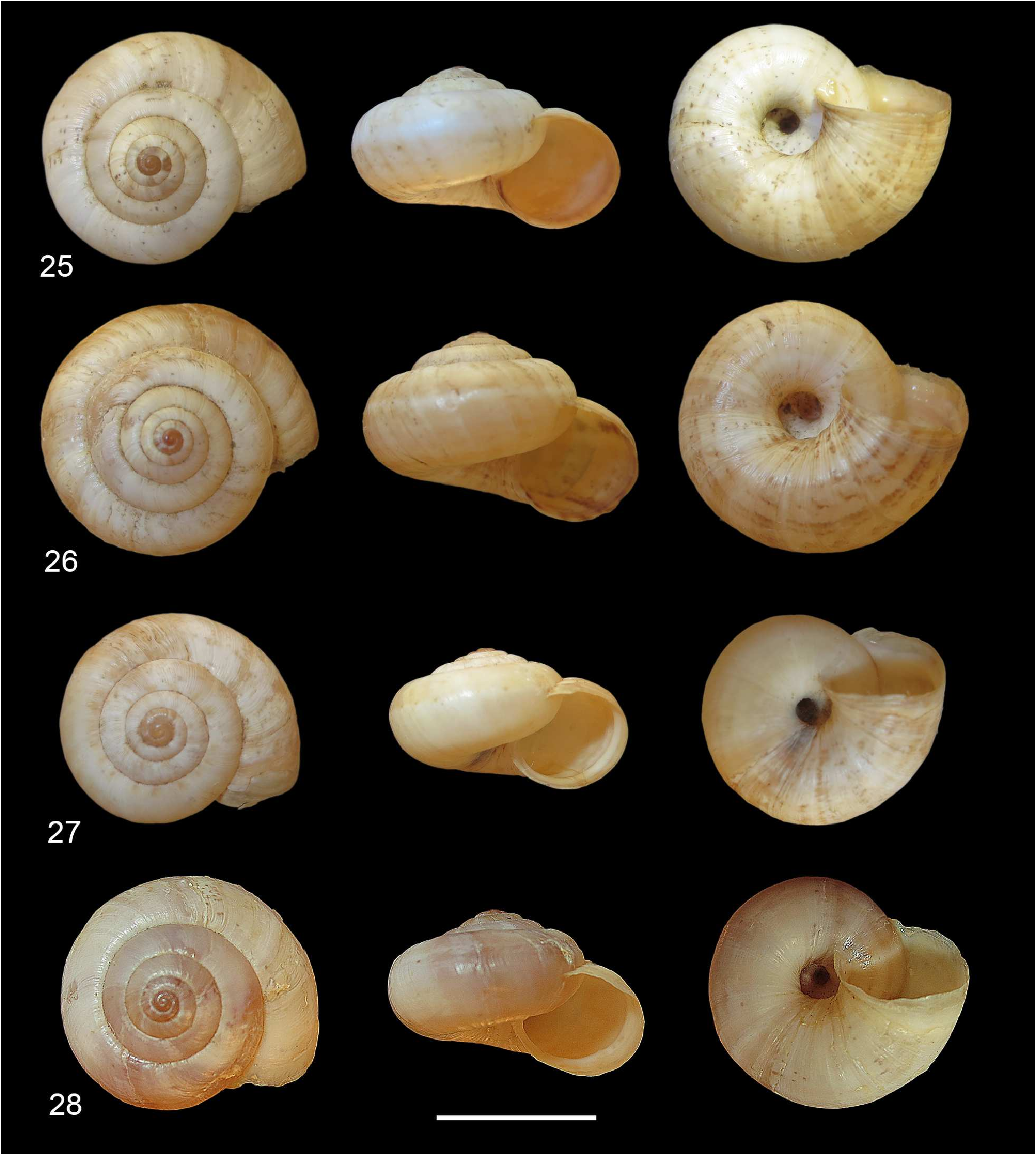
Shell ( Figs. 2526 View Figures 2528 ). Dextral, subglobose to depressed, moderately robust; protoconch light brown; teleoconch creamy to light yellowish in colour, with six fine, interrupted bands visible only on lower side of the last whorl; a white continuous band is present along the last whorl; external surface of teleoconch waxy with welldefined, irregularely spaced growth lines; spire conical to slightly raised, with 4⅓ 4 ¾ regularly growing, convex whorls, last whorl large and swollen, only moderately descending near aperture; umbilicus open, wide about ¼ of the maximum shell diameter; sutures deep; aperture rounded to moderately elliptical, periscope interrupted, never reflected at external, seldom with a thin whitish thickening on the palatal inner margin.
Shell dimensions diameter 15.2± 0.8 mm (range 14.3–16.3 mm); height 9.2± 0.9 mm (range 8.6–9.4 mm) (n=6). Ratio D/H 1.6.
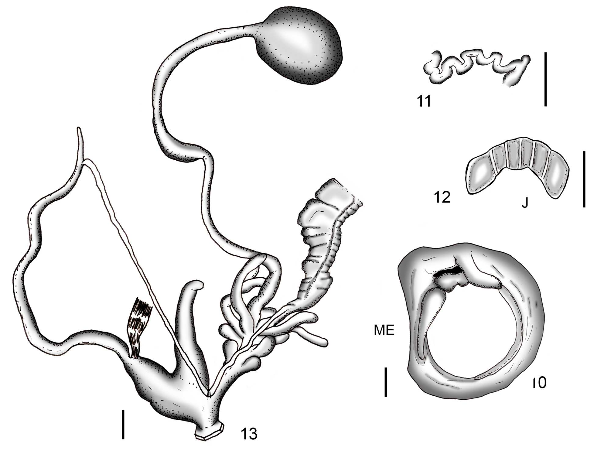
Genitalia ( Figs. 112 View Figures 19 View Figures 1013 ). The proximal part of the vagina has 1014 digitiform (sometimes branched) glands, disposed in four tufts of 24 units. The dartsac complex, entering the distal vagina on opposite sides (2+2 dartsac complex), consists of two proximal, clublike, longer, dartbearing, and two distal, clublike, shortest, dartless stylophores. Cavities of each of the 4 stylophores open almost independently into the vagina. The penial flagellum is moderately long and slender. The epiphallus is roughly three times longer than the flagellum. The penis is approximately half as long as epiphallus, without any penial sheath. It has a slender, smooth, penial papilla with a circular apical opening. The penial papilla has an outer sheath and presents a large, extended corpus cavernosus ( Fig. 5 View Figures 19 ). The genital atrium contains a slender and conical penial appendage, which is usually twice as long as the penis. A large crestlike structure develops from the tip of this appendage and extends to the atrial aperture while gradually increasing in height (fig. 3). A second smaller, lobated crestlike structure can be found in this appendage, parallel to the main one, situated on its right.
Greek population
Figures 26 and 27 View Figures 2528 depict the shell of a specimen of X. krynickii from Livadia (Sterea Ellada, Mainland Greece). In all investigated specimens from Greece (n=18) the width of the umbilicus is about one fifth that shell. This ratio can also be seen in, amongst others, De Mattia (2007: 11) and WelterSchultes (2012: 571). In the genital anatomy, some differences from Montenegrin specimens can be detected as regards the morphology of the penial papilla and the 2+2 stylophores. Greek specimens constantly show a penial papilla with lateral opening and much shorter proximal stylophores ( Figs. 4, 7 View Figures 19 , 23 View Figures 1424 , and 24).


No known copyright restrictions apply. See Agosti, D., Egloff, W., 2009. Taxonomic information exchange and copyright: the Plazi approach. BMC Research Notes 2009, 2:53 for further explanation.