
Costabolus baculus, Golovatch & Vandenspiegel, 2022
|
publication ID |
https://doi.org/10.11646/zootaxa.5150.1.5 |
|
publication LSID |
lsid:zoobank.org:pub:2F6FD286-5E1E-40F7-B658-490EFD453BDF |
|
DOI |
https://doi.org/10.5281/zenodo.6610031 |
|
persistent identifier |
https://treatment.plazi.org/id/F33487D0-FFDD-FFFC-FF78-FF56FE0518AE |
|
treatment provided by |
Plazi |
|
scientific name |
Costabolus baculus |
| status |
sp. nov. |
Costabolus baculus sp. nov.
Figs 4–7 View FIGURE 4 View FIGURE 5 View FIGURE 6 View FIGURE 7
Material examined. Holotype male (with exposed gonopods, body broken into two pieces) ( VNHM 198 ), Nigeria, 27 km NE of Calabar , 20 m inside native forest, 12.VIII.1984, collector unknown.
Paratypes: 2 males ( VNHM 198 ), same data as holotype ; 1 female ( VMNH 154 View Materials ), Nigeria, 27 km NE Calabar (Ekang road), under logs on fairly dry soil in new canopy forest farmed to young maize, 28.IV.1984, collector unknown ; 1 male, 1 female ( VMHN 199 ), Nigeria, 27 km N of Calabar , between trees, native forest, 20 m off logging road, 12.VIII.1984, collector unknown .
Name. Baculus in Latin means “crutch”, referring to the long coxal processes on male legs 2 and 3; a noun in apposition.
Diagnosis. Basically as in the genus. Body short, up to 27 mm long, with 38 body rings plus telson, metazonae with simple longitudinal crests/ribs, epiproct slightly projecting caudally. Male with tarsal pads and prominent coxal projections on leg-pairs 2 and 3.
Description. Measurements: male holotype with 39 body rings (38 + telson), ca 27 mm long, 3 mm wide. Paratypes, both male and female, same as holotype.
Coloration faded in alcohol, but a darker pattern still visible dorsally and laterally, yellowish ventrally. Head, ommatidia and anterior part of collum dark brown, antennae, legs and telson yellowish ( Fig. 4 View FIGURE 4 ).
Head capsule with an axial suture, this being especially distinct on labrum; ca 22 ommatidia located in an irregular oval cluster ( Fig. 5A View FIGURE 5 ).
Labrum as usual, with three irregular teeth and a single row of 12 stout marginal setae. Clypeus with two setiferous foveolae on each side ( Fig. 5E View FIGURE 5 ). Incisura lateralis closed ( Fig. 5A, D View FIGURE 5 ). Antennal cavity/groove present, length of antennae ca 1.3 mm; antennae shorter than body diameter. Relative lengths of antennomeres: 3=4=5<1<6<2 ( Fig. 5D View FIGURE 5 ). Terminal antennomere with four large sensory cones clustered together inside a membranous area ( Fig. 5C View FIGURE 5 ). Antennomeres 5 and 6 each with an apicolateral field of specialized sensilla ( Fig. 5D View FIGURE 5 ).
Gnathochilarium as usual, of spirobolidan conformation. Stipites separated in basal portion, each bearing three apical setae, but no setae on ventral surface. Lamellae linguales separated by anterior portion of mentum, each with two usual setae located behind one another; mentum without a large swollen area apically between both lamellae linguales, subdivided basally by a well-marked suture ( Fig. 5H View FIGURE 5 ).
Mandible: external tooth ( et) prominent, inner tooth ( it) with three smaller inner teeth, a thin lateral tooth ( lt), six rows of pectinate lamellae ( pl), molar plate ( mp) with five transverse furrows ( Fig. 5F, G View FIGURE 5 ).
Collum with a prominent groove along anterior margin, lateroventral margin broadly rounded, not extending ventrad as far as ventrolateral corner of body ring 2 ( Fig. 5I View FIGURE 5 ).
Body rings: Metazonae heavily crested ( Fig. 5J–L View FIGURE 5 ); mesozonae with longitudinal impressions, these being especially distinct ventrally ( Fig. 5K View FIGURE 5 ), caudal fringe/limbus membranous, with very small and regularly spaced spinicles ( Fig. 5L View FIGURE 5 ). Pre-anal ring with a small dorsal tip of epiproct overhanging the paraprocts ( Figs 4A View FIGURE 4 , 5M View FIGURE 5 ); hypoproct rounded; paraprocts (anal valves) well-rounded and devoid of spines or setae ( Fig. 5M View FIGURE 5 ). Posterior margin of paraprocts in lateral view without distinct lips ( Fig. 5M View FIGURE 5 ). Ozopores ( oz) starting with body ring 6.
Legs: Midbody legs ca 1.4 mm long, with prefemur longer than coxa and as long as the other podomeres; with small tarsal pads ( pa) ( Fig. 5O, P View FIGURE 5 ). Male tarsi 3 up to midbody legs with a small tarsal pad not protruding past base of claw, and with a pair of setiform apical spines ( Fig. 6D View FIGURE 6 ). Male leg-pairs 1, 2 and 3 modified, coxae 1 fused medially at base only, each prefemur 2 with a well-developed, unciform, basal projection ( hp) ( Fig. 6B, C View FIGURE 6 ), each coxa 3 with a long and spoon-shaped coxal process ( cp), this being almost as long as the leg itself ( Fig. 6B, D View FIGURE 6 ).
Anterior gonopods stout and relatively simple ( Fig. 6E–I View FIGURE 6 ); sternite ( st) produced into a wide, broadly rounded lobe, bearing clear traces of axial fusion ( Fig. 6E View FIGURE 6 ); coxite ( cx1) subtending much of anterior gonopods; telopodites basically 2-segmented, each consisting of a smaller, roundish, apically flattened, basal telopoditomere ( t1) with a very small, accessory, subtriangular segment at base ( Fig. 6H, I View FIGURE 6 ), and a much larger, lobe-shaped, distal telopoditomere ( t2).
Posterior gonopods ( Fig. 6J, K View FIGURE 6 , pg in H) in situ almost completely concealed inside anterior gonopods, clearly divided into a slender coxite ( cx2) and a shorter telopodite ( tl), both being slender ( Fig. 6J View FIGURE 6 ) and connected by a tiny triangular sternite. Telopodite consisting of two components: (1) a median membranous area, and (2) a lateral sclerotized area. Sperm channel running at mesal margin of coxite through telopodite’s membranous area, ending up subapically on a rudimentary solenomere ( sl) ( Fig. 6J, K View FIGURE 6 ).
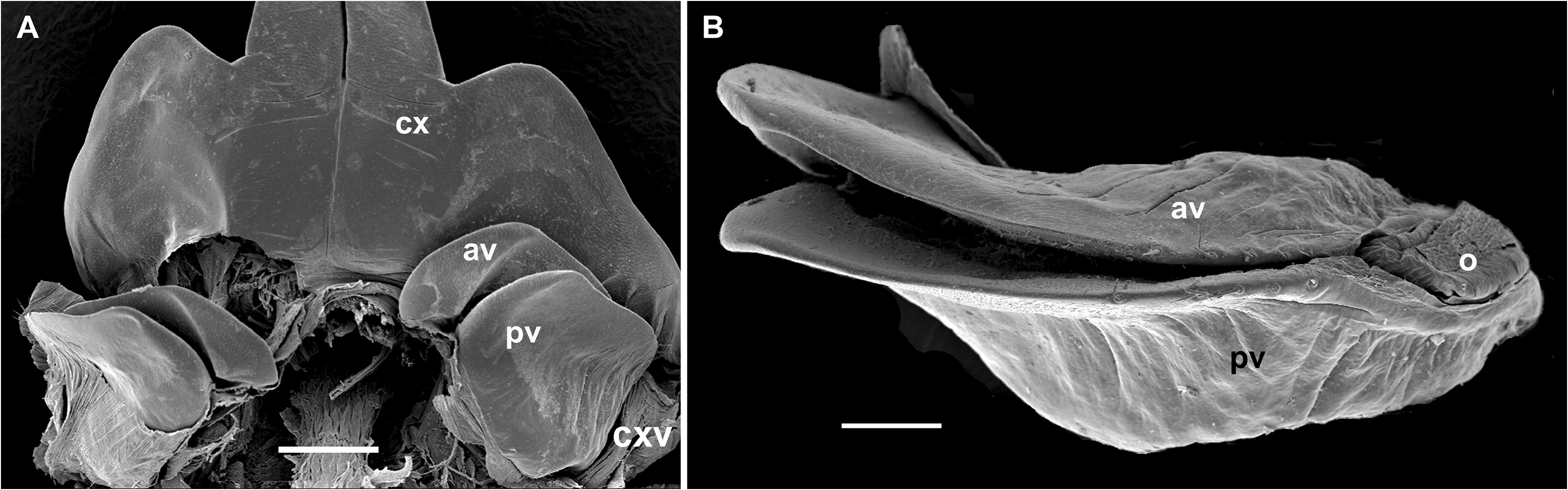
Paratypes. Males as holotype. Female similar in size and appearance to males, but devoid of prefemoral or coxal projections on leg-pairs 2 and 3, respectively. Vulva simple, consisting of two simple, sclerotized plates, bivalve-like and subequal in size ( Fig. 7 View FIGURE 7 ). Both coxite ( cxv) and operculum ( o) of vulva very small and inconspicuous. Both valves only basally with one row of setae directed towards the opening. Anterior valve ( av) slightly larger than posterior one ( pv) ( Fig. 7 View FIGURE 7 ).


No known copyright restrictions apply. See Agosti, D., Egloff, W., 2009. Taxonomic information exchange and copyright: the Plazi approach. BMC Research Notes 2009, 2:53 for further explanation.
|
Kingdom |
|
|
Phylum |
|
|
Class |
|
|
Order |
|
|
Family |
|
|
Genus |