
Filchneria olgae ( McLachlan, 1875 )
|
publication ID |
https://doi.org/10.11646/zootaxa.5277.2.3 |
|
publication LSID |
lsid:zoobank.org:pub:BB5236B9-095A-491F-B016-6F04FEAAAEB1 |
|
DOI |
https://doi.org/10.5281/zenodo.7889745 |
|
persistent identifier |
https://treatment.plazi.org/id/F24EE760-FFE5-8501-9189-7FF5C81FF4E4 |
|
treatment provided by |
Plazi |
|
scientific name |
Filchneria olgae ( McLachlan, 1875 ) |
| status |
|
Filchneria olgae ( McLachlan, 1875) View in CoL
Figs. 49–83 View FIGURES 49–50 View FIGURES 51–54 View FIGURES 55–58 View FIGURES 59–64 View FIGURES 65–68 View FIGURES 69–74 View FIGURES 75–76 View FIGURES 77–83
McLachlan, 1875: 49, Table IV, figs. 10 a, b, Dictyopteryx Olgae. Lectotype, male and paralectotypes ( 1 male 4 females) deposited in ZMMU, Moscow, Russia; Klapálek, 1912: 23, Figs. 19 A, B View FIGURES 16–19 ( Skobeleva Olgae ); Ricker, 1952: 140, figs. 86, 88, 89, ( Skobeleva olgae ); Illies, 1966: 390, Skobeleva olgae ; Raušer, 1968: 362, ( Skobeleva olgae ); Zhiltzova, 1971: 1035, ( Dictyopteryx olgae ); Zwick, 1973: 228, 230, ( Filchneria olgae ); Zwick, 1997: 494, figs. 6 (b‒d), Teslenko & Zhiltzova, 2009: 24, 102–104, ( Filchneria olgae ).
Diagnosis. Males of F. olgae have an obtuse angled (angle about 120°) posterior margin of tergum 10. The paraproctal sclerite is short, wide, and strongly sclerotized, resembling a rectangle with a Ⅽ-shaped notch along the inner margin; the apices of the notch are sharp and short. The aedeagus bears unpaired anterodorsal and posteroventral lobes, and two pairs of lateral lobes with one pair of lateral lobes being fingerlike. Females of F. olgae are distinguished by a short, wide subgenital plate with a straight or slightly wavy posterior margin, the lobes being weakly expressed, and the lateral edges are slightly truncated. Eggs of F. olgae bear clear longitudinal and transverse ridges, a stalked collar, and a rim that is slightly triangular with a wavy outer edge, and a smooth longitudinal carina present. The chorion is rough and evenly covered with flat rounded tubercles that have a distinct groove at the base. Micropyle orifices lack lip-like extensions.
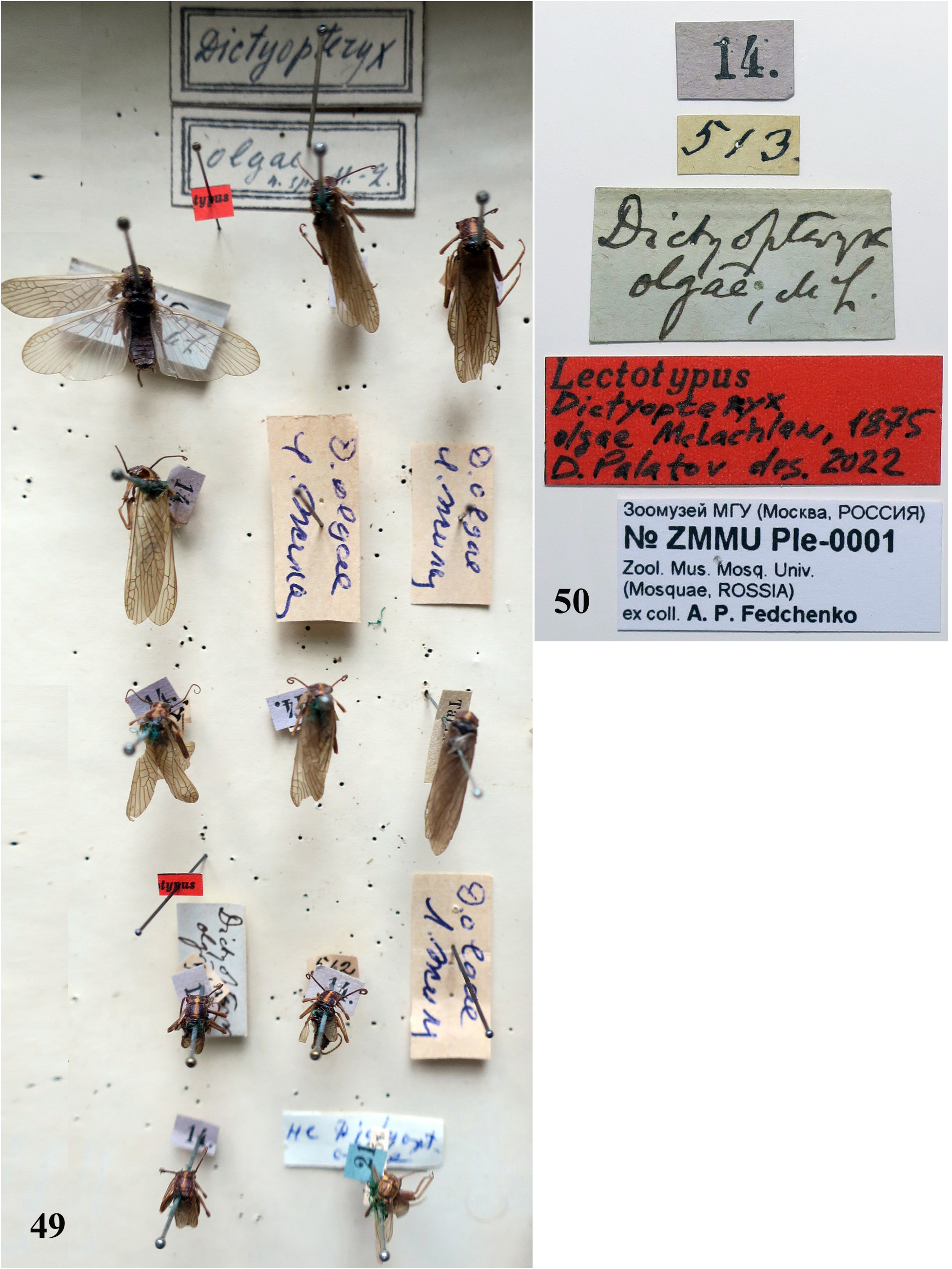
Complementary description. Adult habitus. Filchneria olgae was firstly described based on male and female by MacLachlan (1875) on the materials by A.P. Fedchenko outstanding explorer of the Central Asia. Type seria of F. olgae specimens is shown below ( Fig. 49 View FIGURES 49–50 ). The original description was carried out on base of the coloration and shape of the female subgenital plate and, in general, is consistent with the studied specimens (McLachlan 1975). Complementary description is given below based on the type material and relatively fresh specimens.
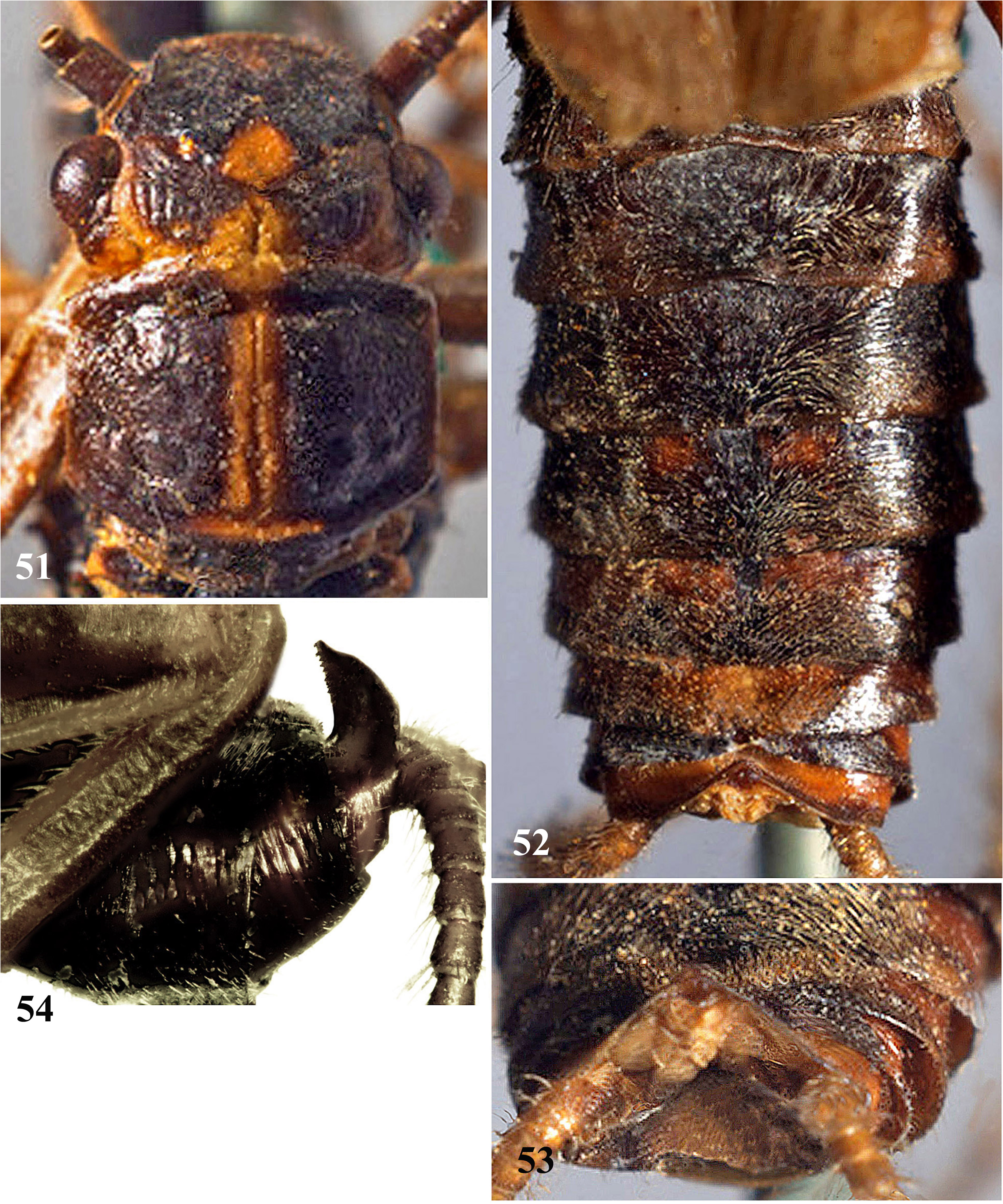
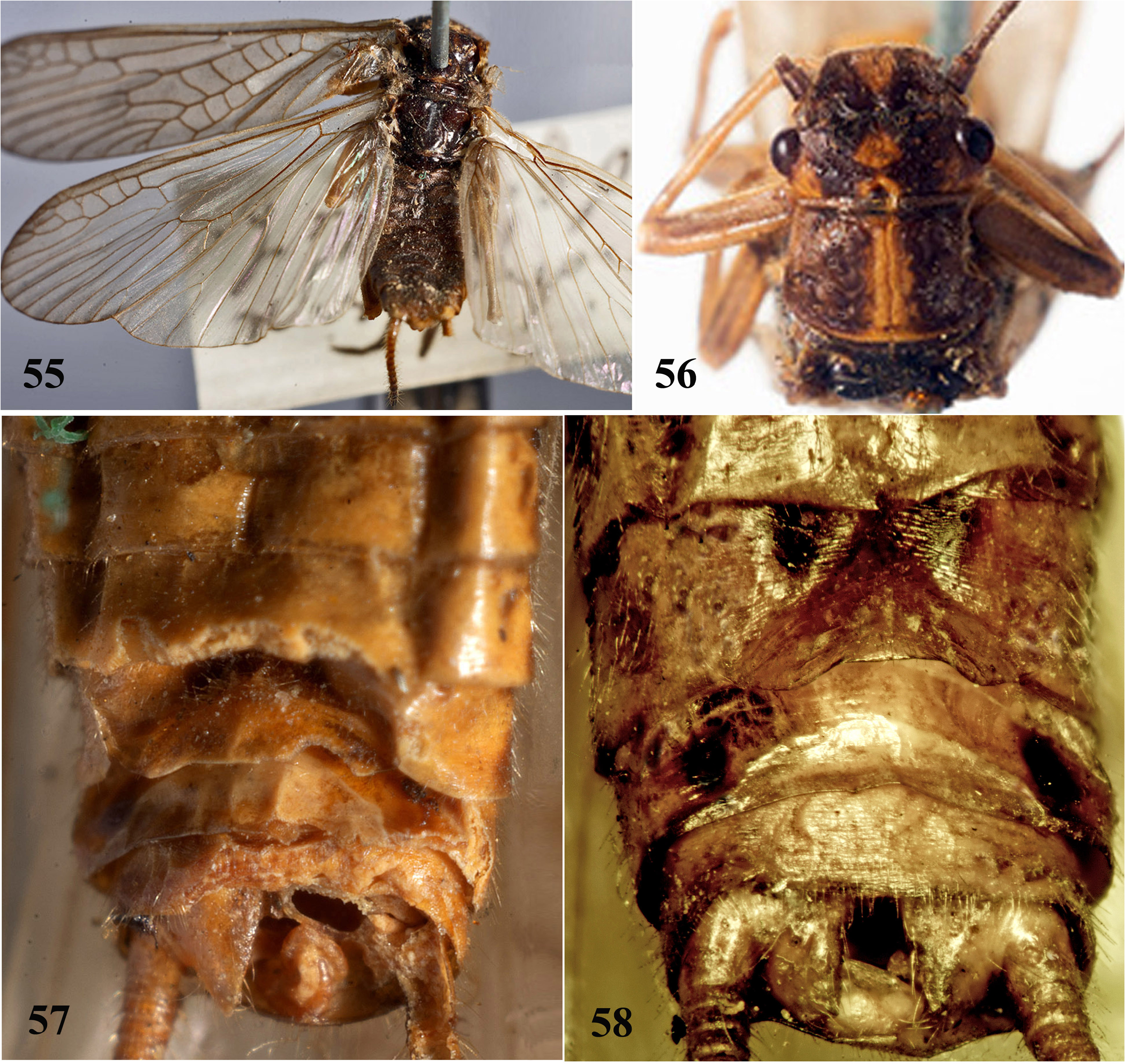
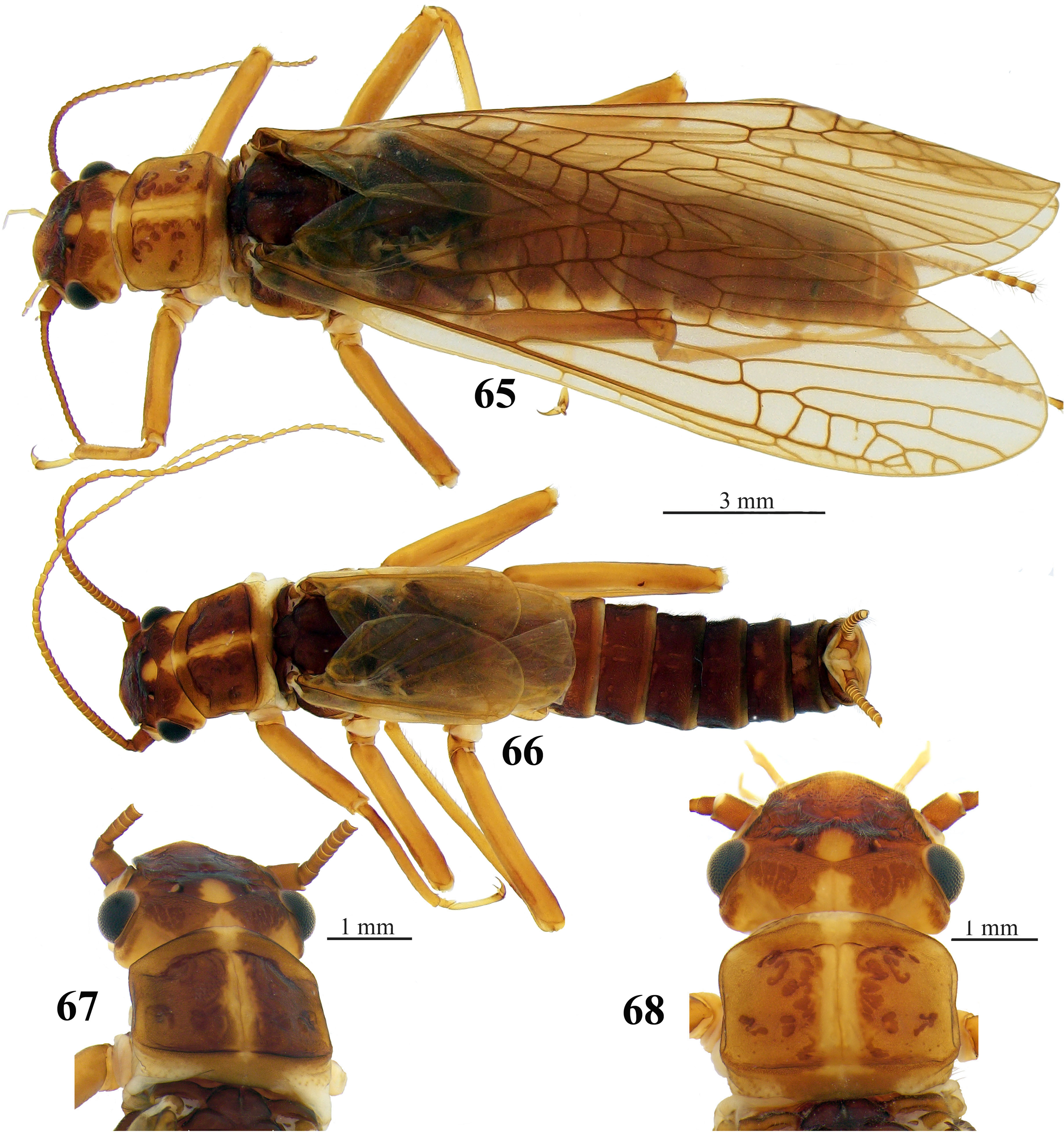
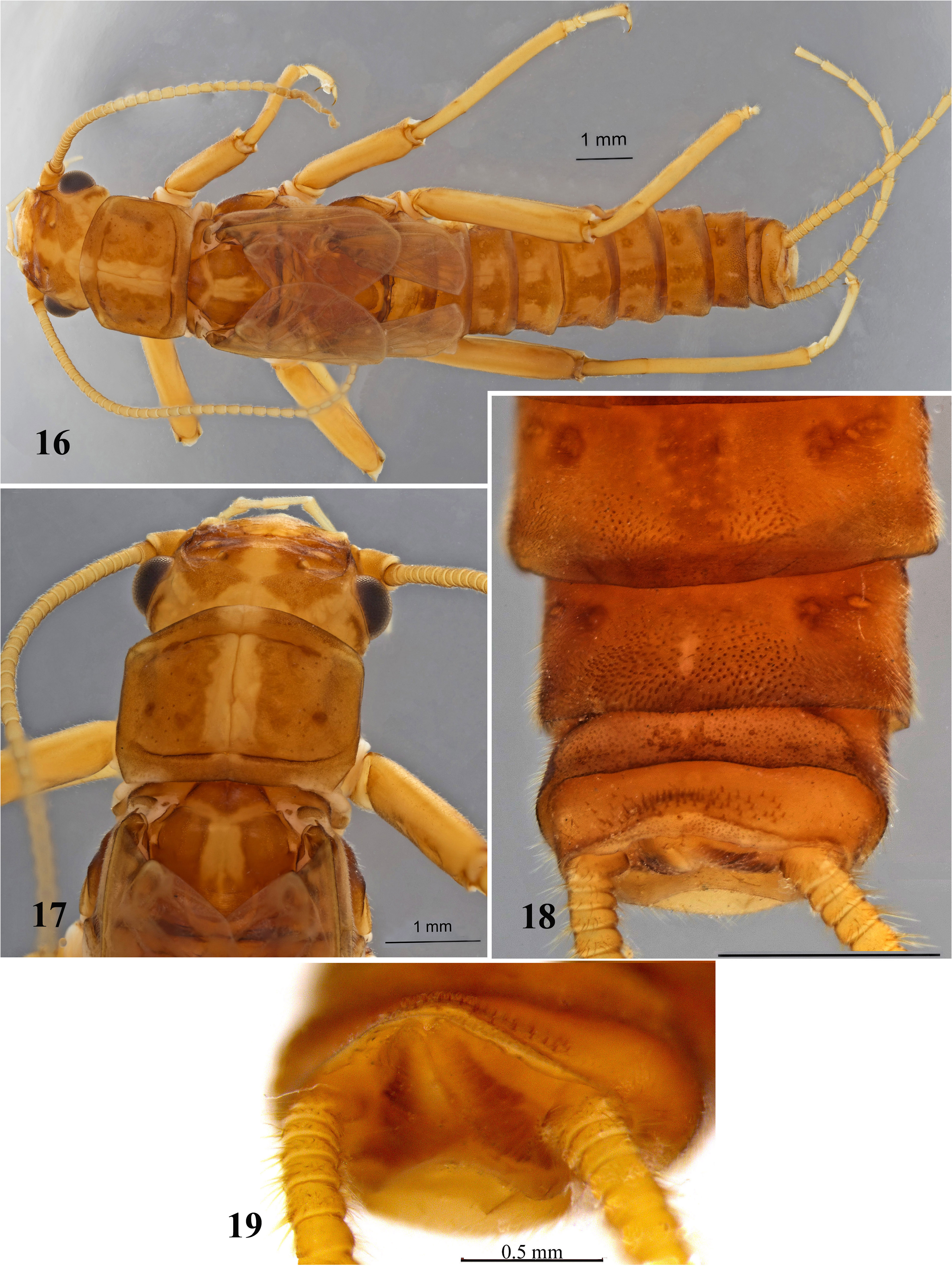
Both sexes may have shortened wings, males may also be brachypterous, females ‒ macropterous; wings are transparent with brown veins; the venation configuration is typical to Filchneria ( Figs. 52 View FIGURES 51–54 , 55 View FIGURES 55–58 , 65, 66 View FIGURES 65–68 ). The general body color brown, males are dark brown ( Figs. 65, 66 View FIGURES 65–68 ). Head brown, M-line brown ( Figs. 51 View FIGURES 51–54 , 56 View FIGURES 55–58 , 67, 68 View FIGURES 65–68 ). The interocellar area carries a small yellow spot slightly widened posteriorly ( Figs. 51 View FIGURES 51–54 , 56 View FIGURES 55–58 , 67, 68 View FIGURES 65–68 ). A yellow Ushaped band extends across the occiput, forming triangular medial projections along the epicranial stem; a brown posterolateral spot behind each compound eye ( Figs. 51 View FIGURES 51–54 , 56 View FIGURES 55–58 , 67, 68 View FIGURES 65–68 ). Antennae are brown, palpi pale. The pronotum brown with relatively thin yellow median band slightly widened posteriorly, the pronotal rugosities dark brown forming X-shaped pattern ( Figs. 65–68 View FIGURES 65–68 ). Legs are brown, with wide brown band on femur and dark brown stripe on tibia basally ( Fig. 66 View FIGURES 65–68 ). Cerci light brown, each cercal segment darker distally ( Fig. 65 View FIGURES 65–68 ).
Male. Body length 13.3‒16.8 mm (n=5). Wings short, not reaching the posterior margin of tergum 3 ( Figs. 52 View FIGURES 51–54 , 66 View FIGURES 65–68 ). Terga 4‒9 humped laterally. Abdominal terga 1–6 brown, sometimes longitudinal row of 8–12 very small pale spots visible; the middle pair of spots is more pronounced than the others ( Fig. 66 View FIGURES 65–68 ). Terga 7–8 with butterfly-shaped pale sport; in tergum 8 a pale spot expanded posteriorly and divided by a median, triangular, and pale spot, widened anteriorly; two submedial swellings densely covered by sensilla basiconica and brownish hairs close to the posterior margin ( Figs. 52 View FIGURES 51–54 , 66 View FIGURES 65–68 , 69 View FIGURES 69–74 ). Tergum 9 is half hidden under tergum 8, sclerotized laterally; posteromedial half membranous with two swellings, covered with sensilla basiconica and brownish hairs posterolaterally ( Fig. 69 View FIGURES 69–74 ). Tergum 10 pale in caudal view up curved ( Figs. 52–54 View FIGURES 51–54 , 66 View FIGURES 65–68 , 69–71 View FIGURES 69–74 ); in dorsal view the posterior margin of tergum 10 with thin brown line, obtuse angled (angle about 120°), bears posteriomedial patch of short and sparse sensilla basiconica distributed to one-thirds of the tergum length in the middle ( Figs. 54 View FIGURES 51–54 , 74 View FIGURES 69–74 ). The paraproctal sclerite is short, wide, and strongly sclerotized, resembling a rectangle with a Ⅽ-shaped notch along the inner margin; the apices of the notch are sharp and short ( Figs. 53 View FIGURES 51–54 , 66 View FIGURES 65–68 , 69–71 View FIGURES 69–74 ). In repose paraproctal sclerite surrounds a triangular membranous lobe rounded at the top, coated by fine sensory scales and small thin spinules dorsally and dorsomedially ( Figs. 69, 71 View FIGURES 69–74 ). Naturally everted aedeagus membranous, short and wide bears unpaired anterodorsal and posteroventral lobes, and two pairs of lateral lobes ( Figs. 71, 73 View FIGURES 69–74 ). The anterodorsal lobe spherical dorsally; a cuticular triangular swelling adjoins the anterodorsal lobe ventrally with anterolateral fingerlike cuticular lobes directed downward, theirs apices are narrowed ( Figs. 71, 72 View FIGURES 69–74 ). At the bases of anterolateral lobes there are additional small lateral swellings ventrally which adjacent to unpaired, large, and transverse posteroventral lobe, bearing large posterolateral swellings resemble auricles; in ventral view the lobes are fan-shaped ( Fig. 73 View FIGURES 69–74 ).
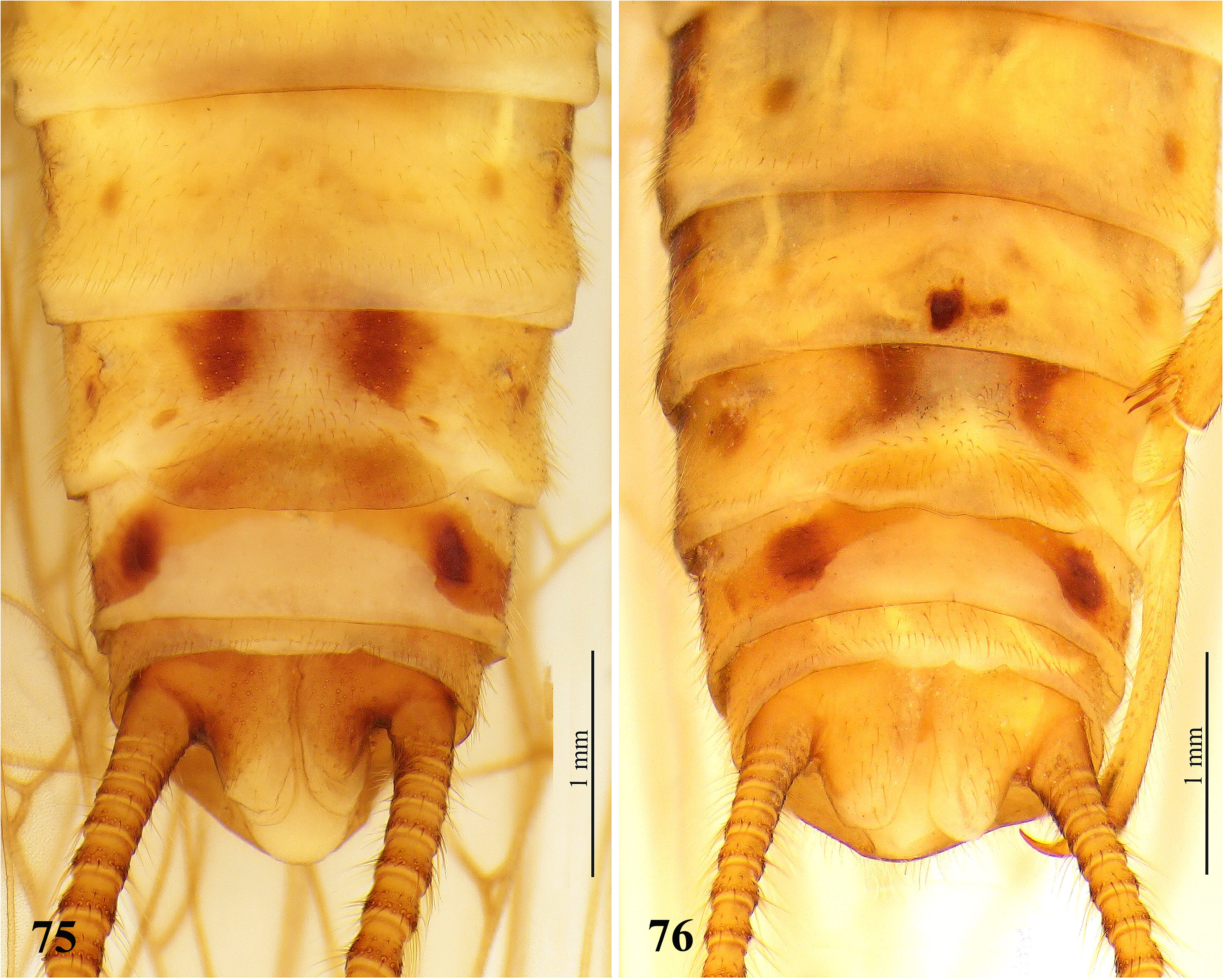
Female. Body length 17.8‒21.5 mm (n=5). Sterna 1‒7 yellow with a thin brownish band anteriorly and a pair of small brownish spots laterally ( Figs. 75, 76 View FIGURES 75–76 ). Sternum 8 pale with a pair of rounded brown spots laterally and a pair of oblique dark brown lateral sclerites surrounding the brownish subgenital plate anterolaterally ( Figs. 57, 58 View FIGURES 55–58 , 75, 76 View FIGURES 75–76 ). Subgenital plate is short and wide, extending about three-quarters of the sternum 8 widths (along posterior margin) and about a quarter of the sternum 9 lengths, with an almost straight or slightly wavy posterior margin; the lobes are weakly expressed; the lateral edges are slightly truncated ( Figs. 58 View FIGURES 55–58 , 75,76). Subgenital plate covered with short brown setae ( Figs. 75, 76 View FIGURES 75–76 ). Sternum 9 medially pale with a pair of brown rounded spots laterally, turning into curved brown stripes directed upwards under the subgenital plate ( Figs. 58 View FIGURES 55–58 , 75, 76 View FIGURES 75–76 ). Sternum 10 is usual, pale. Paraprocts are dark brown at the base of cerci; tips are pale.
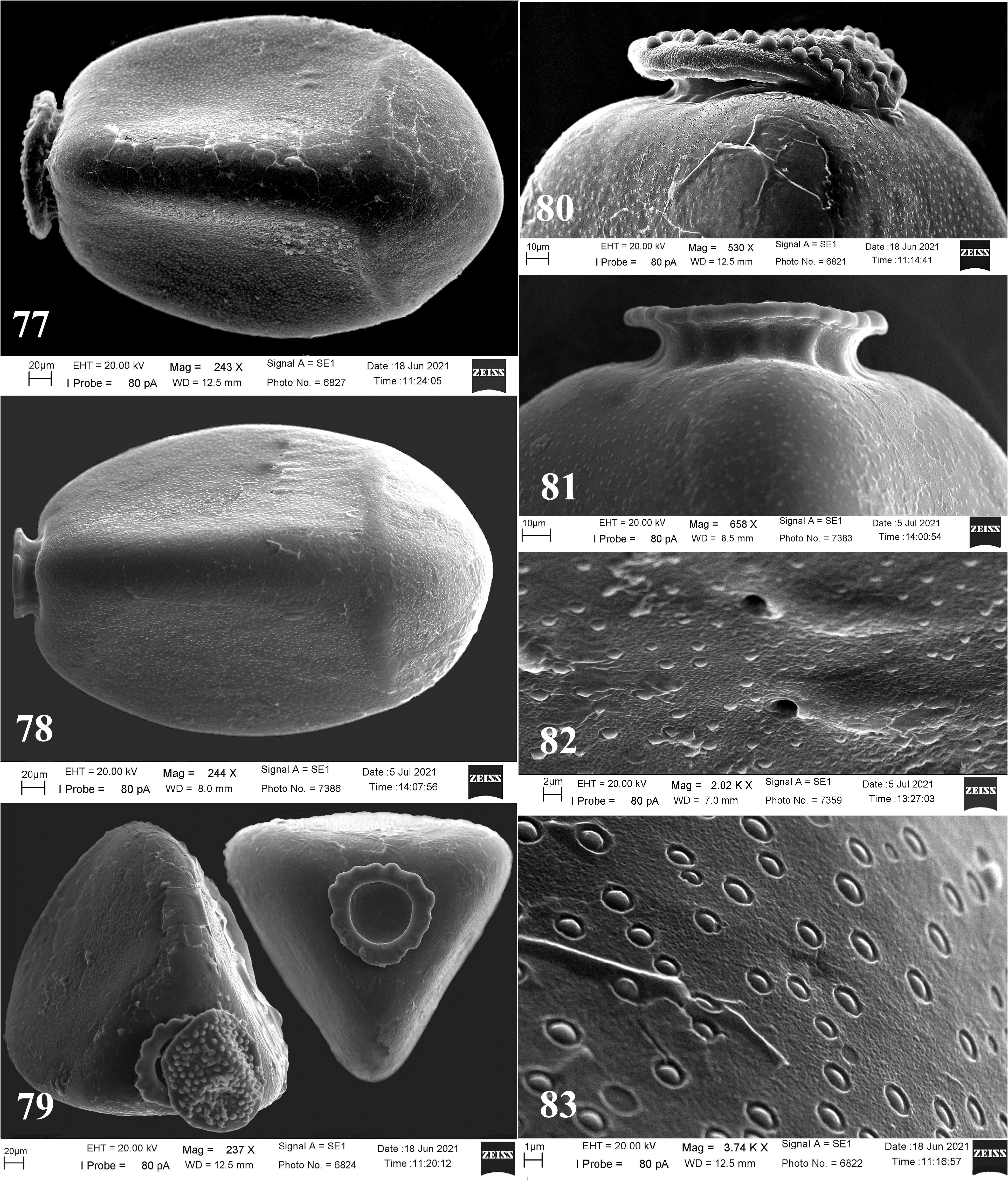
Egg. Medium size, trilateral, with mean dimensions of 416×274 μm (n=11). The transverse and longitudinal ridges are clearly visible, the longitudinal ridges are rather thick and bulged ( Figs. 59, 60, 64 View FIGURES 59–64 , 77, 78 View FIGURES 77–83 ). The collar is stalked, rim slightly triangular with a wavy outer edge, dorsally ( Figs. 61–63 View FIGURES 59–64 , 78, 79, 81 View FIGURES 77–83 ). The sides of the collar bear smooth longitudinal carinae ( Fig. 81 View FIGURES 77–83 ). The anchor plate mushroom-shaped or pan-shaped evenly covered with large globular bodies ( Figs. 77, 79, 80 View FIGURES 77–83 ). A transverse row of 4‒6 micropyles subequatorial, slightly closely to transverse ridge ( Figs. 59, 60 View FIGURES 59–64 , 77, 78 View FIGURES 77–83 ); micropyles clearly visible, their orifices without lips, some set close to micropyles mounds ( Fig. 82 View FIGURES 77–83 ). The chorion surface is rough with numerous flat rounded tubercles, located relatively evenly over the entire surface ( Figs. 59, 60 View FIGURES 59–64 , 77, 78, 82 View FIGURES 77–83 ). The base of the tubercles has a distinct groove ( Fig. 83 View FIGURES 77–83 ).
Material examined: Lectotype male (present designation), paralectotypes 1 male, 4 females, Uzbekistan, Samarkand city, vicinity, Zeravshan River Basin (authors note), 14 March, 1869, coll. Fedchenko A.P., det. McLachlan. Lectotype: № ZMMU Ple-0001, Zool. Mus. Mosq. Univ. ( Mosquae , ROSSIA) ( Fig. 50 View FIGURES 49–50 ). Uzbekistan, Western Tian-Shan , Talas Alataw , 3 males 3 females, Chirchiq River in the area of construction of the Charvan power station ( Syr-Darya River Basin ), 28.03.1968, ( ZIN). Tajikistan, Pamir-Alay mountain system, 2 males 2 females, Ramit Nature Reserve, Sardan-Miyona River, 17.03.1987, coll. L. Zhiltzova ( ZIN) .
Distribution. Filchneria olgae has an early spring emergence in March. It inhabits mountain watercourses of interfluves of the large Central Asian Amu-Darya and Syr-Darya Rivers at elevations of 700 m and above, the upper limit of the habitat has not been established. In Uzbekistan the species was found in the vicinity of Samarkand city, located in the middle reaches of the Zeravshan River (Amu-Darya R. Basin) and in the Chirchiq River (Syr-Darya R. Basin) in West Tian Shan, Talas Alataw. Also F. olgae was found in Tajikistan (State Natural Reserve "Ramit"), in the Sardai-Miyona River which flows down from the southern slopes of the Gissar Ridge of the Pamir-Alai Mountains, stretching over 200 km from the east to the west direction across Tajikistan and Uzbekistan.


No known copyright restrictions apply. See Agosti, D., Egloff, W., 2009. Taxonomic information exchange and copyright: the Plazi approach. BMC Research Notes 2009, 2:53 for further explanation.
|
Kingdom |
|
|
Phylum |
|
|
Class |
|
|
Order |
|
|
Family |
|
|
Genus |