
Brissopsis persica Mortensen, 1940
|
publication ID |
https://doi.org/10.11646/zootaxa.4624.2.13 |
|
publication LSID |
lsid:zoobank.org:pub:62BB8BB4-1059-4FDD-B7C8-9C4F9C088734 |
|
persistent identifier |
https://treatment.plazi.org/id/F2301C32-FFD6-FFCB-F2A1-9869529D7E44 |
|
treatment provided by |
Plazi |
|
scientific name |
Brissopsis persica Mortensen, 1940 |
| status |
|
Brissopsis persica Mortensen, 1940 View in CoL (transferred to genus Metalia by Coppard 2008)
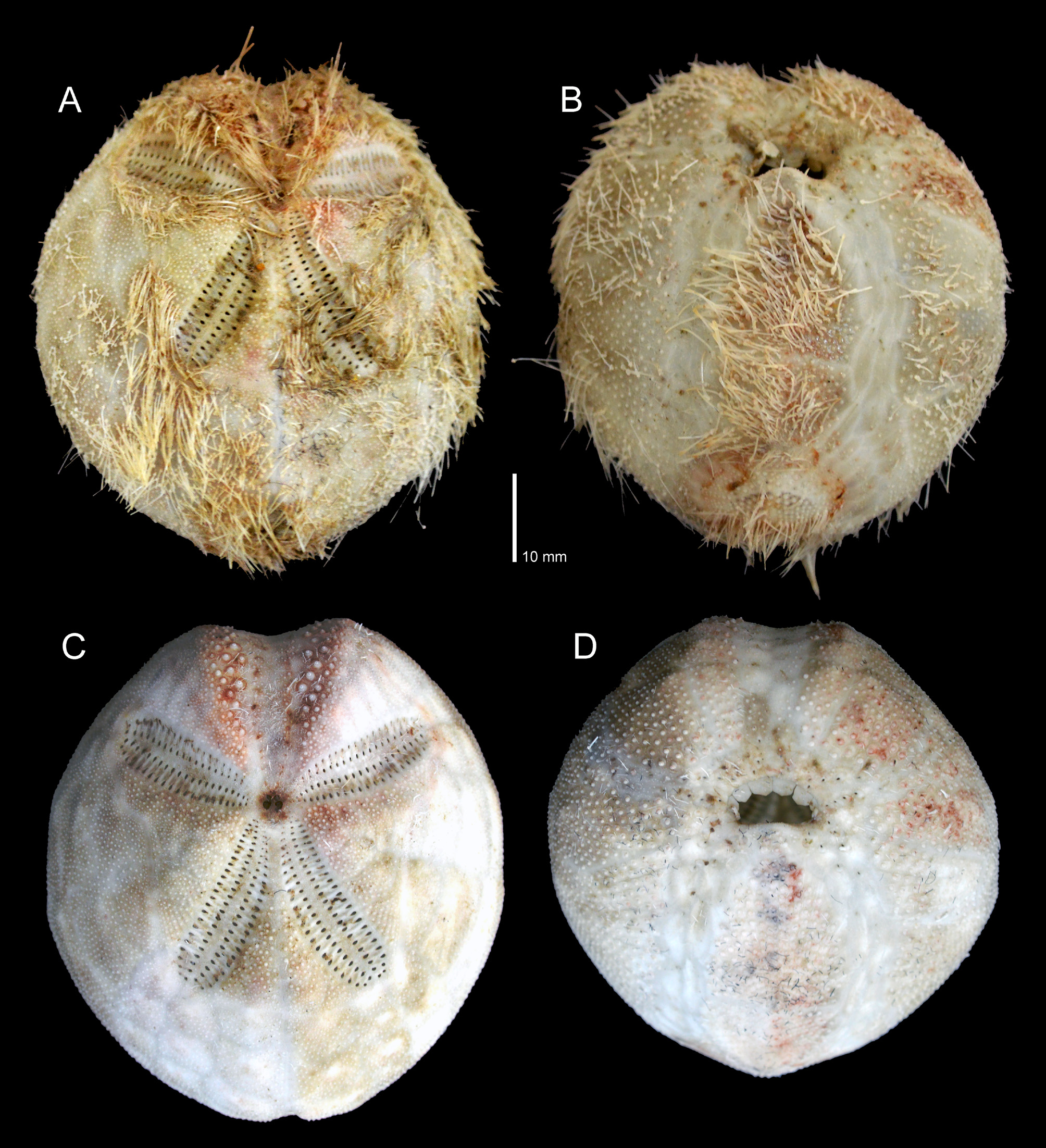
Figures 1 View FIGURE 1 , 2
Material examined: Single specimen from Chinna Neelankarai, Chennai Coast collected on April 5, 2018 ( Figs. 1 View FIGURE 1 , 2). Depth unknown, since this was not recorded for the bycatch that yielded the echinoid specimen.
Size: Test length 50 mm; test width 45 mm.
Colour: Specimen with spines light ochre/yellow-brown, reddish areas particularly associated with tube feet or musculature at bases of largest spines; cleaned test white/pale cream.
Description: Extremely fragile, broadly ovate, high test. Apical system slightly anterior, with four gonopores, anterior pair of gonopores slightly smaller than posterior pair. Anterior unpaired petal strongly sunken, lacking respiratory tube feet, unipores uniserial (Fig. 2C). Shallow notch where anterior unpaired ambulacrum passes around ambitus. Anterior paired petals slightly shorter than posterior petals, with approximately 21 pore-pairs in each column, each petal bending slightly toward the anterior end of the test ( Fig. 1C View FIGURE 1 ). Posterior paired petals with approximately 30 pore-pairs in each column. Posterior paired petals not coalescing adapically, though adapically, pore pairs of more medial column in each petal reduced relative to those of outer column (Fig. 2C). Respiratory pore pairs conjugate, outer pore more elongate than inner, reduced in size adapically (Fig. 2C). All paired petals slightly sunken.
Peristome situated anteriorly on oral surface, crescent-shaped ( Figs. 1B, D View FIGURE 1 , 2C). Labrum broad, in contact posteriorly with plastronal plates ( Fig. 1D View FIGURE 1 ), and in contact only with the first plate of the ambulacrals on each side. Peristomial membrane with five to six large plates along anterior border ( Fig. 1D View FIGURE 1 ), plates reduced in size proximal to mouth. Plastron amphisternous, bordered laterally on each side by narrow, laterally convexly curved ambulacra ( Fig. 1B View FIGURE 1 ). Periproct ovate, larger than peristome, situated high on vertical surface of posterior, not visible in oral view, elongated along anterior-posterior axis ( Figs. 1C View FIGURE 1 , 2D). Periproctal membrane containing plates bearing spines (Fig. 2D). Test convex along plastron. Two downward pointing, unpaired projections along “keel” of oral surface, one at junction of plastronal and sternal plates, and another at posterior edge of sternals.
Large, well-muscled locomotory spines on plastron, and on oral ambulacral regions lateral to ambulacra. Long, curved spines bordering and arching partly over sunken petaloids, strongly developed adapically over proximal part of unpaired anterior ambulacrum ( Fig. 1A View FIGURE 1 ). Oral ambulacral regions with sparse spination, particularly close to peristome ( Fig. 1B View FIGURE 1 ).
Narrow but conspicuous peripetalous fasciole ( Fig. 1C View FIGURE 1 ) running distal to each petal, and at the ambitus where it crosses unpaired anterior ambulacrum, fasciolar path curving adapically in paired interambulacra, but not in unpaired posterior interambulacrum. Well-developed, shield-shaped, subanal fasciole enclosing 5 pore-pairs on right side, 6 on the left. Branches of anal fasciole extend from aboral part of subanal fasciole on either side of periproct aboral about half-way up periproct (Figs. 2A, B).
We were able to find and trace the anal fasciole (Figs. 2A, B), considered to be a distinguishing feature of Metalia by some authors ( Mortensen 1951; Guevara-Plunkett & Mooi 2014), but not described for this species by Coppard (2008). The anal fasciole is readily apparent, though narrow, in the types of M. persica . Coppard (2008) placed Brissopsis persica in Metalia stating that the type species of Metalia , M. sternalis , lacks an anal fasciole. However, the second author of the present work has never seen a specimen of M. sternalis in which the anal fasciole (sensu Mortensen 1951; Clark & Rowe 1971; inter alia) was lacking, so the source of this statement is somewhat puzzling. In fact, all Metalia have an anal fasciole, a feature long used to identify members of that genus. In any case, Metalia persica does have an anal fasciole, as does the specimen described herein (Figs. 2A, B), so this is consistent with what Coppard (2008) determined for the various taxonomic rearrangements he suggested in his analysis, including the placement of Brissopsis persica in Metalia .
Distribution: Persian Gulf, Gulf of Kuwait, and India from depths of 10 to 40 metres.
No known copyright restrictions apply. See Agosti, D., Egloff, W., 2009. Taxonomic information exchange and copyright: the Plazi approach. BMC Research Notes 2009, 2:53 for further explanation.
|
Kingdom |
|
|
Phylum |
|
|
Class |
|
|
Order |
|
|
Family |
|
|
Genus |